R E S E A R C H
Open Access
HIVBrainSeqDB: a database of annotated HIV
envelope sequences from brain and other
anatomical sites
Alexander G Holman
1, Megan E Mefford
1, Niall O
’
Connor
2, Dana Gabuzda
1,3*Abstract
Background:The population of HIV replicating within a host consists of independently evolving and interacting
sub-populations that can be genetically distinct within anatomical compartments. HIV replicating within the brain causes neurocognitive disorders in up to 20-30% of infected individuals and is a viral sanctuary site for the development of drug resistance. The primary determinant of HIV neurotropism is macrophage tropism, which is primarily determined by the viral envelope (env) gene. However, studies of genetic aspects of HIV replicating in the brain are hindered because existing repositories of HIV sequences are not focused on neurotropic virus nor annotated with neurocognitive and neuropathological status. To address this need, we constructed the HIV Brain Sequence Database.
Results:The HIV Brain Sequence Database is a public database of HIV envelope sequences, directly sequenced
from brain and other tissues from the same patients. Sequences are annotated with clinical data including viral load, CD4 count, antiretroviral status, neurocognitive impairment, and neuropathological diagnosis, all curated from the original publication. Tissue source is coded using an anatomical ontology, the Foundational Model of Anatomy, to capture the maximum level of detail available, while maintaining ontological relationships between tissues and their subparts. 44 tissue types are represented within the database, grouped into 4 categories: (i) brain, brainstem, and spinal cord; (ii) meninges, choroid plexus, and CSF; (iii) blood and lymphoid; and (iv) other (bone marrow, colon, lung, liver, etc). Patient coding is correlated across studies, allowing sequences from the same patient to be grouped to increase statistical power. Using Cytoscape, we visualized relationships between studies, patients and sequences, illustrating interconnections between studies and the varying depth of sequencing, patient number, and tissue representation across studies. Currently, the database contains 2517 envelope sequences from 90 patients, obtained from 22 published studies. 1272 sequences are from brain; the remaining 1245 are from blood, lymph node, spleen, bone marrow, colon, lung and other non-brain tissues. The database interface utilizes a faceted interface, allowing real-time combination of multiple search parameters to assemble a meta-dataset, which can be downloaded for further analysis.
Conclusions:This online resource, which is publicly available at http://www.HIVBrainSeqDB.org, will greatly
facilitate analysis of the genetic aspects of HIV macrophage tropism, HIV compartmentalization and evolution within the brain and other tissue reservoirs, and the relationship of these findings to HIV-associated neurological disorders and other clinical consequences of HIV infection.
* Correspondence: [email protected]
1Department of Cancer Immunology and AIDS, Dana-Farber Cancer Institute,
Dana-Farber Cancer Institute, 44 Binney Street, Boston, Massachusetts, 02115, USA
Full list of author information is available at the end of the article
Introduction
The population of HIV replicating within a host consists of independently evolving and interacting sub-popula-tions, as demonstrated by the various degrees of phylo-genetic compartmentalization seen across and within anatomical compartments and various rates of decay in viral load during HAART therapy [1,2]. Several factors contribute to this genetic compartmentalization: (i) viral target cell tropism–HIV infects CD4+ T cells and macrophages in the periphery, and primarily infects macrophages and microglia (and rarely, astrocytes) in the brain [3]; (ii) viral adaptation in response to immune selection pressures that differ between anatomical com-partments [3,4]; (iii) physical barriers such as the blood-brain barrier [5]; and (iv) variable antiretroviral drug penetration into different tissues [6,7]. An important viral sub-population is HIV replicating within the brain [8-10]. HIV replicating in the brain causes neurocogni-tive and neuropathological disorders in up to 20-30% of infected individuals, particularly in later stages of dis-ease; in the era of HAART, HIV-associated neurocogni-tive disorders (HAND) have emerged as a significant cause of mortality and morbidity [4,6]. Additionally, the brain is a sanctuary site for the development of drug resistance, because poor antiretroviral drug penetration into the CNS leads to sub-therapeutic drug concentra-tions and incomplete suppression of viral replication [6]. The primary determinant of HIV neurotropism is macrophage tropism, which is primarily determined by genetic variation in the viral envelope (env) gene [8]. Phylogenetically related populations of tro-pic virus are found across brain and other macrophage-rich tissues, such as lung and bone marrow [11,12]. Thus, studies of the genetics of HIV replicating in the brain are pertinent to important clinical aspects of HIV, as well as the biology of the virus replicating within spe-cific anatomical compartments.
There are several excellent existing repositories of HIV sequences in the public domain, two of the most widely used being Genbank at the NCBI [13] and the HIV Sequence Database at the Los Alamos National Laboratory (LANL) (http://hiv.lanl.gov). However, neither is focused on neurotropic virus nor contains clinical annotations of neurocognitive and neuropatholo-gical diagnosis. Though more than 20 publications have clonally sequenced HIVenvfrom the brain, assembling a meta-dataset of these sequences presents significant technical challenges. To address these challenges, we constructed the HIV Brain Sequence Database (HBSD), the first comprehensive database of HIV envelope sequences clonally sequenced from brain and non-brain tissues, which is publicly available at http://HIVBrain-SeqDB.org
The HIV Brain Sequence Database
The HBSD contains 2517 envelope sequences from 90 patients. Sequences were obtained from 22 published studies (Table 1) ranging in publication date from 1991 to 2009 and in number of sequences per publication from 1 to over 700. 1272 of these sequences are brain-derived; the remaining approximately 1245 are derived from blood, lymph node, spleen, bone marrow, colon, lung and other non-brain tissues. 44 independent tissue types are represented within the database. These tissue types are grouped into 4 categories: (i) brain, brainstem, and spinal cord; (ii) meninges, choroid plexus, and CSF; (iii) blood and lymphoid; and (iv) other (bone marrow, lung, liver, etc) (Table 2). Figure 1 shows the database sequence content aligned to the envgene of HXB2. V3 region and near full-length gp120 region sequences comprise the majority of the database, with approxi-mately 1100 and 800 sequences, respectively. There are also approximately 200 near full-lengthenv sequences, 150 V4-V5 region, and 100 V1-V2 region. As new publi-cations emerge, facilitated by new sequencing technolo-gies, we expect the size of the HBSD to follow the
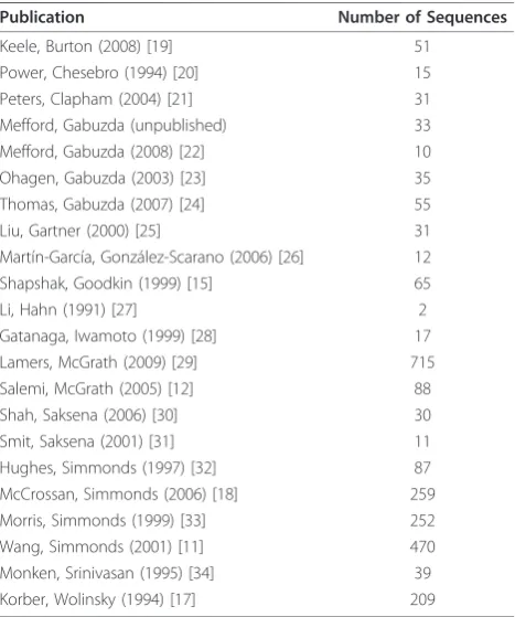
Table 1 Publications describing the cloning of sequences included in the HBSD
Publication Number of Sequences
Keele, Burton (2008) [19] 51
Power, Chesebro (1994) [20] 15
Peters, Clapham (2004) [21] 31
Mefford, Gabuzda (unpublished) 33
Mefford, Gabuzda (2008) [22] 10
Ohagen, Gabuzda (2003) [23] 35
Thomas, Gabuzda (2007) [24] 55
Liu, Gartner (2000) [25] 31
Martín-García, González-Scarano (2006) [26] 12
Shapshak, Goodkin (1999) [15] 65
Li, Hahn (1991) [27] 2
Gatanaga, Iwamoto (1999) [28] 17
Lamers, McGrath (2009) [29] 715
Salemi, McGrath (2005) [12] 88
Shah, Saksena (2006) [30] 30
Smit, Saksena (2001) [31] 11
Hughes, Simmonds (1997) [32] 87
McCrossan, Simmonds (2006) [18] 259
Morris, Simmonds (1999) [33] 252
Wang, Simmonds (2001) [11] 470
Monken, Srinivasan (1995) [34] 39
Korber, Wolinsky (1994) [17] 209
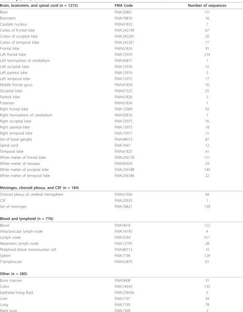
Table 2 Classification of tissues represented in the database, with their respective Foundational Model of Anatomy (FMA) codes
Brain, brainstem, and spinal cord (n = 1272) FMA Code Number of sequences
Brain FMA:50801 171
Brainstem FMA:79876 16
Caudate nucleus FMA:61833 7
Cortex of frontal lobe FMA:242199 67
Cortex of occipital lobe FMA:242205 20
Cortex of temporal lobe FMA:242201 77
Frontal lobe FMA:61824 91
Left frontal lobe FMA:72970 214
Left hemisphere of cerebellum FMA:83877 1
Left occipital lobe FMA:72976 12
Left parietal lobe FMA:72974 5
Left temporal lobe FMA:72972 17
Middle frontal gyrus FMA:61859 10
Occipital lobe FMA:67325 25
Parietal lobe FMA:61826 3
Putamen FMA:61834 1
Right frontal lobe FMA:72969 43
Right hemisphere of cerebellum FMA:83876 1
Right occipital lobe FMA:72975 16
Right parietal lobe FMA:72973 18
Right temporal lobe FMA:72971 15
Set of basal ganglia FMA:84013 87
Spinal cord FMA:7647 12
Temporal lobe FMA:61825 41
White matter of frontal lobe FMA:256178 111
White matter of neuraxis FMA:83929 29
White matter of occipital lobe FMA:256188 140
White matter of temporal lobe FMA:256186 22
Meninges, choroid plexus, and CSF (n = 184)
Choroid plexus of cerebral hemisphere FMA:61934 44
CSF FMA:20935 1
Set of meninges FMA:76821 139
Blood and lymphoid (n = 776)
Blood FMA:9670 122
Infraclavicular lymph node FMA:14193 4
Lymph node FMA:5034 417
Mesenteric lymph node FMA:12795 28
Peripheral blood mononuclear cell FMA:86713 15
Spleen FMA:7196 129
T-lymphocyte FMA:62870 61
Other (n = 285)
Bone marrow FMA:9608 31
Colon FMA:14543 135
Epithelial lining fluid FMA:276456 5
Liver FMA:7197 34
Lung FMA:7195 78
exponential expansion seen by other sequence databases [13].
Collection and assembly of HIV sequences
The HBSD attempts to contain all available, published HIV sequences meeting stringent inclusion criteria. For inclusion in the HBSD, sequences must meet the follow-ing criteria: (i) be deposited in Genbank; (ii) include some portion of the HIV env region; (iii) be clonal, amplified directly from tissue; and (iv) be sampled from the brain, or sampled from a patient for which the HBSD already contains brain sequences. We identified sequences for inclusion both by searching the public
sequence databases–Genbank and the LANL
HIV sequence database–and by identifying publications that sequenced HIV from the brain. In several cases, we communicated directly with study authors to encourage deposition of sequences that had not been previously submitted to Genbank. Additionally, BLAST alignment was used to screen for possible contamination with commonly used lab strains (i.e., ADA, HXB2, JR-CSF, NL4-3, SF2, BaL, IIIB, MN, SF162, and JR-FL)
Annotation Structure
The HIV Brain Sequence Database contains three categories of annotations: publication references, patient and sampling information, and sequence properties (Table 3). The publication annotations include biblio-graphic information identifying the study that generated the sequences. Patient sampling annotations contain information describing the individual patients, as well as clinical information at the time of sampling. This infor-mation was obtained by manual curation of the original publications and in some cases direct communications with the study authors. In cases where multiple studies examined tissue samples from the same patient, the resulting sequences are linked to the same patient code to increase statistical power. Sample timepoint annota-tions describe the patient’s clinical health status, neuro-cognitive, neuropathological status, CD4 counts, viral load, and anti-retroviral treatment history at the time of sampling. Clone and sequence annotations describe the individual sequences, the tissue from which they were cloned, and the method of PCR amplification and clon-ing. This includes the sequence start and end locations numbered based on alignment to the HXB2 reference genome, and tissue source coded using terms from a formal anatomical ontology. Alignment to HXB2 was performed using the HIV Sequence Locator tool located at the LANL HIV Sequence Database (http://hiv.lanl. gov). Currently, amplification and cloning methods included in the database are: bulk PCR then cloning (1736 sequences) and limiting-dilution PCR then cloning (781 sequences). As new sequencing projects are
completed, we hope to expand the database to include significant numbers of sequences cloned via single gen-ome amplification.
Annotation of Tissue Type
Annotation of tissue source presented several challenges. First, the granularity of tissue annotation varied by pub-lication–we encountered tissue type annotations as gen-eral as “Brain” and as specific as “White matter of occipital lobe”. However, within the HBSD a search for a more general tissue type, such as cerebrum should also return sequences from sub-parts of the cerebrum, such as caudate nucleus and putamen. Second, publica-tions utilize non-standard tissue names that are human-readable but difficult to parse in a database search. To address these challenges, we utilized a formal anatomical ontology, the Foundational Model of Anatomy (FMA) to code tissue source [14]. The FMA defines terms for approximately 75,000 human anatomical structures, ran-ging in scale from biological macromolecules to whole organ systems. These terms are linked by ontological relationships defining subpart relationships, allowing the calculation of transitive closure within the database. In addition, we assigned sequences into one of four classes: (i) Brain; (ii) Meninges, choroid plexus, and CSF; (iii) Blood and lymphoid; and (iv) Other. Meninges, choroid plexus, and CSF were grouped separately from Brain because phylogenetic evidence suggests that the CSF represents an intermediate compartment, contain-ing virus from both the brain and periphery [8].“Other” includes organs such as lung, liver, stomach and pros-tate, bone marrow, and fluid samples such as lung epithelial lining fluid.
Annotation of Neurocognitive and Neuropathological Diagnosis
Diagnosis for patient 196 stated: “insufficient informa-tion for patient 196 for the diagnosis of HAD, though there was evidence for neuropsychiatric disease.”[15]. Given that we lacked the further information to meet the strict criteria for an ANI or MND diagnosis, we chose the more general NPI: unknown defined in Woods et al. 2004 [16]. Diagnoses for patients 1 through 6 stated,“Clinical material was obtained from six HIV-1 infected patients with significant neurological signs and symptoms requiring image-guided stereotactic brain biopsy for definitive diagnosis. ... Neurological signs and symptoms were consistent with the onset of global neu-rological dysfunction, with clinical evidence supporting acute rather than chronic HIV-1-associated neurological disease.”[17]. As an acute diagnosis, this does not fit the criteria for HAD, so it was annotated in the database as acute HIV encephalopathy [17].
Design and Implementation
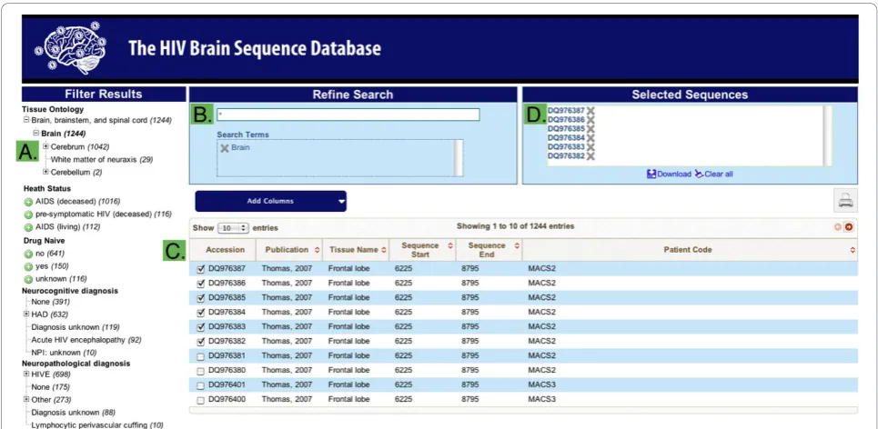
The HBSD structure is sequence-centric and uses NCBI GI and Genbank accession numbers as identifiers, sim-plifying correlations with other databases. The database exists in two forms. The master version is kept intern-ally as a relational SQL database utilized for sequence management and curation. This is replicated to an external interface that uses the Apache Solr search plat-form to optimize for flexible search and data retrieval. The search interface (Figure 2) is based on a filtering paradigm; the user begins with the set of all sequences and narrows by applying filtering criteria to the sequence annotations. Filtering criteria are specified by two means. A faceted search interface presents all values
for categorical annotations, such as tissue class or neu-rocognitive status. Clicking on a value adds it to the search criteria and filters for matching sequences. Addi-tionally, a global search box allows direct entry of search terms. Multiple searches in the global search box sequentially add filtering criteria, allowing the construc-tion of complex searches. Sequences are initially pre-sented with a default set of annotations, however, users can select to add or remove columns from the set of all annotations available. The final filtered set of sequences and annotations can be downloaded for local analysis in tab-separated and FASTA formats.
Visualization of the contents of the database
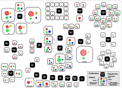
To better understand the highly complex network of publications, patients, and sequences, we used Cytoscape to visualize the connections between patients and the publications that sequenced virus from those patients (Figure 3). This network visualization demonstrates that, while most publications examine a unique set of patients, there is an emerging network of patients from the Edinburgh MRC HIV Brain and Tissue Bank (coded as NA#) that are shared among multiple publications. Additionally, Figure 3 illustrates the dramatic differences in sequencing depth between patients, and in number of patients between studies.
Many experimental designs examining compartmenta-lization or tissue specific effects depend on overlap in the viral regions sequenced and matched tissue source. In order to quantify the power of the database to make these comparisons, we visualized the total number of across-tissue and within-tissue comparisons possible with
6225 8795
0
500
1000
1500
2000
2
500
HXB2 numbering
S
equence number
gp120 gp41
V1 V2 V3 V4 V5 HR1 HR2 MSD Region
Coverage
99
)XOOOHQJWKHQY
99 99 9 9JS 9 99 9 99 JS RWKHU
Sequence Count
Figure 1 Sequence coverage of the HIVenvgene, numbered according to HXB2. Start and end coordinates are represented, but sequences are not internally aligned so gaps are not represented. The x-axis shows HXB2 nucleotide numbering with a schematic of theenv
the current database content (Figure 4). Panel A visua-lizes, for each tissue pair, how many patients contain overlapping sequences. Each comparison is ontologically inclusive–for example entries under Frontal lobe also consider sequences from White matter of frontal lobe, Cortex of frontal lobe, etcetera. This visualization reveals structures within the dataset useful for experimental design. For example, while a large number of patients contain overlapping sequences from lymph node and another tissue, in 8, 11, and 7 patients, respectively, it is possible to compare frontal lobe to occipital, temporal, or parietal lobes. Figure 4B is a complementary visualization counting the number of pairwise patient to patient
comparisons possible within each tissue type. This illus-trates, for example, that while many patients have over-lapping sequences from the cerebrum, frontal lobe is a particularly well-represented tissue. Conversely, though the database contains sequences from the cerebellum, there are no across patient comparisons that can be made. The numbers in both A and B of Figure 4 do not represent simple sums or permutations, because each considers sequence overlap. If hypothetical patients A, B, and C contained full-length env, V3 region, and V5 region sequences, respectively, then only 2 pair-wise comparisons would be possible (A to B and A to C), not the 3 given by a simple permutation.
Table 3 Annotation categories
Patient Column Definition
Patient code patient code
Sex gender
Risk factor HIV risk factor
Tissue bank tissue bank distributing samples
Patient year of death patient year of death
Sampling timepoint
Sampling geo-region patient geo-region at time of sampling
Sampling country patient country at time of sampling
Sampling city patient city at time of sampling
Patient age patient age at sampling
Health status patient health status at sampling
Subtype predominant subtype at time of sampling
Drug naïve (ART) has patient had ART
Antiretroviral treatment (ART) patient ART history
Viral load plasma (copies/mL) plasma viral load
Viral load brain (copies/million cells) brain viral load
Viral load lymphoid (copies/million cells) lymphoid viral load
CD4 count (cells/uL) CD4 count
Neurocognitive diagnosis neurocognitive diagnosis
Neuropathological diagnosis neuropathological diagnosis
Giant cells were giant cells present in the brain
Sequence
Genbank accession Genbank accession number
GI Genbank GI number
PubMed ID Pubmed ID for original publicaiton
Sequence length sequence length
Clone name publication assigned clone name
Cloning strategy methods of genome amplification and cloning
Sample tissue class global tissue class (Brain, Blood & Lymphoid, etc...)
Sample tissue name tissue source
Sample tissue FMA code tissue FMA code
Nucleic acid type was proviral DNA or viral RNA sequenced
Start and end coordinates sequence start and end referenced to HXB2
Discussion
The HBSD is a public database designed to facilitate the assembly of a large meta-dataset of HIVenv sequences that will be invaluable to investigations into the different patterns of viral evolution in the brain and other tissue reservoirs, and the relationship of these findings to each other and to clinical consequences of HIV infection, particularly development of HAND. The database con-tains 2517 envsequences cloned from 90 patients and 44 tissues sources. 1272 of these sequences are brain-derived; the remaining 1245 are derived from blood, lymph node, spleen, bone marrow, colon, lung, and other non-brain tissues. The majority of these sequences are from the V3 region (45%) or near full-length gp120 region (31%), with the remainder being near full-length
env (9%), V4-V5 region (6%), V1-V2 region (4%) and others (5%) (Figure 1). The HBSD is unique compared to other sequence databases, such as the LANL HIV Sequence Database or Genbank, because of its specific focus on HIV in the brain, its stringent inclusion of only clonal sequences from patients with brain sequences, and its rigorous curation with detailed clinical, patient, and HAND annotations.
An HIV env meta-dataset annotated with detailed clinical information will allow studies that previously have not been feasible. Combining datasets to increase the number of sequences and tissue-types increases the statistical power available. This increased statistical power can be used to examine questions such as the genetic variations withinenvimportant for macrophage tropism, which is the primary requirement for HIV replication in the brain, and nucleotide positions within
envunder positive genetic selection during HIV replica-tion in the CNS. Annotareplica-tion of neurocognitive status, neuropathological status, and AIDS progression will facilitate correlation of viral genotype to clinical pheno-types, and may help to reveal how viral genotypes affect the development of HAND.
During the assembly and annotation of the HBSD, we encountered a number of challenges. Non-uniform tis-sue coding made consistent database annotation diffi-cult. To overcome this obstacle, we utilized the FMA anatomical ontology to convert various tissue source descriptions into a set of defined terms with ontological linkages. We encountered several instances of ambigu-ous patient coding. Because tissue samples are shared
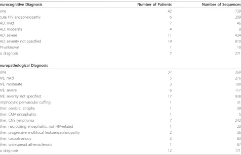
Table 4 Neurocognitive and neuropathological annotations in the database
Neurocognitive Diagnosis Number of Patients Number of Sequences
None 42 739
Acute HIV encephalopathy 6 209
HAD: mild 7 46
HAD: moderate 4 8
HAD: severe 11 424
HAD: severity not specified 19 810
NPI-unknown 1 10
No diagnosis 7 271
Neuropathological Diagnosis
None 37 369
HIVE: mild 5 276
HIVE: moderate 3 100
HIVE: severe 6 117
HIVE: severity not specified 17 938
Lymphocytic perivascular cuffing 1 31
Other: cerebral atrophy 1 39
Other: CMV encephalitis 1 5
Other: CNS lymphoma 7 242
Other: necrotizing encephalitis, not HIV-related 1 23
Other: progressive multifocal leukoencephalopathy 2 36
Other: toxoplasmosis 3 83
Other: widespread atherosclerosis 1 87
No diagnosis 12 171
within laboratories, and tissue banks distribute samples from the same patient to multiple laboratories, viruses from one patient may be sequenced in multiple publica-tions. By examining patient annotation data and corre-sponding with study authors, we identified 3 patients that were coded differently by multiple studies (NA118_p5, NA420_p6 and NA21_UK1) and 2 cases of separate patients that were coded identically by different studies (NA20 and NA234). Combining sequences from multiple publications and grouping by patient can increase the diversity of tissue types and the depth of sequencing available, while carefully tracking patient coding can avoid incorrect grouping of non-identical patients. Many publications included in the HBSD con-tain duplicate sequences cloned from the same tissue sample. These duplicate sequences could result either from PCR resampling in studies utilizing bulk PCR before cloning, or could represent valid cloning of copies of a majority viral variant. Fifteen publications utilized bulk PCR then cloning, 5 utilized limiting dilu-tion then cloning, and 2 used both approaches, based on patient. The database contains 490 repeated
sequences in 161 groups. However, 217 of these repeated sequences were obtained by limiting dilution PCR and therefore are unlikely to represent PCR resam-pling. Comparison of the distribution of the percentage of duplicated sequences between bulk PCR and limiting dilution demonstrated that studies utilizing bulk PCR then cloning did not show a higher rate of sequence duplication than those utilizing limiting dilution (data not shown). Thus duplicated sequences in the database likely represent appropriate cloning of majority viral variants.
The HBSD includes several unique datasets, which, though previously available in the public domain, are now collected in a standardized annotation format for meta-analysis. 15 patients included in McCrossan, 2006 [18] are pre-symptomatic, having died from HIV-unre-lated causes [alcohol/drug overdose (n = 11), cirrhosis (n = 2), suicide (n = 1), and bronchopneumonia (n = 1)]. During late-stage AIDS, declining CD4 counts lead to immune deficiency and reduced selection pres-sure, allowing viral population expansion that may alter the distribution of sequence variants. Based on
treatment history and year of death, the majority of patients in the HBSD died prior to the HAART era. 49 out of 90 patients have annotations for antiretroviral treatment history. Of these 49 patients, 19 are drug naïve and 30 received antiretroviral drugs. The majority of antiretroviral treated patients were on pre-HAART regimens, and 9 received only AZT. Different ART drugs have differing CNS penetration, affecting selection pressures on virus replicating in the brain [6]. Addition-ally, the majority of neurocognitive diagnoses occurred before the 2007 HNRC consensus document [4] that defined criteria for asymptomatic neurocognitive impair-ment (ANI). Future improveimpair-ment of the quality and relevance of the database to the current epidemic requires generating more sequences sampled from the brains of pre-symptomatic patients at earlier stages of disease and HAART-treated patients.
Our laboratory will continue to maintain the HBSD as new sequences are deposited in the public domain. We expect the HBSD to expand in several ways. New deep sequencing projects will increase the number of sequences and expand the diversity of patients, sampling a wider spectrum of stages of disease and HAART treatment regimens. Curation of patient coding may allow us to identify longitudinal sets of sequences sampled from the periphery, which can be paired with brain sequences sampled from the same patient at autopsy. Finally, we chose to focus on envfor the initial database release because it plays a key role in brain infection and provides a tractable scope for develop-ment of a highly curated database. As we consider further database additions, we will continue to weigh the benefits of inclusion against the resources required to maintain our high standards of database curation.
Tat and nefare two logical next steps, as these genes influence brain infection and development of neurocog-nitive disorders. Drug resistance mutations in pol and RT would also be a useful addition that will be consid-ered in the future.
Conclusions
The HBSD is a unique resource for the research com-munity investigating unique genetic and biological char-acteristics of HIV in the brain. Though nearly all the sequences and annotations included were previously
2 2 1 2 1 1 1 1 2 2 2
11 1 11 2 10 10 1 2 1 1 1 2 1 1 1 1 1 1 1 1 1 1 1 2 1 11 1 1 4
5 4 1 1 2 1 2 3 2 1 1 1
1 1 1 1
11 1 10 3 2 7 1 5 1 3 1 1 4 1 1 3 1 1 1 1 1 1 1 2 1 1 6 2 1 2 1 1 4
3 1 3 1 1 1 1 1 1 2 1 3 1 1 1
1 1 1 1 1 1
4
13 1 2 1 1 1 1 1 1 2 1 1 2 1 1 1
1 1 1 1 1 1 1 1 1 1
3 3 3 3 2
38 2 38 5 1 21 2 1 13 3 1 19 16 2 3 6 1 2 3 3 3 1 3 1 1 1 3 6 4 5 2 1 3 6 11 2
12 10 4 10 3 4 2 2 2 2 1 5 3 2 1 2 2 2 1 1 1 2 4 1 5 1 1 2
1 1 1 1 1 1
5 2 5 4 2 1 2 2 2 3 1 2 2 4 1 2 2 3 1 2 1 1 1 2 2 1 4 1 1 1 2 2
8 2 8 4 1 6 4 2 1 2 1 1 5 1 3 1 2 4 6 1 1 2 1 1 2
1 1 1 1 1 1 1 1
4 4 3 4 4 4 4 3 3 4 4 3 4 2 4 1 1 1 2 2 3 1 1
1 1 1 1 1 1 1 1 1 1 1 1 1 1 1 1 1 1 1 1 1 1
1 1 1 1 1 1 1 1 1 1 1 1 1 1 1 1 1 1 1 1 1 1 1
1 1 1 1 1 1 1 1 1 1 1 1 1 1 1 1 1 1 1 1 1 1
6 6 3 6 6 6 6 4 5 4 4 3 4 1 1 1 4 2 2 3 1 1
5 5 2 4 4 4 4 3 3 3 3 1 4 4 1 1 1 2 1 2 1 1 1
10 10 5 2 1 7 6 6 8 4 5 8 5 3 4 1 1 1 4 3 2 3 3 2 1
3 3 2 3 3 3 3 2 3 3 3 1 3 1 1 1 3 2 1 3 1 1
4 4 3 4 4 4 4 3 3 3 3 5 3 4 1 1 1 4 2 2 3 1 1
2 2 4 4 3 2 3 3 2 1
1 1 1 1 1 1 1 1
9 1 9 10 2 1 11 4 2 4 4 8 1 1 3 3 3 8 3 4 1 1 1 4 1 5 4 5 6 4 3 2 1
5 5 2 5 5 5 3 3 3 3 5 3 5 1 1 1 3 2 1 3 1 1
4 4 2 4 4 4 3 3 3 3 2 4 3 4 1 1 1 3 2 2 2 1 1
1 1 1 1 1
1 1 1 1 1 1 1 1 16 1 1
9 1 9 5 1 8 1 6 6 3 8 1 4 3 8 4 6 1 1 1 4 1 2 3 2 19 1 3 2 2
1 1 1
1 1 3 1 6 6 4 5 4 4 3 6 4 6 1 1 1 4 2 2 3 1 1
8 8 3 8 6 6 4 5 4 4 3 6 4 6 1 1 1 4 2 2 13 3 1 1 5 10
2 2 2 2 2 4 1 1
4 4 2 2 2 1 1 4 3 1 4 1 3 2 1 1 1
3 3 7 1 2 8 1 8 1 4 5 11 4 4 3 7 4 6 1 1 1 4 1 6 2 1 10 21 3 1 1 1 7 2 10 2
1 1 1 1 1 1 1
2 1 2 1 1 1 2 2 1 2 1 1 1 1 2 1
10 2 10 1 7 2 2 3 3 5 1 2 2 10 1 4 3 2 5 2 3 1 1 1 3 4 4 4 5 1 1 1 3 1 2 1 2 10 2 1 3 4 8 1 9 1 4 5 9 2 4 3 10 5 6 1 1 1 4 1 8 5 1 10 38 3 1 2 1 3 10 4 11 2
2 2 2 1 1 1 1 1 2 2 2 1 1 1 1 1
2 10 2 1 3 4 8 1 9 1 4 5 9 2 4 3 10 5 6 1 1 1 4 1 8 5 1 12 38 3 1 13 4 1 3 11 1 5 11 2 %UDLQ
:KLWHPDWWHURIQHXUD[LV &HUHEUXP 6HWRIEDVDOJDQJOLD &DXGDWHQXFOHXV 3XWDPHQ )URQWDOOREH :KLWHPDWWHURIIURQWDOOREH &RUWH[RIIURQWDOOREH /HIWIURQWDOOREH 5LJKWIURQWDOOREH 0LGGOHIURQWDOJ\UXV 2FFLSLWDOOREH :KLWHPDWWHURIRFFLSLWDOOREH &RUWH[RIRFFLSLWDOOREH /HIWRFFLSLWDOOREH 5LJKWRFFLSLWDOOREH 7HPSRUDOOREH :KLWHPDWWHURIWHPSRUDOOREH &RUWH[RIWHPSRUDOOREH /HIWWHPSRUDOOREH 5LJKWWHPSRUDOOREH 3DULHWDOOREH /HIWSDULHWDOOREH 5LJKWSDULHWDOOREH &HUHEHOOXP /HIWKHPLVSKHUHRIFHUHEHOOXP 5LJKWKHPLVSKHUHRIFHUHEHOOXP %UDLQVWHP 6SLQDOFRUG 6HWRIPHQLQJHV &KRURLGSOH[XVRIFHUHEUDOKHPLVSKHUH &6) 6SOHHQ /\PSKQRGH 0HVHQWHULFO\PSKQRGH ,QIUDFODYLFXODUO\PSKQRGH %ORRG 3HULSKHUDOEORRGPRQRQXFOHDUFHOO 7O\PSKRF\WH %RQHPDUURZ /XQJ 5LJKWOXQJ (SLWKHOLDOOLQLQJIOXLG &RORQ /LYHU %UDLQ :KLWHPDWWHURIQHXUD[LV &HUHEUXP 6HWRIEDVDOJDQJOLD &DXGDWHQXFOHXV 3XWDPHQ )URQWDOOREH :KLWHPDWWHURIIURQWDOOREH &RUWH[RIIURQWDOOREH /HIWIURQWDOOREH 5LJKWIURQWDOOREH 0LGGOHIURQWDOJ\UXV 2FFLSLWDOOREH :KLWHPDWWHURIRFFLSLWDOOREH &RUWH[RIRFFLSLWDOOREH /HIWRFFLSLWDOOREH 5LJKWRFFLSLWDOOREH
7
H
PSRUDOOREH
:KLWHPDWWHURIWHPSRUDOOREH &RUWH[RIWHPSRUDOOREH /HIWWHPSRUDOOREH 5LJKWWHPSRUDOOREH 3DULHWDOOREH /HIWSDULHWDOOREH 5LJKWSDULHWDOOREH &HUHEHOOXP /HIWKHPLVSKHUHRIFHUHEHOOXP 5LJKWKHPLVSKHUHRIFHUHEHOOXP %UDLQVWHP 6SLQDOFRUG 6HWRIPHQLQJHV &KRURLGSOH[XVRIFHUHEUDOKHPLVSKHUH &6) 6SOHHQ /\PSKQRGH 0HVHQWHULFO\PSKQRGH ,QIUDFODYLFXODUO\PSKQRGH %ORRG 3HULSKHUDOEORRGPRQRQXFOHDUFHOO
7
O\PSKRF\WH
%RQHPDUURZ /XQJ 5LJKWOXQJ (SLWKHOLDOOLQLQJIOXLG &RORQ /LYHU
1 55 10 55 3 6 78 3 703 66 10 28 6 15 10 45 3 10 10 136 10 6 120 300 15 120 17 190 1338 3 55 2714 1 4005
$
&RPSDULVRQVDFURVVWLVVXHVZLWKLQSDWLHQWV%
&RPSDULVRQVDFURVV SDWLHQWVZLWKLQWLVVXHVavailable in the public domain, the data did not exist in a well-annotated and accessible format and its assembly and curation represented a significant hurdle. The HBSD will be an invaluable resource for studying the viral genetics of HIV evolution within the brain and other tissue reservoirs, and the relationship of these findings to each other and to the development of HIV-associated neurocognitive disorders.
Acknowledgements
The authors wish to thank Mick Correll and Yaoyu Wang of The Center for Cancer Computational Biology, Dana-Farber Cancer Institute, Boston, MA for assistance with developing the database website and interface. We also thank the National NeuroAIDS Tissue Consortium (NNTC) for providing missing clinical data for some cases. The tissue source annotation is based on the FMA developed at the University of Washington by the FMATM Research Project and is provided under license from the University of Washington.
This work was supported by an ARRA supplement NIH/NIMH #3ROI MH83588-12S1 and the parent grant MH83588. MEM was supported in part by NIH fellowship 1F31NS060611-01. Core facilities were supported by the Harvard Center for AIDS Research and DFCI/Harvard Center for Cancer Research grants. The NNTC was supported by NIH funding through the NIMH and NINDS Institutes by the following grants: Manhattan HIV Brain Bank U01MH083501, R24MH59724 Texas NeuroAIDS Research Center U01MH083507, R24 NS45491 National Neurological AIDS Bank 5U01MH083500, NS 38841 California NeuroAIDS Tissue Network U01MH083506, R24MH59745, Statistics and Data Coordinating Center U01MH083545, N01MH32002. The funders and NNTC had no role in study design, data analysis, or preparation and submission of the publication.
Author details
1Department of Cancer Immunology and AIDS, Dana-Farber Cancer Institute,
Dana-Farber Cancer Institute, 44 Binney Street, Boston, Massachusetts, 02115, USA.2Center for Cancer Computational Biology, Dana-Farber Cancer Institute, Dana-Farber Cancer Institute, 44 Binney Street, Boston, Massachusetts, 02115, USA.3Department of Neurology, Harvard Medical School, 25 Shattuck Street, Boston, Massachusetts, 02115, USA.
Authors’contributions
AH designed the sequence database, assembled and curated sequences, performed all bioinformatic analysis, and drafted the manuscript. MM assembled and curated sequences and clinical data. NO designed and implemented the database interface. DG conceived of the study, participated in its design and coordination, and helped to draft the manuscript. All authors read and approved the final manuscript.
Competing interests
The authors declare that they have no competing interests.
Received: 9 November 2010 Accepted: 14 December 2010 Published: 14 December 2010
References
1. Simon V, Ho DD:HIV-1 dynamics in vivo: implications for therapy.Nature Reviews Microbiology2003,1:181-190.
2. Frost SD, Dumaurier MJ, Wain-Hobson S, Brown AJ:Genetic drift and within-host metapopulation dynamics of HIV-1 infection.Proc Natl Acad Sci USA2001,98:6975-6980.
3. Stevenson M:HIV-1 pathogenesis.Nature medicine2003,9:853-860. 4. Antinori A, Arendt G, Becker JT, Brew BJ, Byrd DA, Cherner M, Clifford DB,
Cinque P, Epstein LG, Goodkin K,et al:Updated research nosology for HIV-associated neurocognitive disorders.Neurology2007,69:1789-1799. 5. Ivey NS, MacLean AG, Lackner AA:Acquired immunodeficiency syndrome
and the blood-brain barrier.J Neurovirol2009,15:111-122. 6. McGee B, Smith N, Aweeka F:HIV pharmacology: barriers to the
eradication of HIV from the CNS.HIV Clin Trials2006,7:142-153.
7. Saksena NK, Potter SJ:Reservoirs of HIV-1 in vivo: implications for antiretroviral therapy.AIDS Rev2003,5:3-18.
8. Dunfee R, Thomas ER, Gorry PR, Wang J, Ancuta P, Gabuzda D: Mechanisms of HIV-1 neurotropism.Curr HIV Res2006,4:267-278. 9. González-Scarano F, Martín-García J:The neuropathogenesis of AIDS.Nat
Rev Immunol2005,5:69-81.
10. van Marle G, Power C:Human immunodeficiency virus type 1 genetic diversity in the nervous system: evolutionary epiphenomenon or disease determinant?J Neurovirol2005,11:107-128.
11. Wang TH, Donaldson YK, Brettle RP, Bell JE, Simmonds P:Identification of shared populations of human immunodeficiency virus type 1 infecting microglia and tissue macrophages outside the central nervous system.J Virol2001,75:11686-11699.
12. Salemi M, Lamers SL, Yu S, de Oliveira T, Fitch WM, McGrath MS: Phylodynamic analysis of human immunodeficiency virus type 1 in distinct brain compartments provides a model for the
neuropathogenesis of AIDS.J Virol2005,79:11343-11352.
13. Benson DA, Karsch-Mizrachi I, Lipman DJ, Ostell J, Sayers EW:GenBank.
Nucleic Acids Res2010,38:D46-51.
14. Rosse C, Mejino JLV:A reference ontology for biomedical informatics: the Foundational Model of Anatomy.J Biomed Inform2003,36:478-500. 15. Shapshak P, Segal DM, Crandall KA, Fujimura RK, Zhang BT, Xin KQ,
Okuda K, Petito CK, Eisdorfer C, Goodkin K:Independent evolution of HIV type 1 in different brain regions.AIDS Res Hum Retroviruses1999, 15:811-820.
16. Woods SP, Rippeth JD, Frol AB, Levy JK, Ryan E, Soukup VM, Hinkin CH, Lazzaretto D, Cherner M, Marcotte TD,et al:Interrater reliability of clinical ratings and neurocognitive diagnoses in HIV.J Clin Exp Neuropsychol
2004,26:759-778.
17. Korber BT, Kunstman KJ, Patterson BK, Furtado M, McEvilly MM, Levy R, Wolinsky SM:Genetic differences between blood- and brain-derived viral sequences from human immunodeficiency virus type 1-infected patients: evidence of conserved elements in the V3 region of the envelope protein of brain-derived sequences.J Virol1994,68:7467-7481. 18. McCrossan M, Marsden M, Carnie FW, Minnis S, Hansoti B, Anthony IC,
Brettle RP, Bell JE, Simmonds P:An immune control model for viral replication in the CNS during presymptomatic HIV infection.Brain2006, 129:503-516.
19. Keele BF, Tazi L, Gartner S, Liu Y, Burgon TB, Estes JD, Thacker TC, Crandall KA, McArthur JC, Burton GF:Characterization of the follicular dendritic cell reservoir of human immunodeficiency virus type 1.J Virol
2008,82:5548-5561.
20. Power C, McArthur JC, Johnson RT, Griffin DE, Glass JD, Perryman S, Chesebro B:Demented and nondemented patients with AIDS differ in brain-derived human immunodeficiency virus type 1 envelope sequences.J Virol1994,68:4643-4649.
21. Peters PJ, Bhattacharya J, Hibbitts S, Dittmar MT, Simmons G, Bell J, Simmonds P, Clapham PR:Biological analysis of human
immunodeficiency virus type 1 R5 envelopes amplified from brain and lymph node tissues of AIDS patients with neuropathology reveals two distinct tropism phenotypes and identifies envelopes in the brain that confer an enhanced tropism and fusigenicity for macrophages.J Virol
2004,78:6915-6926.
22. Mefford ME, Gorry PR, Kunstman K, Wolinsky SM, Gabuzda D:Bioinformatic prediction programs underestimate the frequency of CXCR4 usage by R5X4 HIV type 1 in brain and other tissues.AIDS Res Hum Retroviruses
2008,24:1215-1220.
23. Ohagen A, Devitt A, Kunstman KJ, Gorry PR, Rose PP, Korber B, Taylor J, Levy R, Murphy RL, Wolinsky SM, Gabuzda D:Genetic and functional analysis of full-length human immunodeficiency virus type 1 env genes derived from brain and blood of patients with AIDS.J Virol2003, 77:12336-12345.
24. Thomas ER, Dunfee RL, Stanton J, Bogdan D, Taylor J, Kunstman K, Bell JE, Wolinsky SM, Gabuzda D:Macrophage entry mediated by HIV Envs from brain and lymphoid tissues is determined by the capacity to use low CD4 levels and overall efficiency of fusion.Virology2007, 360:105-119.
25. Liu Y, Tang XP, McArthur JC, Scott J, Gartner S:Analysis of human immunodeficiency virus type 1 gp160 sequences from a patient with HIV dementia: evidence for monocyte trafficking into brain.J Neurovirol
26. Martín-García J, Cao W, Varela-Rohena A, Plassmeyer ML, González-Scarano F:HIV-1 tropism for the central nervous system: Brain-derived envelope glycoproteins with lower CD4 dependence and reduced sensitivity to a fusion inhibitor.Virology2006,346:169-179. 27. Li Y, Kappes JC, Conway JA, Price RW, Shaw GM, Hahn BH:Molecular
characterization of human immunodeficiency virus type 1 cloned directly from uncultured human brain tissue: identification of replication-competent and -defective viral genomes.J Virol1991, 65:3973-3985.
28. Gatanaga H, Oka S, Ida S, Wakabayashi T, Shioda T, Iwamoto A:Active HIV-1 redistribution and replication in the brain with HIV encephalitis.Arch Virol1999,144:29-43.
29. Lamers SL, Salemi M, Galligan DC, de Oliveira T, Fogel GB, Granier SC, Zhao L, Brown JN, Morris A, Masliah E, McGrath MS:Extensive HIV-1 intra-host recombination is common in tissues with abnormal histopathology.
PLoS ONE2009,4:e5065.
30. Shah M, Smit TK, Morgello S, Tourtellotte W, Gelman B, Brew BJ, Saksena NK:Env gp120 sequence analysis of HIV type 1 strains from diverse areas of the brain shows preponderance of CCR5 usage.AIDS Res Hum Retroviruses2006,22:177-181.
31. Smit TK, Wang B, Ng T, Osborne R, Brew B, Saksena NK:Varied tropism of HIV-1 isolates derived from different regions of adult brain cortex discriminate between patients with and without AIDS dementia complex (ADC): evidence for neurotropic HIV variants.Virology2001, 279:509-526.
32. Hughes ES, Bell JE, Simmonds P:Investigation of population diversity of human immunodeficiency virus type 1 in vivo by nucleotide sequencing and length polymorphism analysis of the V1/V2 hypervariable region of env.J Gen Virol1997,78(Pt 11):2871-2882.
33. Morris A, Marsden M, Halcrow K, Hughes ES, Brettle RP, Bell JE, Simmonds P:Mosaic structure of the human immunodeficiency virus type 1 genome infecting lymphoid cells and the brain: evidence for frequent in vivo recombination events in the evolution of regional populations.J Virol1999,73:8720-8731.
34. Monken CE, Wu B, Srinivasan A:High resolution analysis of HIV-1 quasispecies in the brain.AIDS1995,9:345-349.
doi:10.1186/1742-6405-7-43
Cite this article as:Holmanet al.:HIVBrainSeqDB: a database of annotated HIV envelope sequences from brain and other anatomical sites.AIDS Research and Therapy20107:43.
Submit your next manuscript to BioMed Central and take full advantage of:
• Convenient online submission
• Thorough peer review
• No space constraints or color figure charges
• Immediate publication on acceptance
• Inclusion in PubMed, CAS, Scopus and Google Scholar
• Research which is freely available for redistribution