R E S E A R C H
Open Access
Mycotoxin binder improves growth rate in
piglets associated with reduction of toll-like
receptor-4 and increase of tight junction
protein gene expression in gut mucosa
Linghong Jin
1,2, Wei Wang
1,2, Jeroen Degroote
1,2, Noémie Van Noten
1,2, Honglin Yan
1,2, Maryam Majdeddin
1,2,
Mario Van Poucke
3, Luc Peelman
3, Anne Goderis
4, Kurt Van De Mierop
4, Ronny Mombaerts
4,
Stefaan De Smet
2and Joris Michiels
1*Abstract
Background:Deoxynivalenol (DON) is a mycotoxin produced byFusariumspecies in the field, commonly found in cereal grains, which negatively affects performances and health of animals. Mycotoxin binders are supposed to reduce the toxicity of mycotoxins.
Method:The effect of a mycotoxin binder (containing acid-activated bentonite, clinoptilolite, yeast cell walls and organic acids) on growth performance and gut health was studied. Hundred and twenty weaning piglets were allocated to 4 treatments, with 5 pens of 6 piglets each, arranged in a 2 × 2 factorial design: control diet; control diet with 1 kg/t binder; control diet with DON; and control diet with DON and 1 kg/t binder. From d0–14, the diet of DON-challenged groups was artificially contaminated with a mixture of DON (2.6 mg/kg), 3-acetyl-deoxynivalenol (0.1 mg/kg) and 15-acetyl-3-acetyl-deoxynivalenol (0.3 mg/kg), after which the total contamination level was reduced to 1 mg/kg, until d37. On d14, one pig from each pen was euthanized and distal small intestinal mucosa samples were collected for the assessment of intestinal permeability, and gene expression of tight junction proteins, toll-like receptor 4, inflammatory cytokines and intestinal alkaline phosphatase.
Results:After 37 d, there were no differences in growth performance between control and DON-challenged groups (P> 0.05). Nevertheless, groups that received diets with binder had a significantly higher average daily gain (ADG) and average daily feed intake (ADFI) for the first 14 d as well as for the whole period, compared to groups without binder (P≤0.05). Groups with binder in the diet also exhibited lower expression of toll-like receptor 4 in distal small intestinal mucosa at d14, compared to groups without binder (P≤0.05). Interestingly, comparing the two DON treatments, piglets fed DON and binder had significantly higher ADFI and ADG compared to those with only DON for the first 14-d (P≤0.05). Addition of binder to DON contaminated diets, also down-regulated the gene expression of toll-like receptor 4 (P≤0.05) and increased mRNA level zona occludens 1 (P≤0.10) as compared to DON.
Conclusions:The present data provide evidence that the binder improves growth rate in piglets associated with reduction of toll-like receptor-4 and increase of tight junction protein gene expression. However, the current study does not allow to assess whether the effects of the binder are mediated by alterations in the toxicokinetics of the mycotoxin.
Keywords:Binder, Deoxynivalenol, Gut barrier, Gut health, Mycotoxin, Pigs
* Correspondence:[email protected]
1Department of Applied Biosciences, Ghent University, Valentin Vaerwyckweg
1, 9000 Ghent, Belgium
Full list of author information is available at the end of the article
Background
The contamination of feedstuffs with mycotoxins is a worldwide issue. Mycotoxins are harmful secondary metabolites of fungi which can cause intoxications at very low dosage. Deoxynivalenol (DON) is a type B
trichothecene mycotoxin, produced by Fusarium
spe-cies. DON is noted for two typical toxicological effects: reduced feed intake and induction of emesis in swine. Under natural conditions, DON is present along with its two major acetylated forms, 3-acetyl-deoxynivalenol
(3A–DON) and 15-acetyl-deoxynivalenol (15A–DON)
at lower concentrations than DON in cereals [1]. DON is physically stable and can easily enter the food chain [2]. Humans and all animal species can exhibit toxic effects after exposure to DON [3], with pigs being the most susceptible species [4].
The intestinal mucosa is constantly challenged by various chemical and biological contaminants, and functions as a vital barrier between the intestinal milieu and the luminal content [5, 6]. After consumption of feed contaminated with DON, the intestine can be ex-posed to high levels of the toxin [7]. Studies show that DON can affect the intestinal histology and morph-ology by affecting intestinal cell viability and prolifera-tion. DON is also able to regulate the production of pro-inflammatory cytokines, increasing the expression
of interleukin 1 beta (IL-1β), interleukin 2 (IL-2) and
interleukin 6 (IL-6) in the jejunum, and IL-1β, IL-6 and
tumor necrosis factor alpha (TNF-α) in the ileum in pigs
[8]. An in vitro study indicated that DON is able to decrease transepithelial electrical resistance (TEER) and increase the permeability of IPEC-1 cells in a dose and time dependent manner [7]. The alterations in these two parameters are related to the decreased expression of spe-cific tight junction proteins (TJPs) 9 [7, 9]. In the intestinal epithelium, the activation of mitogen-activated protein kinase (MAPK) by DON and its acetylated derivatives suppresses the expression of TJPs, which is responsible for the loss of barrier function [10, 11].
In order to solve the problem caused by mycotoxico-sis, various strategies have been developed, including physical, chemical, and biological methods [12, 13]. The most common approach is the addition of mycotoxin binders to feeds [14]. Mycotoxin binders are large weight molecules, capable of binding to mycotoxins in animal feeds. These binder-mycotoxin complexes pass through the gastrointestinal tract without dissociating, preventing mycotoxin uptake [15]. The complex passes through the GIT and is excreted via the faeces, thereby helping to minimize absorption of mycotoxins by target organs and alleviating the adverse effects of mycotoxins.
DON was the most prevalent single mycotoxin found in all feedstuffs all over the world in 2015 [16]. However, there are few publications about the effects of toxin
binders on gut health in piglets. This study was con-ducted to assess the effect of addition of a mycotoxin binder to the feed on gut health and performance in pigs following a 37-d dietary exposure to DON.
Methods
Animals and dietary treatments
An animal feeding experiment in a 2 × 2 factorial design with either or not addition of DON, and either or not addition of binder to the feed was performed. A total of 120 weaning (24 d of suckling period) piglets with an average weight of 7.3 kg were used in this study. They were provided with water and feed ad libitum through-out the experiment. Animal experimental procedures were in accordance to the guidelines of the Ethical Committee of the Faculty of Veterinary Sciences, Ghent University, Belgium.
Piglets were randomly allocated to 4 dietary treatments. Each treatment contained 5 pens with 6 piglets per pen. The 4 treatments were as follows: CON, negative control diet (uncontaminated basal diet); CON + BIN, negative control diet with 1 kg/t mycotoxin binder; DON, negative control diet with DON; DON + BIN, negative control diet with DON and 1 kg/t mycotoxin binder. The mycotoxin binder was a blend of indigestible adsorbents that bind mycotoxins in the GIT (Free-Tox, Nutrex NV, Belgium). It contains acid-activated bentonite, clinoptilolite, yeast cell walls and organic acids. From d 0 until d 14 (sampling on d 14) of the experiment, the diet of DON and DON + BIN was artificially contaminated with a mixture
of DON (2.6 mg/kg), 3A–DON (0.1 mg/kg) and 15A–
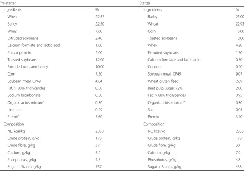
DON (0.3 mg/kg), after which the DON contamination level was reduced to 1 mg/kg from d 14 until d 37. The composition of the negative control wheat-barley-soybean based diets is given in Table 1. The DON-challenge diets were artificially contaminated with a fungal culture con-taining DON and its metabolites. DON was produced in
vitro by F. graminearum. After growing up of the mold,
the amount of DON was quantified by ELISA assay on the medium. The result was verified by LC/MS/MS on the certified standard blank wheat [17]. Results showed that the medium contained 240 mg/kg total DON metabolites
(87.5% DON, 2.7% 3A–DON and 9.8% 15A–DON). All
other mycotoxins were under the detection limit. Based on the concentration in the medium, the amount of medium needed for 3 mg/kg and 1 mg/kg in pre-starter and starter diets, respectively, was calculated. After homogenization, the medium was mixed into the basal diet. Three mg/kg for pre-starter period (from d 0 until d 14) was chosen because typically feed contamination with
2–5 mg/kg DON is required to induce reduction of feed
Sampling
After 14 d of feeding, one pig out of each pen was eutha-nized by intra-peritoneal pentobarbiturate overdose. The GIT was removed and the small intestine was exposed for sample collection. A 10-cm segment from the 75% length of the small intestine (distal small intestine) was collected for Ussing chamber measurements following flushing with saline to remove residual content. Another 20 cm segment from the same region was flushed with saline, placed on a cold plate and slit longitudinally. Then, mucosa was harvested by scraping with a glass-slide followed by snap
freezing and storage at −80 °C pending gene expression
analysis.
Growth performance
The weight of piglets as well as the feed intake per pen were determined at d0, d14 and d37. Average Daily Gain (ADG, g/d), Average Daily Feed Intake (ADFI, g/d) and Feed to Gain ratio (F:G) were calculated for periods
d0-d14, d14-d37 and d0-d37. Diarrhoea and mortality were daily checked and recorded.
Ex vivo measurement of intestinal permeability
Permeability was assessed ex vivo in Ussing chambers by measuring the permeability for the macromolecular marker fluorescein isothiocyanate-dextran 4 (FD4, mo-lecular weight 4 kDa) across sheets of mucosa as described by Wang et al. [19]. Briefly, fresh segments of mucosa samples from the distal small intestine (75% of the total small intestinal length) were separated from the seromus-cular layer and mounted in the Ussing chamber system. Intestinal sheets were bathed in 6.5 mL Ringer buffer solu-tion with 6 mmol/L glucose and 6 mmol/L mannitol in the serosal and mucosal sides, respectively. The system
was maintained at 37 °C and oxygenated (95% O2and 5%
CO2). After a 20-min equilibration period, 0.8 mg/mL
FD4 (Sigma-Aldrich, Bornem, Belgium) was added to the mucosal side. Samples from the serosal compartment
Table 1Composition of the negative control diet (CON) for pre-starter (d0-d14) and starter (d14-d37) periods
Pre-starter Starter
Ingredients % Ingredients %
Wheat 22.57 Barley 25.00
Barley 22.50 Wheat 22.93
Whey 7.00 Corn 15.00
Extruded soybeans 2.40 Toasted soybeans 12.00
Calcium formiate and lactic acid 1.00 Whey 4.20
Potato protein 2.00 Extruded soybeans 1.70
Toasted soybeans 12.00 Calcium formiate and lactic acid 0.50
Extruded oats and barley 10.00 Coconut 0.20
Corn 7.50 Soybean meal, CP49 9.07
Soybean meal, CP49 4.04 Wheat gluten feed 2.69
Fat, > 88% triglycerides 0.50 Beet pulp, sugar 72% 2.00
Sodium bicarbonate 0.30 Fat, > 88% triglycerides 0.95
Organic acids mixturea 0.30 Organic acids mixturea 0.30
Lime fine 0.29 Salt 0.05
Premixb 7.60 Premixc 3.40
Composition Composition
NE, kcal/kg 2350 NE, kcal/kg 2350
Crude protein, g/kg 173 Crude protein, g/kg 178
Crude fibre, g/kg 37 Crude fibre, g/kg 38
Calcium, g/kg 5.2 Calcium, g/kg 7.9
Phosphorus, g/kg 4.5 Phosphorus, g/kg 4.8
Sugar + Starch, g/kg 457 Sugar + Starch, g/kg 438
a
Organic acids mixture: contains formic acid, phosphoric acid and citric acid b
Providing per kg of complete diet: Vitamin A, 15,000 IU/kg; Vitamin D32000 IU/kg; Vitamin E, 200 IU/kg; Vitamin K3, 4.0 mg; Vitamin B1, 3.0 mg; Vitamin B2, 8.0 mg; Vitamin B3, 20 mg; Vitamin B6, 6.0 mg; Vitamin B12, 50.0μg; niacinamide, 40.0 mg; folic acid, 2.0 mg; biotin, 0.3 mg; Cu, 155 mg/kg; Fe, 150 mg/kg; Mn, 49 mg/kg; Zn, 104 mg/kg; I, 1.55 mg/kg; Se, 0.40 mg/kg
c
were taken at 20 min intervals for 80 min to monitor mucosal-to-serosal fluxes of FD4. Fluorescence inten-sity of FD4 was determined by fluorescence spectro-photometry (Thermo Fisher Scientific, Marietta, OH, USA). The flux over the 100 min period was calculated and expressed as an apparent permeability coefficient as described before [19].
RNA isolation and reverse-transcription quantitative real-time PCR
Relative mRNA expression of TJPs (ZO-1,ZO-2,OCLN,
CLDN-1, CLDN-2, CLDN-5, CLDN-7) and
pro-inflam-matory cytokines (TNF-α, IFN-γ, IL-1β,IL-8), toll-like
re-ceptor 4 (TLR-4) and a brush border enzyme intestinal
alkaline phosphatase (IAP) were determined by reverse
transcription quantitative real-time PCR (RT-qPCR) and performed according to the MIQE guidelines. Briefly, mu-cosal total RNA was extracted using the Bio-Rad Aurum Total RNA Fatty and Fibrous Tissue Kit (Bio-Rad Labora-tories, Inc., Hercules, CA, USA) according to the
man-ufacturer’s instructions, including an on-column DNase
I treatment to remove genomic DNA (gDNA). The
concentration and purity (OD260/280) of RNA were
measured with the NanoDrop ND-1000 (NanoDrop Technologies, Thermo Scientific, Wilmington, DE, USA).
1μg RNA was analyzed by 1% agarose gel electrophoresis
to check RNA integrity (28S and 18S rRNA bands). In addition to this assessment, a minus-RT control PCR was
performed usingYWHAZas primer to verify the absence
of any gDNA contamination. Following this,1μg of high
quality DNA-free RNA was reverse transcribed in the
20 μL reverse-transcription reaction with the ImProm-II
cDNA synthesis kit (Promega, Madison, WI, USA), con-taining both oligo dT and random primers. The obtained cDNA was diluted 10 times with molecular grade water
and a control PCR using 2 μL cDNA was performed to
verify the reverse-transcription reaction.
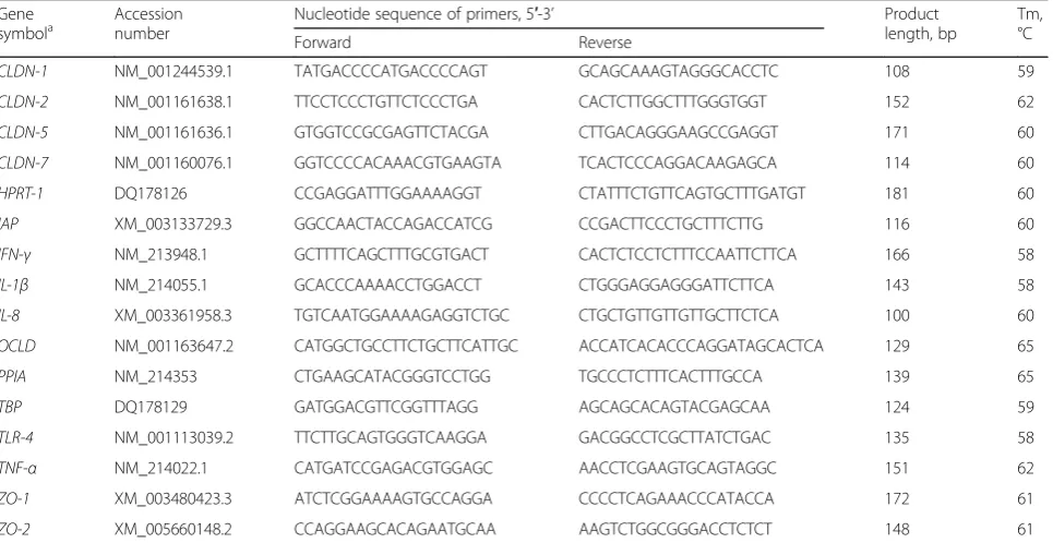
Primers (Table 2) used for genes in the study were designed with Primer3Plus. The repeats, the secondary structure and single nucleotide polymorphism in target sequence were checked with RepeatMarker, mfold and dbSNP, respectively. All these primer sequences were gene isoform specific as they were designed based on cer-tain exon-exon boundaries of published pig gene sequences corresponding to the accession number. Primers were then purchased from IDT (Integrated DNA Technologies, Leuven, Belgium).
The RT-qPCR was performed on the CFX96 Touch Real-Time PCR Detection System (Bio-Rad
Laborator-ies, Inc.). Briefly, 2 μL cDNA template, 5 μL 2× KAPA
SYBR FAST qPCR Kit Master Mix (Kapa Biosystems,
Inc., Wilmington, MA, USA), 2 μL molecular grade
water, 0.5μL forward primer and 0.5μL reverse primer
(5μmol/L each) were added to a total volume of 10μL.
The amplification conditions were as follows:1) enzyme activation and initial denaturation (95 °C for 3 min); 2) denaturation (95 °C for 20 s) and annealing/extension and data acquisition (annealing temperature depending
Table 2Primer sequences used for reverse-transcription quantitative real-time PCR
Gene
symbola Accessionnumber Nucleotide sequence of primers, 5′-3’ Productlength, bp Tm,°C
Forward Reverse
CLDN-1 NM_001244539.1 TATGACCCCATGACCCCAGT GCAGCAAAGTAGGGCACCTC 108 59 CLDN-2 NM_001161638.1 TTCCTCCCTGTTCTCCCTGA CACTCTTGGCTTTGGGTGGT 152 62 CLDN-5 NM_001161636.1 GTGGTCCGCGAGTTCTACGA CTTGACAGGGAAGCCGAGGT 171 60 CLDN-7 NM_001160076.1 GGTCCCCACAAACGTGAAGTA TCACTCCCAGGACAAGAGCA 114 60 HPRT-1 DQ178126 CCGAGGATTTGGAAAAGGT CTATTTCTGTTCAGTGCTTTGATGT 181 60
IAP XM_003133729.3 GGCCAACTACCAGACCATCG CCGACTTCCCTGCTTTCTTG 116 60
IFN-γ NM_213948.1 GCTTTTCAGCTTTGCGTGACT CACTCTCCTCTTTCCAATTCTTCA 166 58
IL-1β NM_214055.1 GCACCCAAAACCTGGACCT CTGGGAGGAGGGATTCTTCA 143 58
IL-8 XM_003361958.3 TGTCAATGGAAAAGAGGTCTGC CTGCTGTTGTTGTTGCTTCTCA 100 60
OCLD NM_001163647.2 CATGGCTGCCTTCTGCTTCATTGC ACCATCACACCCAGGATAGCACTCA 129 65
PPIA NM_214353 CTGAAGCATACGGGTCCTGG TGCCCTCTTTCACTTTGCCA 139 65
TBP DQ178129 GATGGACGTTCGGTTTAGG AGCAGCACAGTACGAGCAA 124 59
TLR-4 NM_001113039.2 TTCTTGCAGTGGGTCAAGGA GACGGCCTCGCTTATCTGAC 135 58
TNF-α NM_214022.1 CATGATCCGAGACGTGGAGC AACCTCGAAGTGCAGTAGGC 151 62
ZO-1 XM_003480423.3 ATCTCGGAAAAGTGCCAGGA CCCCTCAGAAACCCATACCA 172 61
ZO-2 XM_005660148.2 CCAGGAAGCACAGAATGCAA AAGTCTGGCGGGACCTCTCT 148 61
a
CLDN-1claudin-1,CLDN-2claudin-2,CLDN-5claudin-5,CLDN-7claudin-7,HPRT-1hypoxanthine phosphoribosyltransferase 1,IAPintestinal alkaline phosphatase,
on primer for 40 s) repeated 40 cycles; and 3) dissoci-ation (melt curve analysis from 70 to 90 °C with 0.5 °C increment every 5 s).
Primers used in this study were first optimized by gradient quantitative real-time PCR. A 5-fold dilution series (5 points, from 1 times to 625 times dilution) of cDNA as standard curve was included at 3 gradient temperatures to determine PCR amplification efficiency and specificity. The standard curve was also included in each run to determine PCR efficiency. In this study, PCR amplification efficiencies were consistently between 90% and 110%. Gene-specific amplification was verified by agarose gel electrophoresis and melting curve analysis. Efficiency was used to convert the Cq value into raw data with the highest expressed samples (lowest Cq value) as a calibrator for the normalization of raw data. The relative expression was expressed as a ratio of the target gene to the geometric mean of three stable expressed reference
genes (PPIA,HPRT1andTBP) [19].
Statistical analysis
After determination of normality and variance homo-geneity, a general linear model with the fixed effects of mycotoxin and binder, and the interaction term was
used with Tukey’s test as a multiple comparison test in
SAS Enterprise Guide 7 (SAS Institute, Cary, NC, USA).
P ≤ 0.05 was considered as significant. All data are
expressed as mean ± standard errors.
Principal component analysis (PCA) as described by Montagne et al. was conducted to work out the vari-ables that contributed most to the variation between subjects [20, 21]. In brief, the data of 17 variables were standardised before the application of PCA. At first, a scree plot was carried out to fix the number of princi-pal components to be maintained. Five principrinci-pal com-ponents were retained with the eigenvalues >1.0. In addition, variables that had a correlation coefficient
between variable and all principal components ≤0.5
were excluded. Then, retained variables were grouped into families to check the correlation. Only the main represen-tative variable with highest principal component loading,
together with high correlation (r> 0.55;P ≤0.05) within
family was retained for the final analysis. Finally, 11 variables entered the final PCA.
Results
Growth performances
Only few pigs from different groups had diarrhoea problems in the first week of the experiment, likely follow-ing weanfollow-ing stress. No case of emesis or mortality were observed. Overall, no clinical signs of toxicity were found. There were no significant differences between control groups (CON and CON + BIN) and DON-challenged groups (DON and DON + BIN) regarding growth
performances (Table 3). In contrast, pigs supplemented with binder (CON + BIN and DON + BIN) consumed more feed (265 g/d vs. 242 g/d) and had a higher growth (197 g/d vs. 170 g/d) for the first 14-d when compared to pigs that received diets with no binder (CON and DON)
(P ≤ 0.05). Similarly, for the whole experimental period
d0-d37, groups receiving diets with binder (CON + BIN and DON + BIN) showed an improved ADG (368 g/d vs. 341 g/d) and ADFI (548 g/d vs. 519 g/d) compared to groups that received diets without binder (CON and
DON) (P ≤ 0.05). Meanwhile, There was a trend that
groups that received diets with binder (CON + BIN and DON + BIN) had a lower F:G compared to groups that re-ceived diets without binder (CON and DON) from d 1
until d 14 of the experiment (P ≤ 0.10). Interestingly,
within DON-challenged piglets, addition of the binder im-proved performance in the first 14-d of the experiment; DON contaminated diet supplemented with binder (DON + BIN) showed higher ADFI compared to diet only contaminated with DON (DON) (272 g/d vs. 227 g/d)
(P≤0.05). This again resulted in a higher growth rate for
treatment DON + BIN than treatment DON for period
d0–14 (205 g/d vs. 159 g/d) (P≤0.05). Also, pigs
supple-mented with binder (DON + BIN) showed higher body weight at d14 compared to pigs that received diets with
no binder (DON) (10.18 kg vs. 9.63 kg) (P≤0.05).
Permeability measurements in distal small intestine Neither mycotoxin level, nor binder addition affected FD4 fluxes across distal small intestinal sheets
(P > 0.05) (Fig. 1). The mean of control groups (CON
and CON + BIN) was 7.5 × 10⁻⁷ cm/s; while the
average value of DON-challenged groups (DON and
DON + BIN) was 7.6 × 10⁻⁷cm/s. On binder level, the
difference of FD4 flux between groups that received diets with the addition of binder (CON + BIN and DON + BIN) and groups that received diets without the addition of binder (CON and DON) was larger compared to the difference between DON-challenged and DON-control groups but still not significant.
mRNA expression of tight junction proteins, inflammatory cytokines and brush border enzyme in distal small intestine
The gene expressions of TJPs (ZO-1, ZO-2, OCLN,
CLDN-1, CLDN-2, CLDN-5, CLDN-7), pro-inflammatory
cytokines (TNF-α, IFN-γ, IL-1β, IL-8),TLR-4 and IAP in
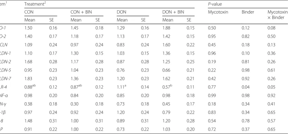
distal small intestine are described in Table 4. Ingestion of diets contaminated with or without DON, did not change the gene expression of TJPs, pro-inflammatory cytokines,
and IAP in distal small intestine (P > 0.05), whereas
add-ing the binder to the diets down-regulated the expression of TLR-4 (0.72 for CON + BIN and DON + BIN vs 1.00
was a trend that groups that received the diet with the addition of binder (CON + BIN and DON + BIN) up-regulated the expression of CLDN-1 compared to groups that received diet without the addition of binder
(CON and DON) (P≤0.10). More specifically,TLR-4gene
expression was down-regulated in the DON contaminated diet supplemented with mycotoxin binder (DON + BIN)
compared to DON (0.57 vs 1.11; P ≤ 0.05). At the same
time, there was a tendency that DON + BIN up-regulated
the expression ofZO-1compared to DON (P≤0.10).
Principal component analysis
BWd0, BWd14, ADG d0-d14, FD4 permeability, ZO-1, ZO-2, OCLN, CLDN-1, CLDN-2, CLDN-5, CLDN-7,
TLR4, TNF-α, IFN-γ, IL-1β, IL-8 and IAP were the 17
variables used in the PCA. After application of a first PCA, 5 principal components were retained following a scree plot. BWd0 was the only variable that did not show high correlation on any principal component and was excluded. Then, BWd14 and ADG d0-d14 were grouped into growth performance family, ZO-1, ZO-2, OCLN, CLDN-1, CLDN-2, CLDN-5 and CLDN-7 were
grouped into TJPs family, and TNF-α, IFN-γ, IL-1βand
IL-8 were grouped into inflammatory cytokines family. FD permeability, TLR-4 and IAP were considered single representatives and were retained for final analysis. Some variables were highly correlated within family. In growth performance family, ADG d0-d14 was highly
correlated with BWd14 (r = 0.966,P ≤ 0.01), yet ADG
d0–14 was not retained. Within the family of TJPS,
OCLN was correlated with ZO-1 (r= 0.762, P ≤ 0.01),
ZO-2 (r = 0.811, P ≤ 0.01) and CLDN-7 (r = 0.683,
P≤0.01). Then, OCLD, CLDN-1, CLDN-2 and CLDN-5
were retained for the final PCA. For the family of
Fig. 1Intestinal permeability for FD4 in distal small intestinal mucosa of piglets fed diets at d14 post-weaning. Data are means ± SE (n= 5). Pfor factor mycotoxin is 0.96, for factor binder 0.21, and for mycotoxin × binder 0.51. CON, negative control diet (uncontaminated basal diet); CON + BIN, negative control diet with 1 kg/t mycotoxin binder; DON, negative control diet with DON; DON + BIN, negative control diet with DON and 1 kg/t mycotoxin binder
Table 3Growth performance (body weight, BW; average daily gain ADG; average daily feed intake, ADFI, and feed:gain ratio, F:G) for periods d0-d14 (pre-starter), d14-d37 (starter) and d0-d37 (total) of piglets fed diets with or without mycotoxins, and with or without binder (n= 5)
Item Treatment1 P-value
CON CON + BIN DON DON + BIN Mycotoxin Binder Mycotoxin
× Binder
Mean SE Mean SE Mean SE Mean SE
Day 0–14 (pre-starter)
BW d0, g 7.30 0.01 7.31 0.01 7.32 0.01 7.30 0.01 0.52 0.85 0.49
BW d14, g 9.82ab 0.13 9.96ab 0.15 9.63b 0.13 10.18a 0.12 0.97 0.02 0.05
ADG, g/d 181ab 10 188ab 11 159b 10 205a 9 0.80 0.02 0.03
ADFI, g/d 256ab 10 257ab 12 227b 10 272a 10 0.50 0.05 0.05
F:G 1.43 0.04 1.38 0.05 1.43 0.04 1.33 0.04 0.64 0.08 0.25
Day 14–37 (starter)
BW d37, g 20.3 0.43 20.4 0.49 19.9 0.43 21.0 0.40 0.95 0.21 0.35
ADG, g/d 446 17 469 17 444 15 466 13 0.86 0.17 0.55
ADFI, g/d 724 21 760 23 716 21 750 19 0.68 0.11 0.44
F:G 1.59 0.03 1.62 0.03 1.62 0.03 1.62 0.03 0.68 0.55 0.83
Day 0–37 (total)
ADG, g/d 344 14 366 14 337 12 370 11 0.90 0.05 0.21
ADFI, g/d 527 14 548 16 511 14 549 13 0.59 0.05 0.19
F:G 1.50 0.03 1.50 0.03 1.51 0.03 1.49 0.02 0.84 0.63 0.92
Means with different superscripts (a, b) within row represent differences among treatments (P≤0.05) 1
CONnegative control diet (uncontaminated basal diet),CON + BINnegative control diet with 1 kg/t mycotoxin binder,DONnegative control diet with DON,
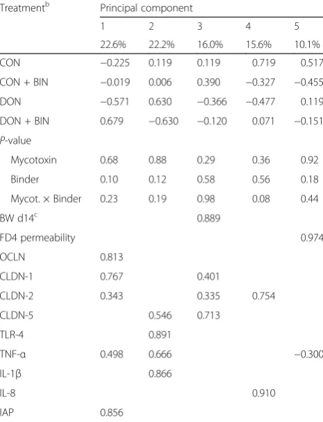
inflammatory cytokines, only IFN-γ was excluded. Finally, 11 variables were kept for this final PCA (Table 5). The 5 principal components explained 85.5% of the variance, of which the first principal component contributing 22.6% and the second principal compo-nent contributing 22.2%. The first principal compocompo-nent grouped the TJPs family members OCLN, CLDN-1,
CLDN-2 as well as TNF-α and brush border enzyme
IAP together. Principal component 1 had higher princi-pal component score in groups with addition of binder (CON + BIN and DON + BIN) compared to groups without addition of binder (CON and DON) (0.330 vs.
−0.398) (P ≤ 0.10). In other words, ingestion of diets
supplemented with binder tended to be associated with
higher gene expression of OCLN, CLDN-1, CLDN-2,
TNF-α and IAP as compared to diet without binder.
This finding is consistent with the gene expression
re-sult of CLDN-1 in Table 4. It supports the finding that
binder may also co-up-regulate the expression of other
TJPs (OCLN, CLDN-2) and IAP. The second principal
component indicates that the high expression ofTLR-4
was associated with high expression of pro-inflammatory
cytokines TNF-α and IL-1β. The third principal
compo-nent denotes that high expression of CLDN-1, CLDN-2
and CLDN-5 was related to higher weight at d14. How-ever, principal components 2 and 3 were not discrimin-atory for treatments.
Discussion
In the current experiment, mycotoxin contamination of piglet diets exhibited no effect on growth and gut health parameters. In contrast, the addition of a mycotoxin binder showed beneficial effects, in particular when diets were contaminated with 3 mg/kg of a mixture of DON and acetylated metabolites. Growth and feed intake were enhanced, in line with improvements of some selected gut health parameters.
Lack of effect of DON addition to feed on performance and gut health
In addition to lack of effect on growth performance, our results did not show an effect of DON on gut health in the distal small intestine, regarding intestinal permeability and mRNA expression of TJPs and pro-inflammatory cytokines, as well as IAP, after 14 d of feeding 3 mg/kg total DON. The lack of effect in the distal small intestine might be associated with the tox-icokinetic properties of DON. In vivo and in vitro studies demonstrated that DON and its acetylated forms are rapidly absorbed from the upper GIT, in-volving stomach until proximal jejunum. After chronic exposure to DON in pigs, a fast and almost complete absorption (> 90%) occurs, with DON appearing within 15 min in the blood and reaching maximal concentrations 1.65 h after oral exposure [22]. Danicke et al. revealed that
Table 4Relative mRNA expression of tight junction proteins, pro-inflammatory cytokines and intestinal alkaline phosphatase in distal small intestinal mucosa of piglets fed diets with or without mycotoxin contamination, and with or without binder at d14 post-weaning (n = 5)
Item1 Treatment2 P-value
CON CON + BIN DON DON + BIN Mycotoxin Binder Mycotoxin
× Binder
Mean SE Mean SE Mean SE Mean SE
ZO-1 1.50 0.16 1.45 0.18 1.29 0.16 1.88 0.15 0.50 0.12 0.08
ZO-2 1.40 0.17 1.18 0.17 1.13 0.17 1.42 0.15 0.95 0.82 0.50
OCLN 1.09 0.24 0.97 0.24 0.83 0.24 1.60 0.22 0.45 0.18 0.13
CLDN-1 1.10 0.17 1.30 0.15 1.03 0.15 1.36 0.15 0.96 0.10 0.36
CLDN-2 1.68 0.28 1.17 0.28 0.87 0.28 1.25 0.25 0.19 0.81 0.26
CLDN-5 0.95 0.23 1.04 0.23 0.76 0.23 0.66 0.21 0.22 0.98 0.61
CLDN-7 1.83 0.23 1.36 0.23 1.20 0.23 1.62 0.21 0.42 0.92 0.26
TLR-4 0.88ab 0.12 0.87ab 0.12 1.11a 0.14 0.57b 0.11 0.77 0.04 0.05
TNF-α 0.98 0.20 0.84 0.20 0.85 0.20 0.98 0.18 0.99 0.98 0.92
IFN-γ 0.38 0.18 0.30 0.18 0.73 0.18 0.45 0.17 0.18 0.34 0.41
IL-1β 0.97 0.24 0.92 0.24 1.20 0.24 0.79 0.22 0.83 0.34 0.65
IL-8 1.48 0.31 1.00 0.31 0.89 0.31 1.20 0.28 0.54 0.78 0.57
IAP 0.91 0.22 1.00 0.22 0.73 0.22 1.03 0.20 0.72 0.37 0.65
Means with different superscripts (a, b) within row represent differences among treatments (P≤0.05) 1
CLDN-1Claudin 1,CLDN-2Claudin 2,CLDN-5Claudin 5,CLDN-7Claudin 7,IAPIntestinal alkaline phosphatase,IFN-γInterferon, gamma,IL-1βInterleukin 1 beta, IL-8Interleukin 8,OCLNOccludin,TLR-4Toll like receptor 4,TNF-αTumor necrosis factor alpha,ZO-1Zona occludens 1,ZO-2Zona occludens 2
2
CONnegative control diet (uncontaminated basal diet),CON + BINnegative control diet with 1 kg/t mycotoxin binder,DONnegative control diet with DON,
88.5% of the DON dose was detected in the stomach whereas only 1.5% in the small intestine [23]. Also, the acetylated derivatives of DON are rapidly hydrolysed to DON in vivo and then absorbed. Thus, the distal small intestine might be less susceptible to DON as the ma-jority of DON is already absorbed in the proximal parts of the GIT [24]. All animal species can exhibit toxic effects when exposed to DON [3], with pigs being the most susceptible species [4]. However, the severity de-pends on various factors, including type and dose of DON, the route and duration of application, as well as the animal status [25]. After application of DON on the apical side or on the basolateral side of IPEC-J2 cells, Diesing et al. found that the apical epithelium seems to be more resistant to DON application while the same
concentration of DON from basolateral side severely impairs barrier integrity [9]. In our case, it can be as-sumed that most DON was absorbed in the upper GIT, reaching the more susceptible basolateral side whereby only a small part of DON was left in the less susceptible
apical side. That’s probably the reason why little effect
of DON administration on gut health was seen in sam-ples from the distal small intestine. Also, it should be taken into account that generally cytokine induction upon DON exposure occurs within hours of exposure [26], and thus differences after chronic exposure as in our study might not always be present.
So far, data about the effects of DON are incomplete, es-pecially due to the lack of in vivo data. At first, feed natur-ally contaminated with DON was used in studies in vivo and in vitro. However, interpretation of data from natur-ally contaminated diets is complicated as co-occurrence with other mycotoxins is commonly found in cereals. Mycotoxicoses may be caused by multiple toxins, making it difficult to unravel the separate effects of the target mycotoxin DON. At present, it is challenging to study the effect of DON and its masked mycotoxins ADONs. Puri-fied mycotoxins are generally used in in vitro experiments. In addition, it is difficult to correlate in vitro exposure with in vivo dosage as the amount of mycotoxin that can be absorbed in vivo does not necessarily correspond to the
amount absorbed by cells in culture. Taken together, it’s
difficult to conclude what dose DON will exhibit toxic properties, as the dose, the type of mycotoxin, the route and duration of exposure can all influence the mode of action.
Binder addition to the feed improves performance and some parameters of gut health
The mycotoxin binder used in this study was a combin-ation of acid-activated bentonite, clinoptilolite, yeast cell walls and organic acids and salt. Acid-activated bentonite increases the adsorption capacity by using a specific acid activation process to increase the surface area and to en-large pores [27, 28]. Clinoptilolite, with a honeycomb like structure, serves to bind a broad range of mycotoxins [29].
Yeast cell walls, which containα-D-mannans andβ-D
-glu-cans, have an active role in reducing mycotoxins in animal feed [30, 31]. Yeast cell walls used in the binder are ex-tracted and harvested in the early stage of the fermenta-tion, during which the network of covalent bonds is less dense, which offers more flexibility and a maximal
accessi-bility of the mycotoxin binding sites [32–34].
In the current study, the ingestion of diets
supple-mented with binder reduced the expression of TLR-4
compared to diets with no binder. TLR-4 plays an important role in recognizing Gram-negative bacteria and activation of the innate immune system. Activation of TLR-4 leads to the release of its downstream
Table 5Loadings and principal component scores for 5 principal components obtained by principal component analysisa(PCA) of 11 variables from piglets fed diets with or without mycotoxin contamination, and with or without binder at d14 post-weaning
Treatmentb Principal component
1 2 3 4 5
22.6% 22.2% 16.0% 15.6% 10.1%
CON −0.225 0.119 0.119 0.719 0.517
CON + BIN −0.019 0.006 0.390 −0.327 −0.455
DON −0.571 0.630 −0.366 −0.477 0.119
DON + BIN 0.679 −0.630 −0.120 0.071 −0.151 P-value
Mycotoxin 0.68 0.88 0.29 0.36 0.92
Binder 0.10 0.12 0.58 0.56 0.18
Mycot. × Binder 0.23 0.19 0.98 0.08 0.44
BW d14c 0.889
FD4 permeability 0.974
OCLN 0.813
CLDN-1 0.767 0.401
CLDN-2 0.343 0.335 0.754
CLDN-5 0.546 0.713
TLR-4 0.891
TNF-α 0.498 0.666 −0.300
IL-1β 0.866
IL-8 0.910
IAP 0.856
a
Rotation method: varimax with Kaiser normalisation; only correlations with |r| > 0.3 are indicated
b
CONnegative control diet (uncontaminated basal diet),CON + BINnegative control diet with 1 kg/t mycotoxin binder,DONnegative control diet with DON,DON + BINnegative control diet with DON and 1 kg/t mycotoxin binder. Principal components scores of subjects were analysed by General Linear model with fixed factor mycotoxin addition, binder addition and the interaction
c
BWbody weight,CLDN-1Claudin 1,CLDN-2Claudin 2,CLDN-5Claudin 5,FD4
FITC-dextran 4,IAPIntestinal alkaline phosphatase,IL-1βInterleukin 1 beta,IL-8
inflammatory modulators, including TNF-α and IL-1 [35]. This mechanism is well known and supported in the present study by the positive association between
TLR-4, TNF-α and IL-1β mRNA expression that was evident in principal component 2 from the PCA. TLR-4 is most well-known for recognizing lipopolysaccharides (LPS), a structural component of the outer membrane of Gram-negative bacteria. LPS induce strong inflam-matory responses in vivo, and are released when the cell is lysed or during bacterial cell division. Supple-mentation of toxin binders has shown reduced
expres-sion of pro-inflammatory cytokines such as IL-1β and
IL-6 and other immune responses in LPS-induced pigs [36]. In other words, the mycotoxin binder might not only bind to mycotoxins, but might also bind other toxins such as bacterial endotoxins. This was not spe-cifically investigated in the present study but might have occurred.
At the same time, there was a tendency that the sup-plementation of binder up-regulated the expression of
CLDN-1. Claudins function as major components of the tight junction strands that regulate the permeability of epithelia. CLDN-1, a member of the claudin family, is an
integral membrane protein. Enhanced CLDN-1
expres-sion can decrease paracellular permeability and tighten the tight junctions. As the mycotoxin binder in the current study may have adsorbed a range of toxins, it could be hypothesized that it adsorbed other xenobiotics which might impair the barrier function by deregulation of TJ assembly.
From the result of PCA, we know that groups with the supplementation of binder tended to have higher scores for principal component 1, which is positively
associated with gene expression of OCLN, CLDN-1,
CLDN-2 andIAP, as compared to diets without binder. This finding is consistent with the gene expression
result of CLDN-1. The PCA suggests that binder may
also up-regulate the expression of other TJPs (OCLN, CLDN-2) and IAP. OCLD, together with the claudin group of proteins, is an important component of the tight junctions. Studies have shown that rather than be-ing important in assembly and maintenance of tight junctions, OCLN is important in stability and barrier
function of tight junctions. As theOCLNgene is
essen-tial and plays a fundamental role in modulating the epithelial tight junctions, OCLN is a respective marker of epithelial barrier and its presence or absence could reflect the permeability of intestinal epithelium [37]. Taken the correlation into consideration, the binder may stimulate the expression of other TJPs as OCLN is highly correlated to ZO-1, ZO-2 and CLDN-7. The family of ZO is a part of the cytoplasmic plaque of the TJPs. The importance of maintenance of gut barrier integrity is further illustrated by the grouping of weight
at d14 and the mRNA levels of claudins within princi-pal component 3.
IAP is a brush border enzyme which is a component of the gut mucosal defence system. IAP is involved in regu-lating secretion of bicarbonate in the duodenum. Failure to neutralize acid environment can lead to acidified chyme injuring epithelial cells, finally increasing inflammation and intestinal permeability. IAP is also known to detoxify LPS and prevent bacterial translocation in the gut [38]. As discussed, LPS will induce strong inflammatory responses in vivo. In other words, IAP can inhibit the inflammatory responses by detoxification of LPS. So, IAP is an import-ant indicator to gut health.
Taken together, the addition of mycotoxin binder could improve the gut health by decreasing the expres-sion of TLR-4 as well as increasing the expresexpres-sion of TJPs and IAP.
Conclusions
The addition of a mycotoxin binder showed beneficial effects for weaned piglets, especially when diets were contaminated with 3 mg/kg of a mixture of DON in the pre-starter period. Growth and feed intake were enhanced. In line with this, reduced toll-like receptor-4 and increase of tight junction protein gene expression might shed light on the mode of action of the binder. However, the current study does not allow to assess whether the effects of the binder are mediated by alterations in the toxicokinetics of the mycotoxin.
Abbreviations
15A–DON:15-acetyl- deoxynivalenol; 3A–DON: 3-acetyl- deoxynivalenol; AC: Activated carbon; ADFI: Average daily feed intake; ADG: Average daily gain; ADONs: Acetylated-deoxynivalenols; AFB1: Aflatoxin B1; CLDN-1: Claudin 1; CLDN-2: Claudin 2; CLDN-5: Claudin 5; CLDN-7: Claudin 7; DON: Deoxynivalenol; F:G: Feed to Gain Ratio; FB1: Fumonisin B1; FD4: FITC-dextran 4; GIT: Gastrointestinal tract; HCK: Hematopoietic cell kinase; HPRT-1: Hypoxanthine Phosphoribosyltransferase 1; HSCAS: Hydrated sodium calcium aluminosilicate; IAP: Intestinal alkaline phosphatase; IFN-γ: Interferon gamma; Ig: Immunoglobulins; IL-1β: Interleukin 1 beta; IL-8: Interleukin 8; LPS: Lipopolysaccharide; MAPK: Mitogen-activated protein kinase; NIV: Nivalenon; OCLN: Occludin; OTA: Ochratoxin A; PCA: Principal component analysis; PPIA: Peptidylprolyl isomerase A; SAPK/JNK: Stress-activated protein kinases/cJun N-terminal kinases; TBP: TATA-binding protein; TJP: Tight junction protein; TLR-4: Toll like receptor 4; TNF-α: Tumor necrosis factor alpha; ZEA: Zearalenone; ZO-1: Zona occludens 1; ZO-2: Zona occludens 2
Acknowledgements
We thank Ms. Tessa Van Der Eecken for her technical support and animal care.
Funding
The research was supported by Nutrex company. The funders had no role in data collection and analysis, or drafting of the manuscript.
Availability of data and materials
All data generated or analysed during this study are included in this published article.
Authors’contributions
WW, MVP and LP were involved in the gene expression analysis. LJ analysed the data and wrote the first draft of the manuscript. All authors read and approved the final manuscript.
Ethics approval
Animal experimental procedures were approved by the Ethical Committee of the Faculty of Veterinary Medicine (Ghent University). All husbandry practices and euthanasia were performed with full consideration of animal welfare.
Consent for publication See consent form.
Competing interests
The authors declare that they have no competing interests, except AG, KVDM and RM. AG, KVDM and RM are employed by the funding company, however they had no role in data collection and analysis, or drafting of the manuscript. They approved the final manuscript.
Author details
1Department of Applied Biosciences, Ghent University, Valentin Vaerwyckweg
1, 9000 Ghent, Belgium.2Laboratory for Animal Nutrition and Animal Product
Quality, Department of Animal Production, Ghent University, Coupure Links 653, 9000 Ghent, Belgium.3Department of Nutrition, Genetics and Ethology,
Faculty of Veterinary Medicine, Ghent University, Heidestraat 19, 9820 Merelbeke, Belgium.4Nutrex, Achterstenhoek 5, 2275 Lille, Belgium.
Received: 28 March 2017 Accepted: 4 September 2017
References
1. EFSA. Opinion of the Scientific Panel on contaminants in the food chain on a request from the commission related to Deoxynivalenol (DON) as undesirable substance in animal feed. EFSA J. 2004;73:1–42. 2. Turner PC, Burley VJ, Rothwell JA, White KL, Cade JE, Wild CP.
Deoxynivalenol: rationale for development and application of a urinary biomarker. Food Add Contam. 2008;25:864–71.
3. Pestka JJ, Smolinski AT. Deoxynivalenol: toxicology and potential effects on humans. J Toxicol Environ Health-Part B-Crit Rev. 2005;8:39–69.
4. Eriksen GS, Pettersson H. Toxicological evaluation of trichothecenes in animal feed. Anim Feed Sci Technol. 2004;114:205–39.
5. Hecht G. Innate mechanisms of epithelial host defense: spotlight on intestine. Amer J Physiology-Cell Physiol. 1999;277:C351–8.
6. Podolsky D. Review article: healing after inflammatory injury–coordination of a regulatory peptide network. Alim Pharmacol Therap. 2000;14(Suppl1):87–93. 7. Pinton P, Nougayrède JP, Del Rio JC, Moreno C, Marin DE, Ferrier L, et al. The
food contaminant deoxynivalenol, decreases intestinal barrier permeability and reduces claudin expression. Toxicol Appl Pharmacol. 2009;237:41–8. 8. Bracarense AP, Lucioli J, Grenier B, Drociunas Pacheco G, Moll WD, Schatzmayr
G, et al. Chronic ingestion of deoxynivalenol and fumonisin, alone or in interaction, induces morphological and immunological changes in the intestine of piglets. Brit J Nutr. 2012;107:1776–86.
9. Diesing AK, Nossol C, Dänicke S, Walk N, Post A, Kahlert S, et al. Vulnerability of polarised intestinal porcine epithelial cells to mycotoxin deoxynivalenol depends on the route of application. PLoS One. 2011;6
10. Pestka JJ. Deoxynivalenol: mechanisms of action, human exposure, and toxicological relevance. Arch Toxicol. 2010;84:663–79.
11. Pinton P1, Braicu C, Nougayrede JP, Laffitte J, Taranu I, Oswald IP. Deoxynivalenol impairs porcine intestinal barrier function and decreases the protein expression of claudin-4 through a mitogen-activated protein kinase-dependent mechanism. J Nutr. 2010;140:1956–62.
12. Doyle MP, Applebaum RS, Brackett RE, Marth EH. Physical, chemical and biological degradation of mycotoxins in foods and agricultural commodities. J Food Prot. 1982;45:964–71.
13. Ramos AJ, Hernandez E. Prevention of aflatoxicosis in farm animals by means of hydrated sodium calcium aluminosilicate addition to feedstuffs: a review. Anim Feed Sci Technol. 1997;65:197–206.
14. Huwig A, Freimund S, Käppeli O, Dutler H. Mycotoxin detoxication of animal feed by different adsorbents. Toxicol Letters. 2001;122:179–88.
15. Döll S, Dänicke S. In vivo detoxification ofFusariumtoxins. Arch Anim Nutr. 2004;58:419–41.
16. Biomin, Biomin mycotoxin survey 2016. 2017. https://www.biomin.net/ ru/blog-posts/2016-biomin-mycotoxin-survey-results-for-swine-feed. Accessed 5 March 2017.
17. Monbaliu S, Van Poucke C, Detavernier C, Dumoulin F, Van De Velde M, Schoeters E, et al. Occurrence of mycotoxins in feed as analyzed by a multi-mycotoxin LC-MS/MS method. J Agri Food Chem. 2010;58:66–71. 18. Haschek WM, Rousseaux CG. Mycotoxins. Chapter 39 In Haschek and
Rousseaux’s Handbook Toxicol Pathol, 3rdEd. 2013;645:699.
19. Wang W, Degroote J, Van Ginneken C, Van Poucke M, Vergauwen H, Dam TM, et al. Intrauterine growth restriction in neonatal piglets affects small intestinal mucosal permeability and mRNA expression of redox-sensitive genes. FASEB J. 2016;30:863–73.
20. Montagne L, Boudry G, Favier C, Le Huërou-Luron I, Lallès JP, Sève B. Main intestinal markers associated with the changes in gut architecture and function in piglets after weaning. Brit J Nutr. 2007;97:45–57. 21. Michiels J, De Vos M, Missotten J, Ovyn A, De Smet S, Van Ginneken C.
Maturation of digestive function is retarded and plasma antioxidant capacity lowered in fully weaned low birth weight piglets. Brit J Nutr. 2013;109:65–75.
22. Goyarts T, Danicke S. Bioavailability of theFusariumtoxin deoxynivalenol (DON) from naturally contaminated wheat for the pig. Toxicol Letters. 2006;163:171–82.
23. Dänicke S, Valenta H, Klobasa F, Döll S, Ganter M, Flachowsky G. Effects of graded levels ofFusariumtoxin contaminated wheat in diets for fattening pigs on growth performance, nutrient digestibility, deoxynivalenol balance and clinical serum characteristics. Arch Anim Nutr. 2004;58:1–17.
24. Awad WA, Aschenbach JR, Setyabudi FM, Razzazi-Fazeli E, Böhm J, Zentek J. In vitroeffects of deoxynivalenol on small intestinal D-glucose uptake and absorption of deoxynivalenol across the isolated jejunal epithelium of laying hens. Poultry Sci. 2007;86:15–20.
25. Bondy GS, Pestka JJ. Immunomodulation by fungal toxins. J Toxicol Environ Health-Part B-Crit Rev. 2000;3:109–43.
26. Zhou HR, Islam Z, Pestka JJ. Rapid, sequential activation of mitogen-activated protein kinases and transcription factors precedes
proinflammatory cytokine mRNA expression in spleens of mice exposed to the trichothecene vomitoxin. Toxicol Sci. 2003;72:130–42.
27. Motlagh MK, Youzbashi AA, Rigi ZA. Effect of acid activation on structural and bleaching properties of a bentonite. Iran J Mat Sci Engineer. 2011;8:50–6. 28. Salem A, Karimi L. Physico-chemical variation in bentonite by sulfuric acid
activation. Kor J Chem Engineer. 2009;26:980–4.
29. Sabet FA, Libre NA, Shekarchi M. Mechanical and durability properties of self consolidating high performance concrete incorporating natural zeolite, silica fume and fly ash. Construct Building Mat. 2013;44:175–84.
30. Yiannikouris A, André G, Poughon L, François J, Dussap CG, Jeminet G, et al. Chemical and conformational study of the interactions involved in mycotoxin complexation withβ-D-glucans. Biomacromolecules. 2006;7: 1147–55.
31. Kogan G, Kocher A. Role of yeast cell wall polysaccharides in pig nutrition and health protection. Livest Sci. 2007;109:161–5.
32. Aguilar-Uscanga B, Francois J. A study of the yeast cell wall composition and structure in response to growth conditions and mode of cultivation. Letters Appl Microbiol. 2003;37:268–74.
33. Klis FM, Boorsma A, De Groot PW. Cell wall construction inSaccharomyces cerevisiae. Yeast. 2006;23:185–202.
34. Klis FM, Mol P, Hellingwerf K, Brul S. Dynamics of cell wall structure in Saccharomyces cerevisiae. FEMS Microbiol Rev. 2002;26:239–56. 35. Telepnev M, Golovliov I, Grundström T, Tärnvik A, Sjöstedt A.Francisella
tularensisinhibits toll-like receptor-mediated activation of intracellular signalling and secretion of TNF-αand IL-1 from murine macrophages. Cell Microbiol. 2003;5:41–51.
36. Collier CT, Carroll JA, Ballou MA, Starkey JD, Sparks JC. Oral administration of Saccharomyces cerevisiaeboulardii reduces mortality associated with immune and cortisol responses toEscherichia coliendotoxin in pigs. J Anim Sci. 2011;89:52–8.
37. Saitou M, Furuse M, Sasaki H, Schulzke JD, Fromm M, Takano H, et al. Complex phenotype of mice lacking occludin, a component of tight junction strands. Mol Biol Cell. 2000;11:4131–42.