Open Access
Case report
A case of sigmoid endometriosis difficult to differentiate from colon
cancer
Philippos Dimoulios
1, Ioannis E Koutroubakis*
1, Maria Tzardi
2,
Pavlos Antoniou
1, Ioannis M Matalliotakis
3and Elias A Kouroumalis
1Address: 1Department of Gastroenterology, University Hospital Heraklion, Crete, Greece, 2Department of Pathology, University Hospital
Heraklion, Crete, Greece and 3Department of Obstetrics and Gynecology, University Hospital Heraklion, Crete, Greece
Email: Philippos Dimoulios - fdimoul@hotmail.com; Ioannis E Koutroubakis* - ktjohn@her.forthnet.gr;
Maria Tzardi - tzardi_maria@yahoo.uk; Pavlos Antoniou - pantoniou_gr@yahoo.com; Ioannis M Matalliotakis - matakgr@yahoo.com; Elias A Kouroumalis - kouroum@med.uoc.gr
* Corresponding author
colon cancerendometriosisintestinal bleeding.
Abstract
Background: Although endometriosis with sigmoid serosal involvement is not uncommon in women of childbearing age, the mucosal involvement is rare and differential diagnosis from colon cancer may be difficult due to the lack of pathognomonic symptoms and the poor diagnostic yield of colonoscopy and colonic biopsies.
Case presentation: We present a case of a young woman with sigmoid endometriosis, in which the initial diagnostic workup suggested colon cancer. Histologic evidence, obtained from a second colonoscopy, along with pelvic ultrasound findings led to the final diagnosis of intestinal endometriosis which was confirmed by laparoscopy.
Conclusion: Colonic endometriosis is often a diagnostic challenge and should be considered in young women with symptoms from the lower gastrointestinal tract.
Background
Extrapelvic endometriosis refers to endometriotic implants located elsewhere in the body including the gas-trointestinal tract, urinary system, pulmonary system, CNS, skin and striated muscle [1,2]. The exact prevalence of extrapelvic endometriosis is unknown. The median age at the time of diagnosis is 34 to 40 years. Periodic hema-tochezia during the menses is a sign usually associated with intestinal endometriosis [3]. Yet, this catamenial character of bleeding and all other accompanying symp-toms, is very often absent, making the clinical history rather misleading [4]. Moreover, the differential diagnosis
of colonic endometriosis from other diseases of the colon is rather difficult due to the lack of pathognomonic symp-toms and the poor diagnostic yield of colonoscopy and colonic biopsies.
In this report we present the case of a young woman with intestinal endometriosis, in which the initial diagnostic workup suggested colon cancer.
Case report
A 35-year-old woman was admitted to the Department of Gastroenterology because of rectal bleeding and bouts of
Published: 07 August 2003
BMC Gastroenterology 2003, 3:18
Received: 15 May 2003 Accepted: 07 August 2003
This article is available from: http://www.biomedcentral.com/1471-230X/3/18
abdominal pain. Nine months before admission the patient began to have rectal bleeding that was related, at first, to the menstrual cycles. She also reported episodes of severe lower abdominal pain that was irrelevant to her menses and were accompanied by abdominal distention and constipation, especially during the last two months. Three weeks before admission she started to have almost daily hematochezia and small-caliber stools.
The patient had her menarche at the age of 13 years. Thereafter she had 27-day to 28-day menstrual cycles and menstrual periods lasting 6 to 7 days with normal blood loss. She had two normal labors at the age of 22 and 25. From her past medical history she reported tonsillectomy at the age of 11 and appendicectomy at the age of 18. She also reported cramping lower abdominal pain accompa-nying menstruation during the last three years. Her father suffered from hypertension. Her mother was healthy without reporting previous history of endometriosis or myomata uteri. Her brother was also healthy.
Physical examination revealed mild lower abdominal ten-derness. No masses were palpated. The bowel sounds were slightly increased. Rectal examination showed bright red blood, but no distinct mass. On the gynecological examination her vulva, vagina and cervix appeared to be normal. Her uterus had normal size and was anteverted. The laboratory work-up revealed a mild anemia (Hb: 11.3 gr/dL, Hct: 34.5%, MCV: 91.7 fL) with an increased white blood cell count (12000/µL: neut: 80.8%, lymph: 12.1%, mono: 4.6%) and ESR: 36 mm. Coagulation parameters, serum urea, creatinine, electrolytes, and liver function tests, were all within normal range.
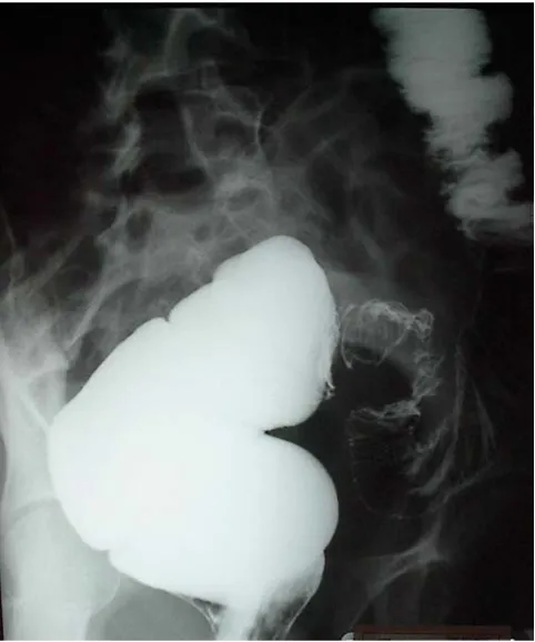
Colonoscopy revealed an extensive polypoid lesion of the mucosa, on the rectosigmoid junction, along with ery-thema, oedema, and ulcerations that resulted in stenosis of the lumen. The endoscope could not be introduced beyond the lesion. The histology of the above lesion dem-onstrated a mild dilation of crypts without goblet cell depletion. The lamina propria was edematous with dilated capillaries and inflammatory infiltrates of lym-phocytes and plasma cells. A barium enema (figure 1), on the following day, showed stenosis of the rectosigmoid region due to both a filling defect and an extrinsic bowel compression. The rest of the colon was normal. An abdominal CT scan, obtained two days later, revealed an eccentric wall thickening of the sigmoid and confirmed Barium enema showing stenosis of the rectosigmoid region
due to both a filling defect and an extrinsic bowel compression
Figure 1
Barium enema showing stenosis of the rectosigmoid region due to both a filling defect and an extrinsic bowel
compression.
Eccentric wall thickening of the sigmoid with filling defect Figure 2
the filling defect. Moreover, it showed ovarian cysts (Fig-ures 2,3).
Because of high suspicion of colon cancer a new colonos-copy was performed, in the 20th day of the menstrual cycle, in order to obtain more biopsy specimens. This time, colonic histology showed stroma and glands of endometrium suggestive of endometriosis (Figure 4).
Serum level of CA-125, that was measured in the 5th day of her menstrual cycle, was 210 IU/ml (normal< 35 IU/ ml), while CEA was 1.7 ng/ml (normal<5) and CA 19-9 was 6.1 IU/ml (normal<37). A pelvic ultrasound was per-formed and showed an anteverted homogeneous uterous. The right ovary had normal size with a small chocolate cyst (3 × 3.5 cm), while the left ovary had also normal size, but a larger chocolate cyst (4 × 4 cm). A laparoscopy performed a week later revealed stage IV endometriosis (American Fertility Society classification). The uterus was anteverted. Both the ovaries and the fallopian tubes were involved. The right ovary was found with a small choco-late cyst and the left ovary with a larger chocochoco-late cyst (4 × 5 cm). The cul-de-sac had extensive adhesions. An endometriotic implant was found on the sigmoid colon. All endometriotic lesions and adhesions were removed. The patient started treatment with a GnRH agonist (leu-proreline acetate, 3.75 mg/month IM).
Five months later a new colonoscopy was performed that showed only mild oedema of the sigmoid mucosa with disappearance of the stenotic lesion and easy introduction of the instrument to the caecum. The rest of the large bowel appeared to be normal. Serum level of CA-125 (7th day of menstrual cycle) was now normal (28 IU/ml). The patient was free of symptoms.
Discussion
The gastrointestinal tract is the most common site of extrapelvic endometriosis, affecting 5–15% of women with pelvic endometriosis [1,2]. Among women with intestinal endometriosis, rectum and sigmoid colon are the most common involved areas (75–90%). Other parts Eccentric wall thickening of the sigmoid with filling defect
Figure 3
Eccentric wall thickening of the sigmoid with filling defect. Note also the ovarian cysts
Large bowel mucosa with a focus of endometriosis in the lamina propria (stroma and glands of endometrium) Figure 4
of the bowel commonly affected are the distal ileum (2– 16%), and appendix (3–18%) [3]. Only the serosa and the muscularis propria are usually involved, while the mucosa is very rarely affected [4].
Intestinal endometriosis may present with rectal bleeding, bowel obstruction and rarely with perforation or malig-nant transformation [5,6]. Symptoms can be cyclical in about 40% of patients, can vary depending on the site and include crampy abdominal pain, distention, diarrhea, constipation, tenesmus and hematochezia [1]. The classic triad of dysmenorrhea, dyspareunia and infertility, as a result of concomitant pelvic disease, may also exist. We should, however notice, that the cyclical character of symptoms does not exclusively appear in endometriosis. It is well established that clinical manifestations in inflammatory bowel disease and irritable bowel syn-drome may aggravate during the menses [7,8].
The clinical, radiological and endoscopic picture may be confused with neoplasms, ischemic colitis, inflammatory bowel disease, post radiation colitis, diverticular disease and infection. Although endoscopic diagnosis of colonic endometriosis has been reported [9] usually the endo-scopic appearance, even if there is mucosal involvement, is not diagnostic. Biopsies obtained endoscopically usu-ally yield insufficient tissue for a definite pathologic diag-nosis [10]. Moreover, endometriotic deposits can induce secondary mucosal changes, which mimic findings of other diseases such as inflammatory bowel disease, ischemic colitis, or even a neoplasm [11,12]. The CT scan or barium enema usually demonstrate an extrinsic bowel compression, stenosis or filling defect. MRI seems to be the most sensitive imaging technique for intestinal endometriosis [13]. Yet, the gold standard for the diagno-sis is laparoscopy or laparotomy.
Treatment options include surgery or hormonal manipu-lations, depending on patient's age and desire to maintain fertility and also on the severity and complications of the disease [14]. Recently, laparoscopic treatment of colorec-tal endometriosis, even in advanced stages, has been proven feasible and effective in nearly all patients [15]. The medications used in the treatment of endometriosis are danazol, high dose progestins and GnRH agonists with almost equivalent efficacy [16]. The choice of which to use is based on side effects and costs. Danazol and GnRH agonists are of equivalent cost, but GnRH agonists are usually better tolerated. In our case, although high dose progestins could have been used, after discussion with the patient she chose to receive GnRH agonists.
Our patient represents a case of symptomatic gastrointes-tinal endometriosis with mucosal involvement, without a previous history of pelvic endometriosis. The symptoms
of abdominal pain, constipation and hematochezia, the presence of anemia in combination with the radiologic and endoscopic findings were suggestive of a neoplasm. On the other hand the patient's long history of dysmenor-rhea, the normal levels of CEA and CA19-9, and the absence of neoplastic infiltration in all biopsy specimens were against the diagnosis of colon cancer. Furthermore, the ovarian chocolate cysts showed by the pelvic ultra-sound and the histologic evidence, obtained from the second colonoscopy, directed us towards the diagnosis of intestinal endometriosis that was finally confirmed dur-ing the laparoscopy. Moreover, this patient had elevated serum levels of CA-125, which has been established as a useful marker for determining the severity of endometrio-sis [17,18]. The colonic mucosal involvement in this case could be explained by the invasion of endometrial cells through the bowel wall. Lymphatic or vascular metastasis could explain rare cases of endometriosis located in pleura, umbilicus, muscle, brain, vagina, cervix and retro-peritoneal space [1,2].
In conclusion, intestinal endometriosis is often a diagnos-tic challenge mimicking a broad spectrum of diseases and should be considered in any young woman with symp-toms from the lower gastrointestinal tract.
List of abbrevations
CEA, carcinoembryonic antigen; CNS, central neural sys-tem; CT, computer tomography; GnRH, gonadotropin-releasing hormone; MCV, mean corpuscular volume; MRI, magnetic resonance imaging;
Competing interests
None declared.Acknowledgements
Written consent was obtained from the patient for publication of the patient's details.
References
1. Jubanyik K and Comite F: Extrapelvic endometriosis. Obstet Gyne-col Clin North Am 1997, 24:411-40.
2. Schwartz JL and Schwartz LB: Extrapelvic endometriosis. In
Endometrium and Endometriosis Edited by: Diamond MP, Osteen KG.
Blackwell Science; 1997:247-254.
3. Miller LS, Barbarevech C and Friedman LS: Less frequent causes of lower gastrointestinal bleeding. Gastroenterol Clin North Am 1994, 23:21-52.
4. Levitt MD, Hodby KJ, van Merwyk AJ and Glancy RJ: Cyclical rectal bleeding in colorectal endometriosis. Aust N Z J Surg 1989, 59:941-3.
5. Varras M, Kostopanagiotou E, Katis K, Farantos Ch, Angelidou-Man-ika Z and Antoniou S: Endometriosis causing extensive intesti-nal obstruction simulating carcinoma of the sigmoid colon: a case report and review of the literature. Eur J Gynaecol Oncol
2002, 23:353-7.
6. Yantiss RK, Clement PB and Young RH: Neoplastic and pre-neo-plastic changes in gastrointestinal endometriosis: a study of 17 cases. Am J Surg Pathol 2000, 24:513-24.
Publish with BioMed Central and every scientist can read your work free of charge "BioMed Central will be the most significant development for disseminating the results of biomedical researc h in our lifetime."
Sir Paul Nurse, Cancer Research UK
Your research papers will be:
available free of charge to the entire biomedical community
peer reviewed and published immediately upon acceptance
cited in PubMed and archived on PubMed Central
yours — you keep the copyright
Submit your manuscript here:
http://www.biomedcentral.com/info/publishing_adv.asp
BioMedcentral
syndrome: a prevalence study. Am J Gastroenterol 1998, 93:1867-1872.
8. Heitkemper MM, Cain KC, Jarrett ME, Burr RL, Hertig V and Bond EF: Symptoms across the menstrual cycle in women with irri-table bowel syndrome. Am J Gastroenterol 2003, 98:420-30. 9. Bozdech JM: Endoscopic diagnosis of colonic endometriosis.
Gastrointest Endosc 1992, 38:568-70.
10. Scully RE, Mark EJ, McNeely WF, Ebeling SH and Ellender SM: Case Records of the Massachusetts General Hospital (Case 13– 2000). N Engl J Med 2000, 342:1272-1278.
11. Barcclay RL, Simon JB, Vanner SJ, Hurlbut DJ and Jeffrey JF: Rectal passage of intestinal endometriosis. Dig Dis Sci 2001, 46:1963-1967.
12. Langlois NEI, Park KGM and Keenan RA: Mucosal changes in the large bowel with endometriosis: a possible cause of misdiag-nosis of colitis? Hum Pathol 1994, 25:1030-4.
13. Brosens J, Timmerman D, Starzinski-Powitz A and Brosens I: Nonin-vasive diagnosis of endometriosis: the role of imaging and markers. Obstet Gynecol Clin North Am 2003, 30:95-114.
14. Urbach DR, Reedijk M, Richard CS, Lie KI and Ross TM: Bowel resection for intestinal endometmetriosis. Dis Colon Rectum
1998, 41:1158-64.
15. Jerby BL, Kessler H, Falcone T and Milsom JW: Laparoscopic man-agement of colorectal endometriosis. Surg Endosc 1999, 13:1125-8.
16. Mahutte NG and Arici A: Medical Management of Endometrio-sis-Associated Pain. Obstet Gynecol Clin North Am 2003, 30:133-50. 17. Harada T, Kubota T and Aso T: Usefulness of CA19-9 versus CA125 for the diagnosis of endometriosis. Fertil Steril 2002, 78:733-9.
18. Matalliotakis I, Makrigiannakis A, Karkavitsas N, Psaroudakis E, Frou-darakis G and Koumantakis E: Use of CA-125 in the diagnosis and management of endometriosis: influence of treatment with danazol. Int J Fertil Menopausal Stud 1994, 39:100-4.
Pre-publication history
The pre-publication history for this paper can be accessed here:

