1
Are Molecular Tools Solving the Challenges Posed by
Detection of Plant Pathogenic Bacteria and Viruses?
María M. López*, Pablo Llop, Antonio Olmos, Ester Marco-Noales, Mariano Cambra and Edson Bertolini
Centro de Protección Vegetal y Biotecnología, Instituto Valenciano de Investigaciones Agrarias (IVIA). Carretera de Moncada a Náquera km 4.5, 46113 Moncada, Valencia, Spain
*For correspondence: [email protected]
Abstract
Plant pathogenic bacteria, phytoplasmas, viruses and viroids are difficult to control, and preventive measures are essential to minimize the losses they cause each year in different crops. In this context, rapid and accurate methods for detection and diagnosis of these plant pathogens are required to apply treatments, undertake agronomic measures or proceed with eradication practices, particularly for quarantine pathogens. In recent years, there has been an exponential increase in the number of protocols based on nucleic-acid tools being those based on PCR or RT-PCR now routinely applied worldwide. Nucleic acid extraction is still necessary in many cases and in practice inhibition problems are decreasing the theoretical sensitivity of molecular detection. For these reasons, integrated protocols that include the use of molecular techniques as screening methods, followed by confirmation by other techniques supported by different biological principles are advisable. Overall, molecular techniques based on different types of PCR amplification and very especially on real-time PCR are leading to high throughput, faster and more accurate detection methods for the most severe plant pathogens, with important benefits for agriculture. Other technologies, such as isothermal amplification, microarrays, etc. have great potential, but their practical development in plant pathology is still underway. Despite these advances, there are some unsolved problems concerning the detection of many
plant pathogens due to their low titre in the plants, their uneven distribution, the existence of latent infections and the lack of validated sampling protocols. Research based on genomic advances and innovative detection methods as well as better knowledge of the pathogens’ lifecycle, will facilitate their early and accurate detection, thus improving the sanitary status of cultivated plants in the near future.
Introduction
the diseases they cause. Moreover, the need for rapid techniques of high accuracy is especially necessary for quarantine pathogens, because the risk of the disease and the spread of the inoculum must be reduced to nearly zero (López et al., 2003).
Here we present the state of the art of molecular detection of plant pathogenic bacteria, phytoplasmas, and viruses. In this review, detection refers to the presence of a particular target organism in plant tissues, vectors, plant products, or environmental samples, with emphasis on symptomless plants, whereas diagnosis is related to the identification of the nature and cause of a disease in plants showing symptoms (Shurtleff and Averre, 1997; Louws et al., 1999; López et al., 2006). The open question that we will try to answer is left hanging in the air: Are molecular methods solving the challenges of the high sensitivity, specificity and accuracy posed by detection of plant pathogens?
From past to present
Traditionally, the available detection and diagnostic techniques for plant pathogenic bacteria have been microscopical observation, isolation, biochemical characterisation, serology (mainly through immunofluorescence and Enzyme-Linked Immunosorbent Assay (ELISA) using polyclonal and/or monoclonal antibodies), bioassays and pathogenicity tests. Biological indexing, electron microscopy and some biochemical and staining tests have been used for testing pathogens of the genus Spiroplasma and phytoplasmas. For viruses and viroids, biological indexing (using herbaceous and/or woody indicator plants), electrophoresis, electron microscopy and ELISA-based techniques have been the choice.
Standard protocols for detection of plant bacteria based on isolation and further identification are time-consuming and not always sensitive and specific enough. Consequently, they are obviously not suited for routine analysis of a large number of samples. Other handicaps are the low reproducibility of identification by phenotypic traits, frequent lack of phylogenetic significance and false negatives due to stressed or injured bacteria, or those in the viable but non culturable state (VBNC), which escape from isolation. The VBNC
state is a survival strategy in which bacterial cells do not form visible colonies on non-selective solid medium, but remain viable according to culture-independent methods (Oliver, 2005). Detection of cells in particular physiological states is important, especially for quarantine organisms, because they can retain pathogenicity and constitute a hazard for plant health. On the other hand, commercially available serology-based kits, which have been developed for the most economically important pathogens, are not suitable for analysing latent infections as they usually have relatively low sensitivity and do not detect low numbers of the target in asymptomatic tissues.
outside the plants’ vegetative period because the titre of some viral pathogens is usually very low.
The present
Nucleic acid-based methods are sensitive, specific and allow genetic relationships to be determined. In plant pathology, the most frequently used molecular techniques have been, first, molecular hybridisation and, afterwards, the polymerase chain reaction (PCR). Compared to traditional methods, PCR offers several advantages, because, organisms do not need to be cultured before their detection, it affords high sensitivity at least theoretically, enabling a single target molecule to be detected in a complex mixture, and it is also rapid and versatile. In fact, the different variants of PCR, have increased the accuracy of detection and diagnosis, and opened new insights into our knowledge of the ecology and population dynamics of many pathogens, providing a valuable tool for basic and applied studies in plant pathology. Detection of DNA provides evidence for the presence/absence of targets, rRNA is an indicator of cell activity or viability, and m-RNA signals specific activity and expression of certain metabolic processes (Chandler and Jarrell, 2005). However, nucleic acids extraction protocols are usually necessary to obtain a successful result when processing plant or environmental samples by molecular methods. This specific aspect, as well as primer design for PCR, will be considered below.
Molecular approaches developed over the last ten years to detect many bacteria, Spiroplasma, phytoplasmas, viruses, and viroids in plant or environmental samples (Louws et al., 1999; Alvarez, 2004) can be grouped as follows, according to Bonants et al. (2005), a) RNA level: RT-PCR, NASBA or AmpliDet RNA; and b) DNA level: hybridisation, FISH, and PCR variants (conventional PCR, nested PCR, co-operative PCR, multiplex PCR, real-time PCR).
Technological advances in PCR-based methods enable fast, accurate detection, quantification and characterization of plant pathogens and are now being applied to solve practical problems. For example, the use of molecular techniques in bacterial taxonomy allows different taxa of etiologically significant bacteria to
be separated (De Boer et al., 2007). Therefore, molecular diagnostics can provide the degree of discrimination needed to detect and monitor plant diseases, which is not always obtained by other types of analysis.
Despite nucleic-acid technology is the only choice when bacteria or phytoplasma have not been cultured up to date, DNA-based methods have not yet completely replaced traditional culture and phenotypic tests in the most common plant pathogens detection, because the information from several methods can be complementary. For this reason, the current trend in the European Union (EU) and European and Mediterranean Plant Protection Organization (EPPO) protocols for the detection of plant pathogens is to combine conventional, serological and molecular techniques in integrated approaches (López et al., 2003 and 2005; Alvarez, 2004). The use of polyphasic or integrated approaches for detection is adviced, especially when the targets are plant quarantine bacteria or viruses (López and Cambra, 1996; López et al., 2003 and 2005; Alvarez, 2004; Janse, 2005). As an example, the recently published new versions of the official EU protocols for Clavibacter michiganensis subsp. sepedonicus (Anonymous, 2006a) and Ralstonia solanacearum (Anonymous, 2006b) incorporate PCR as screening test in an integrated protocol, including also serological techniques, isolation and bioassays, for higher accuracy of the detection of these quarantine pathogens. This approach, not only increases our ability to detect plant pathogens but also can provide new insights into their ecology and epidemiology (Martin et al., 2000; Alvarez, 2004). For quarantine bacteria, the isolation and proof of pathogenicity is required in the EU and EPPO current protocols. This could be substituted, after appropriate validation, by real-time PCR based on detection of m-RNA of selected target genes, which correlates with cell viability and pathogenicity. The methodology for selecting and validating a test for routine diagnosis has also been discussed (Janse, 2005).
From present to future
specificity could be attained with new molecular techniques, it will be still necessary to obtain additional information on some other features of many diseases, regarding the sources of inoculum and the hidden life of pathogens in outside-plant reservoirs and vectors. Furthermore, sampling methodology and sample processing need to be improved, given the uneven distribution of most bacteria and viruses in plots, orchards, and nurseries or inside plants.
Information resulting from detection by improved molecular methods could be used to optimize disease control through more rational decisions about the choice and use of control measures. Besides optimization of PCR and real-time PCR protocols, the advances in microarray, microchip or biochip technology will allow to test simultaneously, the prospect of a wide variety of pathogenic microorganisms, and the potential of this tool will open new fields of studies in plant pathology. Since cultivated plants can be affected by diseases caused by many types of organisms (nematodes, fungi, bacteria, phytoplasmas, viruses and viroids), a method able to detect several pathogens simultaneously would be ideal for testing plant material, especially for quarantine pathogens. Protocols based on PCR have already been developed for the most important pathogens and they should be optimised soon, looking for multiplex detection, trying to simplify the RNA or DNA extraction without decreasing the robustness of the methods. Analyses for comparison, validation, and standardization are strictly necessary for molecular methods to be accepted and widely used in routine diagnosis (Martin et al., 2000; Alvarez, 2004).
Furthermore, we are at the age of genomics, in which entire DNA or RNA sequences of organisms and their genetic mapping are being determined, providing the data needed to generate microarrays for the detection and identification of plant pathogens (Lévesque, 2001). Thus, we are immersed in this fascinating era, with a fast-developing present and a hopeful future full of new possibilities. However, whether current or new molecular methods will be used for accurate detection of plant pathogens in the future as yet remains unanswered.
Plant sample preparation: the tricky step
laboratory protocols have also been developed with few steps and minimal handling, reducing the risk of cross-contamination, cost and time, with similar results to those of longer and more expensive protocols (Llop et al., 1999; López et al., 2006). In addition, several commercial automated systems allow the extraction and analysis of nucleic acids from plant and microorganisms and even equipment performing automatic separation has also been developed (QIAcube, QIAgen, CA, USA; DNA extractor, Applied Biosystems, USA; X-Tractor Gene, Corbett, USA).
Another step in the development of sample-preparation systems is leading to the integration of treatment and purification in the so-called LOC devices (Lab-On-A-Chip). Several methods have been investigated, such as sonication (Taylor et al., 2001), heating (Abolmaaty et al., 1998) or electrical devices (Gascoyne and Vykoukal, 2004), with promising results. Nevertheless, they are not always effective with all types of plant material and need to be evaluated for each combination of pathogen and plant or substrate before being adopted in routine detection (López et al., 2003). The latest systems developed employ miniaturized devices, to achieve the DNA extraction in a microchip with different approaches such as laser irradiation (Liu et al., 2007) or capillary electrophoresis (Lin et al., 2007). They have the advantage of enabling subsequent detection by real-time PCR (Lee et al., 2006), facilitating the procedure, with the option of being automated. The choice of one or another system for nucleic acid purification relies in practice on the pathogen to be detected and the nature of the sample, the experience of the personnel, the number of analyses to be performed per day, and the type of technique. As there are no universally validated nucleic-acid extraction protocols for all kinds of material and plant pathogens, those available should be compared before selecting one method for routine.
Currently, no standardised protocols are recommended for detection of plant pathogenic bacteria in soil samples, because they can be very complex and DNA yields could be variable, affecting the diagnostic efficiency. Direct extraction methods of total microbial DNA from soils and sediments have been reviewed
(Johnston et al., 1999; van Elsas and Smalla, 1999) and these protocols can be applied for detection of soil borne plant pathogenic bacteria like Agrobacterium tumefaciens or R. solanacearum. They can include lysozyme or another chemical treatment, bead beating lysis (or freeze-thaw lysis), DNA extraction and precipitation. In other protocols, the target DNA can be “fished out” from the lysate by mixed phase hybridisation with a biotin labelled probe linked to streptavidin coated magnetic beads (Jacobsen, 1999). Furthermore, soil samples can also be previously enriched in the target organism by incubation in solid (Pulawska and Sobiczewski, 2005) or liquid semi selective-medium, facilitating the amplification.
Methods to extract DNA from freshwater and sediments generally share the common feature of cell concentration on micropore membrane, removing biological particles from water by prefiltering and sometimes vacuum filtering using 0.22 - 0.45 µm filters, centrifugation or use of immunomagnetic beads, and lysis of the cells on the filters (Bej, 1999; Pickup et al., 1999); however, this is not always necessary for detection of some plant bacteria. For example, direct amplification with or without previous DNA extraction has been successful in many cases for R. solanacearum detection in freshwater by Co-PCR (Caruso et al., 2003 and 2005). DNA extraction from other sources as insects that act as vectors of important bacterial pathogens (Xylella fastidiosa, sharpshooters; Erwinia amylovora and E. stewartii, bees; Candidatus Liberibacter spp., psyllids), can require specific protocols (Bextine et al., 2005; Meyer et al., 2007).
reactions. Then, several attempts have been undertaken to overcome these problems, such as the use of a previous immunocapture phase (Wetzel et al., 1992), the preparation of crude extracts and subsequent dilution in the same extraction buffer or in special one (Kim et al., 2006), the immobilisation of viral targets on membranes by spotting few amounts of plant crude extracts (Olmos et al., 1996; Osman and Rowhani, 2006), and the purification of total RNA through commercially available kits. An interesting method to prepare samples without extract preparation is the direct printing or squashing of the sample on membranes (Bertolini et al., 2008). The use of previously immobilized targets on filter paper, nitrocellulose, nylon or other materials is advised because they can be stored for long time before being used or even mailed, thus allowing their direct preparation in the field if necessary (Olmos et al., 1996). These systems are simpler and much faster than extractions, and allow the manipulation of quarantine viruses without risks (Cambra et al., 1997; Bertolini et al., 2008). The drawback of techniques that use immobilised plant targets lies in the small amount of sample that can be loaded on the support. This disadvantage could be solved by coupling these easy sample-preparation methods to a high-sensitivity detection technique, such as real-time RT-PCR.
In general, when a new PCR protocol or any other molecular method is developed for pathogen detection or disease diagnosis, pilot experiments are advisable to ensure that inhibitory compounds are excluded from the sample (Louws et al., 1999).
Primers and probes: how to design specific ones The molecular methods for detection of plant pathogens are based on the use of specific sequences (oligonucleotides/probes), and their accuracy is basic for designing a good protocol. As PCR is the most frequently used molecular technique to detect bacteria and viruses, here we will only discuss the design of primers and probes for its use in the different formats of this technique. Partial or complete nucleotide sequences of each DNA or RNA target can be found in the Nucleotide Sequence Search program located in the Entrez Browser program provided by the National Center
for Biotechnology Information (NCBI) (http://www.ncbi.nlm.nih.gov/Genbank/) (Bethesda, MD, USA). Conserved regions for each target can be studied using the similarity search Advanced BLAST 2.2.18, with the blastn program designed for analysis of nucleotides (http://blast.ncbi.nlm.nih.gov/Blast.cgi) (Altschul et al., 1997). Specific nucleotide regions should be selected and, by using this methodology, appropriate PCR primers to different DNA or RNA targets can be easily and properly designed for bacteria and viruses.
Bacteria
involved in the virulence system of different bacterial families (hrp, pth, and vir genes). The utility of PCR protocols that employ specific primers from known pathogenicity genes has been demonstrated for a wide range of bacterial species, although there are also examples of the need to design new primers for some strains that lack some previously considered universal pathogenicity genes (Rivas et al., 2005).
The ribosomal operon has also been employed as a source of primers in many models. The advantage of this target is the universality of the ribosomes in all bacteria, and a size (1,600-2,300 bp), which enables the whole operon to be sequenced quickly and suitable primers to be selected. Databases of ribosomal sequences are available and specific primers can thus be designed for detection purposes (http://rdp.cme.msu.edu/index.jsp). Genus-specific rDNA sequences of phytobacteria are now available, and many primers based on those sequences have been developed to detect many plant pathogens (Li and De Boer, 1995; Louws et al., 1999; Walcott and Gitaitis, 2000; Botha et al., 2001; Wang et al., 2006). The drawback of choosing this region is the relatively low complexity of the sequences, mainly in bacteria belonging to the same genus or species. This can be solved using sequences from the Internal Transcribed Spacer (ITS), which is more variable in its nucleotide composition. Then, this strategy employs primers from conserved regions of the 16S and 23S ribosomal genes to amplify the ITS region, which can include several tRNA genes and noncoding regions. In this case, the identification of bacteria may be based on the differences in the number and length of the amplified PCR products (Jensen et al., 1993). Despite this universality, several primer sets have been developed for the specific detection of some plant pathogenic bacteria from the DNA sequence comparison of the ITS region, as those for C. michiganensis subsp.
sepedonicus (Li and De Boer, 1995), E. amylovora
(Maes et al., 1996) or R. solanacearum (Fegan et al., 1998), but some of them have shown specificity problems later (Roselló et al., 2007). The increasing use of array technology has led to more widespread use of primers and probes from these regions, because of the high number that can be spotted onto the chip
(Franke-Whittle et al., 2005), providing higher accuracy and reliability, but yet without validation in routine testing.
Plasmid DNA is also widely employed in the design of primers for important bacterial diseases. The plasmid genes amplified may be associated to pathogenicity, as indicated above, or be of unknown function. One problem to be addressed is the stability of the target plasmids, except in cases where the plasmid chosen provide special fitness or pathogenicity traits, as in this event they can be probably more stable (Louws et al., 1999). In some cases, primers targeting plasmid genes have not been found useful for the universal detection of a pathogen, due to the lack of the plasmid in some strains. As an example, primers have been used from pEA29 plasmid sequences for sensitive and specific detection of E. amylovora (Bereswill et al., 1992; McManus and Jones, 1995), because this plasmid was considered universal; nevertheless, recently some strains have been found without it (Llop et al., 2006) indicating the risk of false negatives. In other cases the reliability of the sequences seems to be consistent up to now (Hartung et al., 1993 and 1996; Verdier et al., 1998; Cubero et al., 2001 and 2005).
Other sources of primers can be anonymous DNA, obtained through molecular analysis by different techniques, such as RAPD analyses to design primers for Pseudomonas corrugata (Catara et al., 2000), Xylella fastidiosa (Ferreira et al., 2000), X. campestris pv.
phaseoli (Toth et al., 1998), and X. fragariae (Manulis et al., 1994; Pooler et al., 1996). Besides, REP-PCR has been employed to obtain primers for X. fragariae (Pooler et al., 1996), and X. hortorum pv. pelargonii (Sulzinski et al., 1996). Genomic subtraction, a powerful non-sequencing approach to find genetic differences between bacterial strains (Agron et al., 2002) can also be useful. The ability to identify nearly all major sequence differences between two closely related bacteria has been used to design specific probes to identify several plant pathogens like R. solanacearum
in long-term routine detection due to the unknown stability of the target sequences (Louws et al., 1999).
So far the list of primers generated to detect bacterial pathogens is increasing exponentially and they can cover several targets of the same pathogen. For example, there were at least 24 different primer pairs designed to detect R. solanacearum, or 11 for C. michiganensis subsp. sepedonicus (Arahal et al., 2004). An important feature to take into account is the reliability of the information available in the sequence databases from which to perform the design of specific primers for detection. Arahal et al. (2004) analysed the specificity of the primers already designed for R. solanacearum and
C. michiganensis subsp. sepedonicus and they found that some of these primers showed discrepancies with the sequences to which they were matching because sequences are not free of errors, or due to other causes. On the other hand, variations in the chemistry composition of the primers can increase important characteristics related to their specificity (PNAs, Wolffs et al., 2001; LNAs, Veedu et al., 2007) and sensitivity (5’ AT-rich overhangs “flap primers”, Afonina et al., 2007), and offer new improvements in the use of the PCR technologies, from conventional to real-time PCR.
An important drawback in using the PCR technology for detection of bacteria and viruses in plant material is the inhibition of amplification due to different compounds, that could be detected by adding internal controls. Several strategies have been developed to desing such primers to confirm that a negative result in a PCR analysis is a true result and not a failure of the amplification reaction, and internal controls can be used. One strategy consists in constructing a plasmid vector with the same region to be amplified in the analysis, but with an increase or decrease in the lenght of the amplicon, to discriminate the two bands to be obtained. The vector with the internal control is added to all the tubes of the analysis, along with the sample to be analysed. The primers will anneal to both targets, giving bands differing in size. The vector has to be amplified in all samples, indicating that the PCR reaction has not been inhibited (Cubero et al, 2002). The negative samples will certainly be negative if the control band is present in these reaction tubes. Another system consists
in designing a duplex analysis with two pair of primers, one targeting the DNA of interest to be detected, and the other a universal plant gene or a gene from the specific host, that should be detected simultaneously than the pathogen (Hyman et al, 2000; Pastrick et al, 2000; Mansoor et al, 2005; Osman et al, 2007; Ma et al, 2008). One of the genes most widely used for internal control is the cytochrome oxidase (COX) (Li et al., 2006) which is universal in plant cells.
Viruses
Primers and probes design is also critical to the success in amplifying RNA targets by conventional reverse transcription (RT)-PCR (see below). The size of the amplified product should be as small as possible to ensure good efficiency of the reaction and high sensitivity (Singh and Singh, 1997). Primers with a broad range of specificity must be designed from highly conserved genome sequences. Primers for amplification of genomic sequences from many members of a virus group have been described for Potyvirus (Gibbs and Mackenzie, 1997), luteoviruses (Robertson et al., 1991) and geminiviruses (Mehta et al., 1994). Degenerate primers must be used for universal detection of RNA targets belonging to a group, although this may severely affect the overall sensitivity of PCR and require specific optimization on a case-by-case basis, including the balancing of the concentrations of the two primers.
Although DNA and RNA sequences in public databases are enriching our knowledge of plant associated bacteria and viruses, it is unlikely that all of them will be sequenced soon. Thus, it is not possible to check in silico the true specificity of the probes and primers employed against all possible sequences of plant microbiota. Only a careful analysis in practice can provide data about the practical accuracy of each protocol.
Available nucleic-acid based techniques: which one to choose?
The most frequently utilised molecular techniques for detection of bacteria and viruses are discussed below and at the end, their most important features are summarised in Table 1.
Molecular hybridisation
Molecular hybridisation-based assays were first utilised in plant pathology to detect Potato spindle tuber viroid (Owens and Diener, 1981) and adapted to virus detection (Hull, 1993). However, certain problems associated with the use of radioactive probes, relatively low sensitivity and complexity of these techniques and the development of amplification-based assays have minimized new improvements and applications.
Today, the most common molecular hybridization format for the detection of viruses is non-isotopic dot-blot hybridization using digoxigenin-labelled probes. This technique has been employed for Apple mosaic virus (ApMV), Prunus necrotic ringspot virus (PNRSV), Prune dwarf virus (PDV), PPV, and Apple chlorotic leaf spot virus (ACLSV) (Pallás et al., 1998). Furthermore, multiple RNA riboprobes or polyprobes have been used to detect different viruses (Ivars et al., 2004; Herranz, et al., 2005) and they can be associated with tissue printed or squashed material in addition to the spotted extracts. The sensitivity, specificity, accuracy and other parameters of these hybridization techniques, for routine analyses of large numbers of samples, has not been reported or compared with other serological or molecular-based detection techniques. For these
reasons, they are not yet included in officially validated EU protocols.
Molecular hybridisation can also be applied to the specific detection of amplicons generated after amplification techniques based on PCR, thereby increasing their sensitivity and specificity levels (Bertolini et al., 2001) and reducing time when a flow-through system is used (Olmos et al., 2007a).
Fluorescence in situ hybridisation
Fluorescence in-situ hybridisation (FISH) combines microscopical observation of bacteria and the specificity of hybridisation (Wullings et al., 1998; Volkhard et al., 2000) and is dependent on the hybridisation of DNA probes to species-specific regions of bacterial ribosomes. In theory, FISH can detect single cells but in practice, the detection level is near 103 cells/ml of plant extract. There is a high affinity and selectivity of DNA-probes because FISH takes place under very stringent hybridisation conditions, where a difference of one nucleotide in a 15-20 oligonucleotide probe is sufficient for discrimination. This technique has been included in official diagnostic protocols in the EU (Directive 2006/63/CE for R. solanacearum) and recommended in the EPPO protocol for the same pathogen (EPPO, 2004). In practice, FISH can reach a relatively low sensitivity levels in some cases, even though has been employed in some recent works (Ercolini et al., 2006).
Conventional PCR and RT-PCR
PCR was developed over 30 years ago, and its use in the diagnosis of plant diseases has become very common in laboratory practice. Its advantages (speed, sensitivity, specificity) are far more important than its drawbacks (risk of contamination, sensitivity to inhibitors, complexity, cost), and several modifications to solve these problems have been performed with success. In general, PCR, with all its variants, is currently a basic tool in diagnosis, alone or preferentially in combination with other techniques.
discussed above. Its efficiency is also related to many parameters such as polymerase type, buffer composition and stability, purity and concentration of dNTPs, cycling parameters as well as the characteristics of the starting template (López et al, 2006). In addition, as above indicated, the quality of the nucleic acid to be amplified is critical. Because PCR can achieve a relatively high sensitivity (1 – 103 cells/ml of plant extract) and good specificity, it is used for routine bacterial detection, although it has been hampered in some cases by a lack of robustness (van der Wolf et al., 2001). However, PCR protocols have been developed for the most important plant pathogenic bacteria (Henson and French, 1993; Louws et al., 1999; López et al., 2003; Alvarez, 2004; Palacio et al., submitted).
RT-PCR is the “gold standard” molecular method used for the detection of plant viruses due to its high sensitivity and specificity. As the majority of them are RNA viruses, an initial step of reverse transcription that converts single strand RNA to cDNA is necessary for PCR-based molecular amplification. When PCR or RT-PCR is applied routinely for detection purposes, the sensitivity usually afforded tends to be similar to ELISA or hybridisation techniques (Olmos et al., 2005).
Nested PCR
Sensitivity and specificity problems associated with conventional PCR and RT-PCR can be reduced by using nested PCR-based methods, based on two consecutive rounds of amplification (Simmonds et al., 1990; Porter-Jordan et al., 1990). Usually, the products of the first amplification are transferred to another tube before the nested PCR is carried out using one or two internal primers (heminested or nested amplification respectively). The potential of nested-PCR in plant pathology has been already reported (Roberts, 1996; Olmos et al., 1997 and 1999), and there are many published examples of its application to bacteria and viruses detection in plants (Pradhanang et al., 2000; Palacio et al., submitted). Sensitivity is increased by two orders of magnitude reaching about 102 bacterial cells/ml of extract. However, the two rounds of
amplification in different tubes also increase the risk of contamination, especially when the method is used on routine in a large scale. To prevent this problem, some authors proposed single-tube nested-PCR protocols for the bacteria E. amylovora (Llop et al., 2000), for Pseudomonas savastanoi pv. savastanoi (Bertolini et al., 2003), and some viruses (Yourno, 1992). The majority of the nested-PCR protocols developed recently focus on phytoplasmas detection, due to the urgent need to increase the sensitivity of the available protocols (Samuitiene and Navalinskiene, 2006; Khan et al., 2006).
One limitation of the nested PCR approach concerns the need to accurately establish the ratio between external and internal primers and the use of limiting amounts of external primers to avoid interference during the second amplification. A simple device based on the use of a compartmentalised Eppendorf tube, which enables RT reaction and nested PCR to be carried out in a single tube and in one-manipulation, has also been described for detection of Citrus tristeza virus (CTV) and PPV (Olmos et al., 1999 and 2003). Coupling nested-PCR variants with squashed or printed samples on paper membranes has allowed the detection of RNA targets from several viruses in plant material and in individual insect vectors (Cambra et al., 2006a; Moreno et al., 2007).
Co-operational PCR
phytoplasmas as “Ca Phytoplasma mali”, “Ca Phytoplasma prunorum” and “Ca Phytoplasma pyri” (Bertolini et al., 2007). Co-PCR requires only one reaction, minimizing manipulation and reducing risk of contamination. However, the small volume of reagents could increase susceptibility to inhibitors, requiring a previous RNA extraction to reach a good sensitivity in detection (Olmos et al., 2002). Coupled with colorimetric detection, the sensitivity observed in virus detection is at least 1000 times higher than that achieved with RT-PCR and is similar to that of nested RT-PCR, which implies in the case of bacteria a detection level of about 1-10 cells/ml of extract.
Multiplex PCR
The simultaneous detection of two or more DNA or/and RNA targets can be afforded by duplex or multiplex PCR in a single reaction with several specific primers included in the PCR cocktail. Multiplex PCR is very useful in plant pathology because different bacteria or viruses frequently infect a single crop or host. This methodology has demonstrated to be a valuable tool for detection and identification purposes (López et al, 2006). There are several examples of simultaneous detection of viruses (Olmos et al., 2007b) and also bacteria and fungi at the same time (Atallah and Stevenson, 2006). Nevertheless, there are still very few examples in which more than three plant viruses are amplified in a single PCR-based assay, probably due to the technical difficulties of a reaction involving so many compatible primers. Two successful examples are the simultaneous detection of the six major characterised viruses affecting olive trees: CMV, CLRV, SLRSV, Arabis mosaic virus (ArMV), Olive latent virus-1 and Olive latent virus-2 (Bertolini et al., 2001) and the simultaneous detection of nine grapevine viruses (ArMV, grapevine fanleaf virus, grapevine virus A, grapevine virus B, rupestris stem pitting-associated virus, grapevine fleck virus, grapevine leafroll-associated virus-1, -2 and -3) (Gambino and Grinbaudo, 2006).
The design of a multiplex RT-PCR is based on the use of compatible primers specific to different targets, which must be evaluated theoretically in silico and empirically tested in vitro. It is worth noting that the use of general and common primers to amplify different
targets, such as those based on 16SrRNA gene sequence, is not appropriate because the targets are competing and the reaction will be displaced to the most abundant target, making detection of the less abundant ones more difficult.
Multiplex nested PCR
Multiplex nested RT-PCR method in a single tube, combines the advantages of the multiplex PCR with the sensitivity and reliability of the nested PCR, saving time and reagent costs because two reactions are sequentially performed using a single reaction cocktail. In addition, it enables simultaneous detection of RNA and DNA targets. The accurate design of compatible primers is necessary to avoid hairpins and primer-dimer formation. Although there are some examples in which multiplex nested PCR has been used for detection of phytoplasmas, fungi and viruses (Clair et al., 2003; Stuckenbrock and Rosendahl, 2005; Dovas and Katis, 2003), only in one case was this technology performed in a single tube for specific detection of CMV, CLRV, SLRSV, ArMV, and the bacterium P. savastanoi pv. savastanoi in olive plant material using 20 compatible primers (Bertolini et al., 2003). The sensitivity achieved for the bacterium P. savastanoi pv. savastanoi by multiplex nested RT-PCR (1 cell/ml) was similar to the sensitivity achieved by applying the monospecific nested PCR, which was demonstrated to be 100-fold more sensitive than conventional PCR (Bertolini et al., 2003). The authors coupled the multiplex nested RT-PCR with colorimetric detection increasing sensitivity and facilitating the interpretation of results.
Real-time PCR
fewer reagents and less time, and also allows additional studies to be performed during detection (quantification of original target population, detection of several variants of a pathogen or point mutations in a gene). Among the different variants of PCR, real-time PCR represents a quantum leap and is a tool that has proven indispensable in a wide range of molecular biology protocols. In the detection field, this high throughput technique has improved the systems in use, achieving very accurate speed, specificity and reliability, with many protocols having been developed in the last years. Real-time PCR exemplifies an advance that overcomes the principal drawbacks of conventional PCR (risk of cross-contamination, no quantification of the sample) and increases the possibilities of analyses (multiplex, quantitative PCR), due to the use of modified primers, different labels primers in combination with probes, etc. with a high sensitivity. The basis of conventional PCR and the majority of the primers designed for detection can be adapted to the peculiarities of real time PCR, adding another important reason for such transfer.
When setting up a real-time PCR protocol for detection, it is necessary to adapt it to the specific conditions of the detection system and instrumentation, and to the characteristics of the concentration of reagents and cycling, which differ to those of conventional PCR. Among them, the most important are primer design, reaction components and conditions. In contrast to standard PCR, which allows amplification products of several hundred bases without compromising the sensitivity and specificity of the diagnosis, real-time PCR works better with small amplicons (from 50-200 bp). Another difference is the MgCl2 concentration, usually higher than in conventional PCR, as well as the primers and dNTPs concentration. Sometimes, it may be necessary to employ an asymetric primer concentration to obtain the best results, in sharp contrast to standard PCR, which requires equimolar concentration of primers. Because the amplified product is generally quite small, it allows shorter cycling conditions, and thus faster analysis of the samples using 0.2 ml tubes included in plates or capillary tubes.
The advances made in the chemistries of primers and probes mean that new approaches using real-time
protocols have been established, with different characteristics depending on the target and assay requirements (quantification, discrimination between closely related subspecies, SNPs, etc). Thus, the primer design and probe type, must first be evaluated in terms of the features required for the assay in order to choose the best ones to fit our specific requirements. In addition to the most widely employed chemistries (SYBRGreen, TaqMan, Scorpion, Molecular Beacons), other new are recently available (Amplifluor; Locked Nucleid Acid (LNA) Probes, Sigma Proligo; Cycling Probe Technology (CPT), Takara; Light Upon eXtension (Lux) Fluorogenic Primers, Invitrogen Corporation; Plexor Technology, Promega), currently reaching at least twenty different chemicals (Lukhtanov et al., 2007; Gasparic et al., 2008; [http://www.gene-quantification.info]). Among the most widely used TaqMan probes (Heid et al., 1996) are oligonucleotides that are longer than the primers (20-30 bases) and contain a fluorescent dye and a quencher. They are extensively applied to pathogen detection and are designed to anneal to an internal region of a PCR product. Fluorescence occurs when the polymerase replicates a template on which a TaqMan probe is bound and the 5’ exonuclease activity cleaves the probe (Varma-Basil et al., 2004). They have been proposed for detection of C. michiganensis subsp. sepedonicus (Schaad et al., 1999; van Beckhoven et al., 2002), R. solanacearum (Weller et al., 2000; Ozakman and Schaad, 2003), E. amylovora (Salm and Geider, 2004), Ca. Liberibacter asiaticum (Liao et al., 2004; Li et al., 2006), Pantoea stewartii subsp. stewartii (Qi et al., 2003), X. fastidiosa (Schaad and Frederick, 2002; Bextine et al., 2005), and X. fragariae (Weller et al., 2007) among others (WenJun et al., 2007).
detection of D and M types of PPV with an artificial mix of RNA transcripts was only possible in samples when the ratio between both types did not exceed 1:1,000 (Capote et al., 2006).
Currently, real-time PCR is taking the place of conventional PCR in detection, with an increasing number of protocols shifting from conventional to real-time and more and more laboratories are using this technique routinely. Moreover, the prospects that this technology offers is leading to faster and more accurate detection assays. The identification of a quarantine pathogen in imported material or in field samples may cause problems, especially for perishable commodities because the time it takes to send the sample to a specialized laboratory, means a delay in taking suitable measures. This can be solved with portable real-time PCR instruments (R.A.P.I.D. system, Idaho Technology; Smart Cycler, Cepheid), which allow rapid on-site diagnosis. These portable systems were first developed to face the threat of bioterrorism with microbial pathogens for human and crop biosecurity (Schaad and Frederick, 2002), but their use has spread to other diagnostic issues (Hollomon, 2003; Levin, 2004; Mavrodieva et al., 2004) and their efficiency in detecting plant pathogens has been evaluated in different laboratories.
Nucleic Acid Sequence Based Amplification (NASBA) NASBA is an isothermal amplification method that can be used to detect RNA targets. The reaction requires the use of three enzymes, AMV-RT for reverse transcription and to obtain double stranded cDNA, RNase H to hydrolize the RNA fragment of the hybrid molecule DNA-RNA and T7 DNA-RNA polymerase to produce a large amount of anti-sense, single strand RNA transcripts corresponding to the original RNA target. It can be achieved by using two specific primers, one of them including at 5’ end the T7 promoter, NTPs and also dNTPs. The entire NASBA process is performed at 41ºC for 60 min and the typical level of amplification is at least a factor of 109. The detection of NASBA products can be assessed by chemi-luminescent or colorimetric detection using an internal specific probe digoxigenin labelled or in a real-time assay using molecular beacons (Amplidet
RNA) (van Beckhoven et al., 2002; van der Wolf, 2004). NASBA-beacon assay yields results in less than 1 h (Robert and Kerst, 2001), and offer the advantages that no contaminating DNA is amplified, is performed at 41ºC without the need of a thermal-cycler, and requires only 60 minutes reaction affording high levels of sensitivity, superior in some cases to real-time PCR (Scuderi et al, 2007).
This technology has been applied for detecting plant viruses such as Apple stem pitting virus (Klerks et al., 2001), PPV (Olmos et al., 2007a), Potato virus Y, ArMV and the bacteria C. michiganensis subsp. sepedonicus and R. solanacearum (Szemes and Schoen, 2003). The sensitivity of this method has proven similar to that obtained by real-time RT-PCR when applied to PPV detection (Olmos et al., 2007a).
Loop-mediated isothermal amplification (LAMP)
Loop-mediated isothermal amplification (LAMP) is another type of isothermal amplification that it is being increasingly used in the diagnostic field offering sensitivity and economic costs (Notomi et al., 2000). The method requires a set of four specifically designed primers that recognize six distinct sequences of the target and a DNA polymerase with strand displacement activity. Theamplification products are stem-loop DNA structures with severalinverted repeats of the target and cauliflower-like structures with multiple loops, yielding >500 !g/ml. The LAMP reaction was enhanced by the addition of loop primers (Nagamine et al., 2002), reducing time and increasing sensitivity. The amplification takes place at 60-65ºC for 60 min. Although it was initially developed for DNA it can be adapted to amplify RNA (RT-LAMP) (Fukuta et al., 2003). The method has only been applied to the detection of some plant viruses such as PPV, with a sensitivity level similar to that obtained by real-time PCR (Varga and James, 2006).
Microarray technology
composed of thousands of specific probes spotted onto a solid surface (usually nylon or glass). Each probe is complementary to a specific DNA sequence (genes, ITS, ribosomal DNA) and hybridisation with the labelled complementary sequence provides a signal that can be detected and analysed. Although there is great potential for microarray technology in the diagnosis of plant diseases, the practical development of this application is still in progress. For example, following the methodology utilised for genetic analysis (Brown and Botstein, 1999) large numbers of DNA probes used in two-dimensional arrays have allowed thousands of hybridisation reactions to be analysed at the same time (Hadidi et al., 2004). Until now, the microarray technology focuses its use in multiplex format of similar or very different pathogens, taking advantage of the number of probes that can be employed in one chip (Bonants et al., 2002; Schoen et al., 2002 and 2003; Fessehaie et al., 2003; Franke-Whittle et al., 2005; Bonants et al., 2005; Boonham et al., 2007; van Doorn et al., 2007; Pasquini et al., 2008).
With the availability of genomic sequences of pathogens and the rapid development of microarray technology, as well as a renewed emphasis on detection and characterization of quarantine pathogens, there is a rush in the European Union to set up this technology and apply it to detection. Several international projects have developed diagnostic microarrays for plant pathogens, but the final results are still under evaluation (http://www.cost853.ch/agendaWaedi02.htm, COST ACTION 853; http://diagchip.co.uk/index.cfm, Diagchip project).
The probes can be prepared in at least three basic formats: a) PCR fragments arrayed on nylon membranes, hybridised against cDNA samples radioactively labelled, called macroarrays (Richmond et al., 1999); b) PCR products spotted onto glass slides and DNA labelled with fluorescent dyes (Richmond et al., 1999; Zimmer et al., 2000; Wei et al., 2001); and c) oligonucleotides of different length (from 18 to 70 bp) arrayed and hybridised with the same type of labelled DNA material (Lockhart et al., 1996; Loy et al., 2002 and 2005; Fessehaie et al., 2003; Peplies et al., 2003). For bacterial detection, the material spotted until now is
almost universally oligonucleotides targeting the 16S-23S rDNA genes (Crocetti et al., 2000; Loy et al., 2002; Fessehaie et al., 2003; Peplies et al., 2003; Loy et al., 2005; Franke-Whittle et al., 2005). The microarrays are analysed either by scanning or by a direct imaging system. Another type of microarray under development is called the nanochip (Sosnowski et al., 1997; Nanogen, Inc., San Diego, CA 92121, USA) based on an electronically addressable electrode array that provides direct electric field control over the transport of charged molecules to selected microlocations and concentration over an immobilized substrate. A particular feature of this system is that biotinylated immobilised molecules can be either oligo capture probes or amplified PCR samples. Hybridisation is detected and analysed by fluorescent oligo probes. By regulating the electric-field strength, hybridisation stringency can be adjusted for homologous interactions. Nano chips have shown high specificity and accuracy to diagnose bacterial and viral pathogens affecting potato, due to their ability to discriminate single nucleotide changes (Ruiz-García et al., 2004).
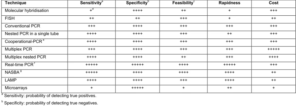
Table 1. Comparison of sensitivity, specificity, feasibility, rapidness and cost of different techniques in detection of plant pathogenic bacteria and viruses
Technique Sensitivitya
Specificityb
Feasibilityc
Rapidness Cost Molecular hybridisation +d
++++ ++ + +++
FISH ++ ++ +++ + ++
Conventional PCR +++ ++++ +++ +++ +++
Nested PCR in a single tube ++++ ++++ +++ ++ +++
Cooperational-PCR e
++++ ++++ +++ +++ +++
Multiplex PCR +++ ++++ +++ +++ +++++
Multiplex nested PCR ++++ ++++ ++ +++ ++++
Real-time PCR f
+++++ +++++ ++++ +++++ +++
NASBA g
+++++ ++++ ++++ ++++ ++
LAMP ++++ ++++ +++ ++++ ++
Microarrays + +++++ + ++ +
a
Sensitivity: probability of detecting true positives.
b
Specificity: probability of detecting true negatives.
c
Feasibility: practicability in routine analysis, execution and interpretation.
d
The number of + symbols indicates how methods rate regarding each considered criterion, from acceptable (+) to optimum (+++++).
e
Coupled with hybridisation and colorimetric detection.
f
Using TaqMan probes.
g
Using Molecular Beacons probes.
Optimization of molecular techniques in routine analysis: relevant issues
Molecular techniques like PCR or RT-PCR, despite their advantages have not been yet widely adopted for routine screening protocols in diagnostic laboratories in many countries (Schaad et al., 2003; Alvarez, 2004) for pathogens detection. One of the reasons is that the low titre of the majority of pathogens in plants outside the vegetative period or in symptomless propagative material with latent infections, and the frequent uneven distribution in the host tissues, make them difficult to detect accurately. This fact is especially relevant in the case of fruit trees, grapevines, and other woody plants that exhibit winter dormancy, or in seeds, insect vectors, water and soil, that usually contain low amounts of the target pathogens. Besides, the size of the sample to be analysed is an important unsolved question and molecular methods prescribe very small-volume samples, hampering accurate detection. Sampling protocols must be improved including concentration of the targets or previous enrichment of the pathogen, to reach realistic orders of scale. According to Alvarez (2004), conclusions drawn from very sensitive methods that require only microliters of sample, often misrepresent the real situation. Furthermore, the way in which samples are collected and handled is also very
important, so care should be taken to avoid contamination among samples, to ensure that it is both appropriate and suitable for molecular testing and specifically, for PCR amplification.
Very often, when conventional PCR or RT-PCR is applied routinely for detection purposes, the sensitivity afforded is often lower than expected due to potential inhibitors of transcriptases and/or polymerases. In this context, the possibility of adding new anti-inhibitors compounds in the amplification cocktail to avoid the need of DNA or RNA purification requires more investigation. As indicated above, the presence of different components as well as specific RT-PCR conditions may inhibit the reverse transcription and amplification. Amplification success can also depend on the growth stage, physiological condition or type of plant tissue assayed (Maes et al., 1996). These problems can be solved by testing different preparation methods of the samples or by inclusion of compounds that reduce inhibition and/or by simple dilution of the samples.
genome of pathogenic bacteria and viruses will certainly enable more primers to be developed that target known pathogenicity and virulence genes (Louws et al., 1999).
Due to the nature of conventional, nested, or multiplex PCR, practical questions regarding the high level of sensitivity (up to 1 target per reaction) and the amplification of an enormous number of copies of the target sequence should be taken into account (Louws et al., 1999). False positives can arise from contamination during sample collection or sample processing and/or from the sequential contamination of consecutive PCR runs from a few molecules of PCR-generated fragments, being the first amplification cycle critical. False positives can also result not only from cross-amplification of nontarget DNA, but from exogenous DNA from one positive sample to another, from cell/cultures or aerosols, or from contaminating DNA originating from carry-over of previous experiments (Louws et al., 1999; van der Wolf et al., 2001), as indicated since this technique was first developed (Kwok and Higuchi, 1989). Although, these risks decrease on using real-time PCR, the use of PCR-based assays for routine analysis in plant pathology requires numerous negative controls, in addition to non-contaminant sampling and sample preparation methods.
Another potential problem with PCR amplification in routine use is the amplification of products other than those predicted, like single-stranded DNA (Valentine et al., 1992) or mis-priming or amplification of primer artefacts (“primer dimerization”). This background amplification can not only confuse test results, but it can interfere with amplification of predicted products by consuming reaction reagents (Henson and French, 1993). Procedures like “hot start” (Chou et al., 1992) or “heat-soaked” (Ruano et al., 1992) were designed to eliminate or reduce background because they ensure initiation of reactions at denaturation temperature.
False negatives in standard PCR protocols can be attributed to several causes, like the presence of compounds that inhibit the polymerases, degradation of the DNA target sequence, or reagent problems (Louws et al., 1999). Then, it is convenient to include one or several positive controls as extra samples and internal PCR controls as co-amplification of host DNA or other
strategies. In any routine use of a PCR protocol, external quality assurance schemes should be applied to contribute to increasing the accuracy of the final result, but to our knowledge there are no freely-available approved guidelines for plant pathologists.
A frequent criticism of PCR results is that DNA from dead or VBNC cells may be amplified and provide a positive result of low biological relevance. This is especially relevant when analysing quarantine organisms, where the positive result of the analysis implies strict eradication measures. Enrichment or BIO-PCR (Schaad et al., 1995 and 2003; López et al., 1997) can circumvent this problem, as it involves a previous enrichment step in liquid or solid medium, favouring detection of living cells harvested from the media prior to PCR amplification. However, neither the standard PCR protocols nor BIO-PCR can differentiate among dead and VBNC cells (Roszak and Colwell, 1987). Risk of plant disease caused by VBNC cells is still controversial, but as an example there are in vitro studies of the ability of VBNC cells of E. amylovora to regain culturability and pathogenicity even after nine months in such a state (Ordax et al., 2006). This justifies the use of molecular techniques for screening plant samples, although the isolation of pathogenic bacteria in pure culture and demonstration of their pathogenicity is currently required.
In plant pathology, no decision has been taken for reliance on any single molecular test in most of the protocols developed by different organizations, despite the great sensitivity, specificity and reliability of PCR. Furthermore, in many laboratories, especially in developing countries, the relatively expensive reagents, equipment, and skilled personnel makes it difficult for molecular techniques to be implemented as routine procedures. Nevertheless, regardless of the practical application of these methods in plant health services, published protocols indicate an increasing development of DNA based reports for diagnostic purposes as well as for etiological and epidemiological studies. The number of laboratories of plant protection services equipped with thermocyclers has increased exponentially in the last five years.
detection methods: they are sensitive, specific enough, rapid, and suitable for high throughput screening, and will be the most widely used by plant pathologists in the near future, especially when direct methods of sample preparation (without the need of nucleic acid purification) will be validated. Besides, isothermal amplifications could also be the method of choice for some specific utilisations.
Regardless of the slow development of microarray technology for plant pathogen detection, especially due to its low current sensitivity, it shows potential features that make it a very promising tool. Also, coupling it with other molecular systems, like the multiplex-PCR (Call et
al., 2001; Panicker et al., 2004) increases the system’s
detection and diagnostic potential. Nevertheless, this technique is still far from being used for routine detection of plant pathogens given the need for a previous amplification reaction, the low level of sensitivity achieved, and the high cost of the reagents and equipment. It is likely that microarrays will follow a path similar to that of PCR, which spent several years as a research tool before being routinely utilised in plant
pathogens diagnosis (López et al., 2003).
Selection of diagnostic methods and validation of protocols: what have we learned?
Molecular techniques for plant pathogen detection are developing rapidly and constantly. However, there are still significant drawbacks to include these tests, due to the lack of appropriate studies and validated methods establishing their reliability and reproducibility for routine analysis. In fact, in plant pathology there is insufficient knowledge and information to demonstrate that adequate risk assessment is afforded by many amplification or PCR-based methods, which detracts from confidence in their results.
Sensitivity, specificity and beyond
Detection and diagnostic tests may be interpreted as a function of several parameters that increase the information about the sanitary status of a plant, strengthen or lessen the probability of infection. Because there is no perfect method, false positive and/or false negative results can be obtained. Consequently, it is
necessary to estimate the operational capacity of each technique or method to minimize uncertainty and improve the interpretation of results. In general, the methods of detection and diagnosis are used to classify plants depending on the presence or absence of one specific pathogen or several. The results of the analyses enable a conclusion to be drawn and facilitate effective decision making. Analyses of diagnostic data can be performed with 2x2 contingency tables, enabling indicators of the operational capacity of each technique
to be calculated based on test results versus sanitary
status. Sensitivity and specificity can be calculated according to Altman and Bland (1994a). Sensitivity is defined as the proportion of true positive of infected plants that the technique or method identifies. The methods affording highest sensitivity must be used to discard the presence of a pathogen supplying an accurate diagnosis of healthy plants, because they give an accurate indication of the pathogen-free status. Specificity is defined as the proportion of true negative (of healthy plants) that the method identifies, supplying an accurate estimation of the real positives. Both indicators constitute one approach to evaluating the diagnostic ability of the test. The highest specific methods can be used to confirm the presence of a pathogen offering an accurate diagnosis of true infected plants.
Sensitivity and specificity do not include false positive and false negative rates to calculate their values and predictive values depend on the prevalence of disease. Do parameters free of these influences exist? Likelihood ratios are not influenced by prevalence and they can be calculated on the basis of sensitivity and specificity, which are stable for each method. The positive likelihood ratio will be applied in the event that the technique diagnoses a sample as positive and the negative likelihood ratio will be applied if the technique diagnoses a sample as negative and all of them give the likelihood of having disease. Likelihood ratios can be calculated according to Deeks and Altman (2004): the positive likelihood ratio is the proportion of true positives that are correctly identified by the technique (sensitivity), divided by the proportion of false positive results the method gives (1-specificity). The negative likelihood ratio is the proportion of false negatives given by the method (1-sensitivity), divided by the proportion of true negatives correctly identified by the technique (specificity). Likelihood ratios are useful in assessing the potential utility of a test and those >10 or <0.1 generate large changes in post-test probability whilst likelihood ratios ranging from 0.5 to 2 have little effect (Sackett et al. 2000). The likelihood that a result correctly indicates the sanitary state of a plant is the post-test probability of infection or disease. Pre-test probability of disease can be compared with the estimated later probability of disease using the information provided by a diagnostic test. The difference between the former probability and the latter probability is an effective way to evaluate the efficiency of a diagnostic method. Post-test probability can be calculated using likelihood ratios of the method and pre-test probability is the estimated prevalence of the disease. Bayes’ theorem is used to translate the information given by the likelihood ratios into a probability of disease. Bayes’ theorem states that the pre-test odds of disease multiplied by the likelihood ratio yields the post-test odds of disease. In addition, likelihood ratios of several methods can be sequentially combined (Neves et al., 2004). Thus, this evidence-based approach modifies the previous criterion obtained only by sensitivity and specificity.
Inter-laboratory validation of molecular methods and protocols
The inter-laboratory evaluations of new detection or diagnostic methods provide essential information on test repeatability and reproducibility, ease of implementation, use and interpretation, giving an indication of the robustness in routine analyses of large numbers of samples. A standard protocol must subsequently be established and optimized based on results. Repeatability refers to within-laboratory agreement between replicate observations of the same test performed by the same observer under similar conditions. Reproducibility refers to between-laboratory agreement. Repeatability and reproducibility can be estimated through the calculation of Cohen's kappa coefficients (Cohen, 1960), which measure the agreement of a classification between repetitions. The Kappa index is calculated dividing the subtraction of (observed coincidence - expected coincidence) by the subtraction of (1 - expected coincidence). This kappa coefficient represents to what extent the agreement is better than what would be the result of chance alone. To interpret the kappa value, the following guidelines are used: 0.00 to 0.20: no agreement; 0.21 to 0.40: weak agreement; 0.41 to 0.60: moderate agreement; 0.61 to 0.80: strong agreement; and 0.81 to 1.00, almost perfect agreement (Landis and Koch, 1977).
post-test probability varies. After post-test probability has been estimated, the next step is to decide if it confirms or rejects diagnosis or an additional diagnostic method is necessary (Aldington et al., 2006).
Olmos et al. (2008), reported how an evidence-based approach modified the previous criteria obtained only by sensitivity and specificity, to use RT-PCR (the most sensitive method) as screening test for PPV diagnosis during the dormant period and DASI-ELISA using monoclonal antibodies (the most specific method) as a confirmation test. For instance, the probability of a negative result in wintertime by DASI-ELISA given a prevalence value ranging from 0.01 to 0.1%, confirmed in practice PPV-free status of a tree in springtime, with similar post-test probability to that afforded by RT-PCR. A positive result by DASI-ELISA in wintertime provided a much higher post-test value than RT-PCR. Thus, the information given by the evidence-based approach indicated that DASI-ELISA should be used as a screening test at very low levels of PPV incidence (0.01-0.1) not requiring confirmation by RT-PCR. In the case of prevalence level ranging from 0.5 to 10% post-test probability of negative results by DASI-ELISA was a little higher than RT-PCR. This information suggests that in general DASI-ELISA using specific monoclonal antibodies could be used as a screening test in wintertime surveys. If a more accurate PPV status of a tree was required, RT-PCR for negative results should be performed. However, a positive result by DASI-ELISA gives a much higher post-test probability of PPV infection, not requiring confirmation by RT-PCR. The last scenario is that one with PPV prevalences ranging from 25 to 90%. The evidence-based approach would suggest that RT-PCR should be used as a screening test due to its lower post-test probability of negative results. When test accuracy is a priority, in the cases where DASI-ELISA and PCR give discordant results, a third complementary test such as NASBA-FH could be very helpful because it improves diagnostic accuracy and consequently improves the assessment of the sanitary status of a plant.
Selection of a diagnosis method
The selection of appropriate diagnostic methods should involve some critical appraisals focusing on the objective pursued: i) eradication, certification of mother plants, sanitation or quarantine programs or ii) large surveys to evaluate incidence, or screening tests for surveillance of the spreading of a disease. In the first cases, the need to use the most sensitive method should be stressed, accepting the risk of false positives. For this reason evaluation of sensitivity and specificity of the techniques to select the most sensitive is the main requirement. It would enable the presence of the pathogen to be discarded most effectively because it affords the most accurate diagnosis of healthy plants with high confidence when the target pathogen is not detected. However, in the case of large-scale surveys or screening tests for surveillance, the selection of one, two or several methods should be based on an evidence-based approach, evaluation of cost per analysis, calculation of post-test probability of disease and consideration of different scenarios with different prevalence.