Abstract
The abnormal expression of microRNAs (miRNAs) plays a key role in colorectal cancer (CRC). The present study attempted to identify the potential miRNA biomarkers of CRC due to the important role of microRNAs within the DLK1-DIO3 genomic region, especially the role of mir-411-5p in other cancers. This prospective study characterized the contribution of mir-411-5p to CRC tumorigenesis. The Real-time quantitative reverse-transcrip-tion –polymerase chain reacreverse-transcrip-tion was used to examine miR-411-5p expression levels prospectively in 40 pairs of samples of CRC sues and adjacent noncancerous tissues (>2 cm from cancer tis-sue). Also, the relationship between miR-411-5p expression levels and clinicopathological characteristics was explored. The capabil-ity of miR-411-5p to function as a tumor marker in CRC was also examined. MiR-411-5p was significantly downregulated in a group of CRC samples compared with matched noncancerous tis-sues. A receiver operating characteristic (ROC) curve also showed ROC area of 68% for miR-411-5p (P value=0.006) with 70% and 65% sensitivity and specificity, respectively. According to the sur-vey results, miR-411-5p might be considered as a tumor marker in
CRC and it might be a promising therapeutic option which may help prevent CRC.
Introduction
Each year, 1.23 million individuals are affected by colorectal cancer (CRC). It should be noted that this type of cancer is placed as the third most common cancer type among males and the second
among females.1Moreover, it is the fourth most predominant cause
of cancer death, universally. In northern America and Europe, the frequency of CRC has been assessed to be 30-50 cases for every 100,000 people. In Middle East, this rate is estimated to be 3 to 7
cases for every 100,000.2Predictably, this cancer type acts as one of
the most fatal disorders among western populations. Prior research findings indicated that the frequency of CRC is assumed to rise each year in developing countries, especially in Asian ones throughout
the next two decades.2,3The growing number of CRC diagnosis in
the previous three decades has proven CRC to be an important
pub-lic health problem in Iran.4Furthermore, a high frequency of cancer
in gastrointestinal tract has been reported in East Azerbaijan
(locat-ed in North West of Iran)5. Consequently, there is a possibility to
decrease the number of CRC affliction by accumulating knowledge about the biology and nature of CRC; therefore, designing effective
prognostic, diagnostic ,and treatment plans.6
MicroRNAs (miRNAs) are defined as tiny, non-coding, sin-gle-stranded RNAs with the approximate length of 18-25; they are nucleotides that are expressed endogenously. Moreover, through binding to 3’ untranslated region (3’ UTR) of target mRNAs,
MicroRNAs can post-transcriptionally control gene expression.7
The importance of MicroRNAs in regulating many biological processes (e.g., cell cycle, proliferation, differentiation, apoptosis,
and invasiveness) cannot be underestimated.8The results of
previ-ous studies demonstrate that miRNAs can suppress tumors and
oncogenes.9,10MiRNAs have the status of attractive molecules in
cancer development diagnostics and therapeutics.11 Therefore,
some of miRNAs identified in this pathway may be used as
diag-nostic and progdiag-nostic markers or therapeutic targets.12
MiR-411-5p is located in the imprinted Dlk1-Dio3 region on
chromosome 14q32.31.13A large miRNA of this cluster is situated
within a parentally imprinted chromosome.14 Various species
highly conserve the Protein-coding genes within the Dlk1-Dio3
region, but only mammals conserve the miRNA cluster.15 The
expression of this cluster has been observed in human cancers
including melanoma,16 ependymoma,17 neuroblastoma,18
osteosarcoma,19gastrointestinal stromal tumors,20 hepatocellular
carcinoma,20 uterine osteosarcoma,21and ovarian cancer.22After
all, its expression arrangement, clinical relevance, and functional role in CRC still remain unknown. Consequently, the researchers in this study tried to examine the miR-411-5p expression levels in CRC tissues. A study in this field demonstrated the over expres-Correspondence: Mohammad Ali Hosseinpour Feizi, Department of
Animal Biology, Faculty of Natural Science, University of Tabriz, 29 Bahman Blvd, Tabriz, East Azerbaijan, Iran.
Tel.: +98.41.33362280 - Fax: +98.41.33362282. E-mail: pourfeizi@eastp.ir
Key words: colorectal cancer, biomarker, microRNA, miR-411-5p.
Contributions: the authors contributed equally.
Conflict of interest: the authors declare no potential conflict of interest.
Acknowledgments: the authors would like to thank the patients, staff and nurses in the Endoscopy and the Pathology Department of Tabriz Imam Reza Hospital who profoundly helped us in conducting this project. Received for publication: 22 December 2016.
Revision received: 4 May 2017. Accepted for publication: 5 May 2017.
©Copyright A. Fateh et al., 2017 Licensee PAGEPress, Italy
Journal of Biological Research 2017; 90:6511 doi:10.4081/jbr.2017.6511
This article is distributed under the terms of the Creative Commons Attribution Noncommercial License (by-nc 4.0) which permits any non-commercial use, distribution, and reproduction in any medium, provid-ed the original author(s) and source are crprovid-editprovid-ed.
Importance of mir-411-5p in colorectal cancer
Alavieh Fateh, Mohammad Ali Hosseinpour Feizi, Reza Safaralizadeh, Shirin Azarbarzin
Department of Animal Biology, Faculty of Natural Science, University of Tabriz, Iran
Non
commercial
use
sion of miR-411 in the lung cancer cells.23In a study on breast can-cer, it was revealed that there were significant differences regard-ing miR-411 expression in metastatic breast cancer patients com-pared to control group; as a matter of fact, it’s down regulation was
observed in metastatic breast cancer.24
Materials and methods
In our study at Imam Reza Hospital (Tabriz, Iran), after colonoscopy and sigmoidoscopy in this hospital, all CRC samples and normal adjacent tissues were collected from 40 patients diag-nosed with CRC. Imam Reza hospital is considered as the first affiliated hospital of Tabriz University of Medical Sciences. All samples studied were collected during a period of time between November 2014 and June 2015. From a piece of resected specimen at the furthest distance from tumor (>2 cm from tumor), the researchers obtained the non-tumor tissue. All study participants were born in East Azerbaijan, Iran. The Research ethics Committee of Imam Reza Hospital accepted this research in line with institutional protocol and all patients signed the informed consents. Consistently, the researchers processed all specimens resected for histopathological assessment.
Sample preparation and RNA isolation
It should be noted that regarding the sample preparation and RNA isolation till RNA Extraction, the researchers instantly flash froze all the tissue samples in liquid nitrogen and stored at –80°C. TRIzol reagent (Invitrogen Carlsbad, CA) was used to execute the Phenol based RNA extraction. To sum it up, 1 mL TRIzol LS reagent was added into homogenized tissue sample, then the mixture was pipetted up and down many times; finally, it was incubated at room temperature for 5 minutes. The next step was adding 200 μL chloro-form and strongly shaking it for 15 minutes; then, the sample was incubated at room temperature for 2-15 minutes. For another 15 minutes the sample was centrifuged at 12,000 rpm at 4°C. The next step was transmitting the aqueous phase into a new Eppendorf tube and adding 500 µL of 100% isopropanol. After that, during the night, the mixture was stored at 20°C; next, the mixture was centrifuged for 10 minutes at 13,000 rpm at 4°C with the aim of pelleting the nucleic acid. Then, after removing the supernatant, the pellet con-taining RNA was washed adding 1 ml of 75% ethanol. The next step was centrifuging the sample at 7500 rpm for 5 minutes at 4°C. After adding 25 µL RNase free water to the RNA pellet, the mixture was pipetted up and down for many times. Finally, for 10 to 15 minutes, the mixture was incubated in a water bath at 55-60°C. The picoDrop 2000 (Bob Batty International [BB], UK) was utilized to quantify the amount of isolated RNA concentration. Until cDNA synthesis, the extracted RNAs were stored at –80°C. Here, a 10 µL DNase I treatment reaction was performed in order to degrade any DNA con-tamination in extracted RNAs (Fermentas, Canada).
Reverse transcription and quantitative Real-time PCR
In a final volume of 10 µL reaction systems, both the Reverse transcription and quantitative Real-time PCR Reverse transcription were executed on 120 ng of total RNA. The 10 μL RT reaction mixture was incubated at 37°C for 60 minutes, 85°C for 5 seconds, and then held at 4°C using the Prime Script(R) miRNA cDNA Synthesis Kit (ParsGenome, Iran) according to the instructions given by the manufacturers. Moreover, in order to dilute the RT product, the researchers added 90 µL of the RNase free water.
After that, SYBR® Green was used to carry out the Real-time PCR and 4 µL diluted RT product was added into a 10 μL PCR reac-tion, which also contained 10 μL SYBR Green, 1 μL primer mix (purchased from ParsGenome, Iran), and 1 μL RNase-free water.MiR-411-5p and 5s rRNA (5srRNA was selected as a house-keeping gene used for normalization and data analysis) primers were also purchased from ParsGenome, Iran. In spite of using Rotor-Gene Q - QIAGEN Real-time PCR Detection System, the researchers administer all PCR reactions, including non-template controls, in triplicate. Finally, using the REST2009 Software, the raw data were examined. It should be mentioned that the researchers processed all samples in triplicate. According to the definition given in the litera-ture, the threshold cycle (CT) is the cycle number at which the fluo-rescence passes the fixed threshold. Each experiment included a control without a template. Polyacrylamide gel electrophoresis (PAGE) validated the final products of real-time PCR).
Normalization and statistical analysis
The relative expression analysis of miR-411-5p was performed by a randomization test using the Relative Expression Software Tool (REST) 2009
(http://gene-quantification.com/rest-2009.html). 2-ΔΔCtmethod was employed to analyze the expression
levels of miR-411-5p in CRC tissues relative to their matched non-tumor counterparts. The threshold cycle (Ct) of fluorescence for each sample was determined. ΔCt indicated the difference in expression levels with the Ct value between miR-411-5p and 5s rRNA (ΔCt=Ct miR-411-5p - Ct 5s). ΔΔCt indicated the difference in the ΔCt value between the cancer tissue and the matched control
(ΔΔCt=ΔCt cancer-ΔCt control). The 2-ΔΔCtvalue (fold value) was
also calculated. It was found that when the fold value was <1, there was a low expression of miR-411-5p in the cancer tissues com-pared to their non-tumorous counterparts. Here, decreased expres-sion was defined as the fold change less than one in expresexpres-sion. SPSS 18.0 software was utilized to carry out all the analyses (Chicago, IL, USA). All P-values cited contained two sides. It should, also, be noted that P-values<0.05 were estimated to be sta-tistically significant. Receiver operating characteristic (ROC) curve was also constructed to evaluate the specificity and sensitiv-ity of predicting CRCs and normal tissues by miR-411-5p expres-sion levels. Moreover, the sensitivity/specificity at various cut off values was calculated using Sigma Plot 12.5. A statistically signif-icant difference was indicated by P values<0.05.
Expression levels of miR-411-5p in CRC
and normal tissue
All the samples’ Ct values were entered in the REST 2009 soft-ware with the aim of comparing the miR-411-5p expression‘s total level in CRC connected to normal tissues. The results of the ran-domization test showed that mir-411-5p expression in tumor sam-ples decreased 6.5 times more than in normal tissues (Figure 1).
Clinicopathological characteristics
and their association with miR-411-5p expression
Considering clinicopathological characteristics, no significant relationship was reported between the miR-411-5p expression and
gender (P=0.703), age (P=0.408), histological grade (P =0.053),
and tumor location (P=0.375). (Table 1)
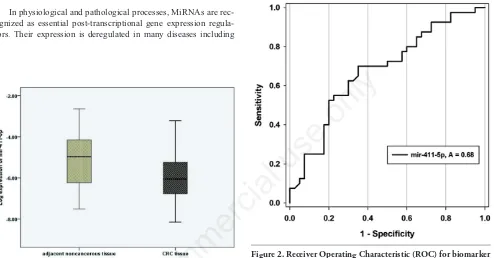
Capability of miR-411-5p to function
as a CRC tumor marker
Receiver operating characteristic (ROC) curve was construct-ed and the area under the curve (AROC) was calculatconstruct-ed to evaluate
Non
commercial
use
the specificity and sensitivity of predicting CRCs and normal tis-sues by miR-411-5p expression levels. Based on the analysis of ROC curves, miR-411-5p showed a ROC area (AROC) of 68%. The expression of miR-411-5p with a value of 0.68, compared with 1.0, conveyed that this microRNA is almost highly sensitive and specific; therefore it has the capability to distinguish tumor sam-ples in CRC; subsequently, it can be viewed as a tumor marker (Figure 2).
Discussion
In physiological and pathological processes, MiRNAs are rec-ognized as essential post-transcriptional gene expression regula-tors. Their expression is deregulated in many diseases including
cancer.13MiRNAs can be used as biomarkers for the diagnosis and
prognosis of several malignancies and can recognize cancer
sub-classes of different clinical management.13The DLK-DIO3 region
was selected because this region hosts 53 miRNAs and the find-ings reported here indicate that these miRNAs are involved in a wide spectrum of human diseases, especially cancers. They may modulate important signaling pathways like MAPK (mitogen-acti-vated protein kinase) and p53; besides, they are related to cytokine
Figure 1. Differential expression of miR-411-5p. miR-411-5p expression in tumor samples showed a significant decrease (6.5 times) compared to normal samples with confidence interval of 95% (CI=95%), P Value=0.006, and P<0.05.
Figure 2. Receiver Operating Characteristic (ROC) for biomarker in detection of CRCs. The ROC curve was automatically generat-ed from 40 points of cutoff values set by the software SigmaPlot 12.5. The area under the ROC curve (AROC) is 0.68 out of 1, P Value=0.006 (P<0.05), with 70% and 65% sensitivity and speci-ficity respectively.
Table 1. Relationships between miR-411-5p expression levels in cancer tissue samples from patients with CRC and clinicopathological characteristics.
Variable N° miR-411-5p relevant expression (2-ΔΔCt) P value Gender 0.70
Male 18 20.31±4.26 Female 22 18.91±3.91
Age 0.05 <60 17 19.51±4.05
≥60 23 19.56±4.19 Histological grade
Well differentiated 19 19.36±3.87 Moderate differentiated 17 20.06±4.13 Poorly differentiated 4 18.18±5.60
Tumor location 0.38 Colon 16 18.66±4.11
Sigmoid 22 20.51±4.28 Rectum 12 19.74±3.93
Data presented as mean±SD; P values obtained using ANOVA test. No significant relationship was found between miR-411-5p expression levels and clinicopathological characteristics.
Non
commercial
use
signaling cascades, DNA methylation, oncogenic kinases
expres-sion, and many others.13
Mir-411-5p is known as a member of the miR-379 family. This family is known to be placed in the miR-379/miR-656 cluster inside the DLK-DIO3 region on human chromosome. Placental mammals conserved the miR-379/miR-656 cluster to a great
extent.25Based on prior research studies, the vital role of
miR-411-5p in different biological processes in various human cancer cells
has been proven.26 MiR-411 was reported to be upregulated in
facioscapulohumeral muscular (FSHD) dystrophy and to suppress
myogenic factors.27
In Another study, it was revealed that miR-411-5p possesses an inverse correlation among TGF-β1/SPRY4 and levels of miR-411-5p. Moreover, it was indicated that Re-expression of miR-411-5p
can prevent in vitrorhabdomyosarcoma (RMS) cell proliferation
as well as in vivotumorigenicity. Here, the researchers accentuated
an anti-oncogene role for miR-411-5p. MiR-411 cluster represents another set of transforming growth factor-beta1 (TGF-β1)-sup-pressed miRNAs in RMS, and miR-411-5p expression was
nega-tively regulated by TGF-β1 in RMS.28Studies also investigated the
effects of TGF-²1 on human CRC. For instance, it has been revealed that TGF-β1 plays an important role in CRC, and TGF-β1 expression might be a complementary mechanism in the onset of CRC; that is to say, it can have a major effect on the prognoses of
patients.29TGF-β1acts both as an inhibitor of tumor growth and as
a promoter of tumor progression.30Furthermore, a negative
regu-latory effect for Sprouty homolog 4 (SPRY4) (i.e., a direct target of miR-411-5p) has been reported on: i) Activation of protein kinase C (PKC) generated by PKCα-mediated Mitogen; ii) Activated protein kinases (MAPKs); iii) Vascular endothelial
growth factor-A26 activation propel to growth arrest of RMS.28In
CRC, the link between MAPK pathway signaling and cell
adhe-sion, angiogenesis, invaadhe-sion, and metastasis is entrenched.31
SPRY proteins have major roles in regulating tubular morpho-genesis, such as angiomorpho-genesis, as well as in lung, placenta, and
kid-ney development.32,33The reason why SPRY proteins are regarded
as tumor suppressors is their ability to be repressed in some malig-nancies. Another study on colon cancer showed that epigenetic silencing and loss-of-function mutations of SPRY4 could lead to
tumorigenesis.34The documents at hand indicate that in RMS there
is an autoregulatory loop among TGF-β1/miR-411-5p/SPRY4 and
MAPK (especially p38MAPK) pathway.28
Further, it was shown that in hepatocellular carcinoma (HCC) cells, the expression of MiR-411 is upregulated. The findings of the previous body of research demonstrated that ectopic expression of miR-411 leads to downregulation of ITCH, which results in cyclin D1 and c-Myc upregulation. This should not be forgotten that this upregulation has a vital role in carcinogenesis of human cancer which results in an increase in the proliferation of HCC
cell.26Overexpression of miR-411 in lung cancer regulates G1/S
transition with cell cycle regulators such as cell cycle inhibitors
p21 and p27; it also upregulates cell cycle regulator cyclin D1.23
Nevertheless, the role of miR-411 in cell cycle in CRC is still unknown.
Various studies in this field proved that another target for
miR-411 was fork head box O1 (FoxO1),23which has a regulating role
for angiogenesis, apoptosis, cell invasion and metastasis, cell metabolism, oxidative stress, immune regulation, and self-renewal and stem cells discrimination. In addition, the findings revealed that FoxO1’s expression can be prevented by the over expression
of miR-411 in lung cancer.23,35,36Expression of FoxO1 correlated
with autophagic capacity and tumor development in human colon
cancer cells.36
Conclusions
In summary, as far as the authors are concerned, this study is the first report on the expression patterns of miR-411-5p in CRC tissues. The results obtained here revealed that compared to the normal tissues, miR-411-5p was noticeably downregulated in the cancerous tissues. Whereas, many additional researches with a larger sample size are needed to fully convey the connection among the microRNA studied here and clinicopathological fea-tures. In the present study, the capability of miR-411-5p expression level to function as a tumor marker to distinguish CRCs from nor-mal counterparts was also assessed suggesting that miR-411-5p has a great sensitivity and specificity; therefore, it can be consid-ered as a tumor marker in diagnosing CRC. The researchers of the present study believe that regarding the importance of miR-411-5p in biology and especially in cancer, various other studies are required in order to understand the other roles played by miR-411-5p in CRC.
References
1. Siegel R, Naishadham D, Jemal A. Cancer statistics, 2012. CA Cancer J Clin 2012;62:10-29.
2. Ansari R, Mahdavinia M, Sadjadi A, et al. Incidence and age distribution of colorectal cancer in Iran: results of a popula-tion-based cancer registry. Cancer Lett 2006;240:143-7. 3. Sung JJ, Lau JY, Goh K, Leung W, Cancer APWGoC.
Increasing incidence of colorectal cancer in Asia: implications for screening. Lancet Oncol 2005;6:871-6.
4. Dolatkhah R, Somi MH, Bonyadi MJ, et al. Colorectal cancer in Iran: molecular epidemiology and screening strategies. J Cancer Epidemiol. 2015;2015.
5. Somi MH, Golzari M, Farhang S, et al. Gastrointestinal cancer incidence in East Azerbaijan, Iran: update on 5 year incidence and trends. Asian Pacific J Cancer Prevent. 2014;15:3945-9. 6. Ferlay J, Soerjomataram I, Ervik M, et al. GLOBOCAN 2012
v1. 0, Cancer incidence and mortality worldwide: IARC CancerBase No. 11. International Agency for Research on Cancer, Lyon, France. 2013. globocan. iarc. fr. 2015.
7. Adachi T, Nakanishi M, Otsuka Y, et al. Plasma microRNA 499 as a biomarker of acute myocardial infarction. Clin Chem 2010;56:1183-5.
8. Traver S, Assou S, Scalici E, et al. Cell-free nucleic acids as non-invasive biomarkers of gynecological cancers, ovarian, endometrial and obstetric disorders and fetal aneuploidy. Human reproduction update. 2014:dmu031.
9. Ai J, Zhang R, Li Y, et al. Circulating microRNA-1 as a poten-tial novel biomarker for acute myocardial infarction. Biochem Biophys Res Commun 2010;391:73-7.
10. Chan E, Prado DE, Weidhaas JB. Cancer microRNAs: from subtype profiling to predictors of response to therapy. Trends Mol Med 2011;17:235-43.
11. Takayama K, Tsutsumi S, Katayama S, et al. Integration of cap analysis of gene expression and chromatin immunoprecipita-tion analysis on array reveals genome-wide androgen receptor signaling in prostate cancer cells. Oncogene 2011;30:619-30. 12. Faltejskova P, Svoboda M, Srutova K, et al. Identification and
functional screening of microRNAs highly deregulated in col-orectal cancer. J Cell Mol Med 2012;16:2655-6.
13. Benetatos L, Hatzimichael E, Londin E, et al. The microRNAs
Non
commercial
use
within the DLK1-DIO3 genomic region: involvement in dis-ease pathogenesis. Cell Mol Life Sci 2013;70:795-814. 14. Lin S-P, Youngson N, Takada S, et al. Asymmetric regulation
of imprinting on the maternal and paternal chromosomes at the Dlk1-Gtl2 imprinted cluster on mouse chromosome 12. Nat Genet 2003;35:97-102.
15. Liu L, Luo G-Z, Yang W, et al. Activation of the imprinted Dlk1-Dio3 region correlates with pluripotency levels of mouse stem cells. J Biol Chem 2010;285: 19483-90.
16. Bartel DP. MicroRNAs: target recognition and regulatory functions. Cell 2009;136:215-33.
17. Costa FF, Bischof JM, Vanin EF, et al. Identification of microRNAs as potential prognostic markers in ependymoma. PloS One 2011;6:e25114.
18. Gattolliat C, Thomas L, Ciafre S, et al. Expression of miR-487b and miR-410 encoded by 14q32. 31 locus is a prognostic marker in neuroblastoma. Br J Cancer 2011;105: 1352-61. 19. Thayanithy V, Sarver AL, Kartha RV, et al. Perturbation of
14q32 miRNAs-cMYC gene network in osteosarcoma. Bone 2012;50:171-81.
20. Haller F, von Heydebreck A, Zhang JD, et al. Localization-and mutation-dependent microRNA (miRNA) expression signa-tures in gastrointestinal stromal tumours (GISTs), with a clus-ter of co-expressed miRNAs located at 14q32. 31. J Pathol 2010;220:71-86.
21. Devor EJ, De Mik JN, Ramachandran S, et al. Global dysreg-ulation of the chromosome 14q32 imprinted region in uterine carcinosarcoma. Exp Ther Med 2012;3:677-82.
22. Zhang L, Volinia S, Bonome T, et al. Genomic and epigenetic alterations deregulate microRNA expression in human epithe-lial ovarian cancer. Proc Natl Acad Sci 2008;105:7004-7009. 23. Zhao Z, Qin L, Li S. miR-411 contributes the cell proliferation
of lung cancer by targeting FOXO1. Tumor Biol 2015:1-10. 24. McGuire A, Brown JA, Kerin MJ. Metastatic breast cancer: the
potential of miRNA for diagnosis and treatment monitoring. Cancer Metastasis Rev 2015;34:145-55.
25. Glazov EA, McWilliam S, Barris WC, Dalrymple BP. Origin,
evolution, and biological role of miRNA cluster in DLK-DIO3 genomic region in placental mammals. Mol Biol Evol 2008;25:939-48.
26. Xia K, Zhang Y, Cao S, et al. miR-411 regulated ITCH expres-sion and promoted cell proliferation in human hepatocellular carcinoma cells. Biomed Pharmacother 2015;70:158-63. 27. Harafuji N, Schneiderat P, Walter MC, Chen Y-W. miR-411 is
up-regulated in FSHD myoblasts and suppresses myogenic factors. Orphanet J Rare Dis 2013;8:55.
28. Sun M, Huang F, Yu D, et al. Autoregulatory loop between TGF-β1/miR-411-5p/SPRY4 and MAPK pathway in rhab-domyosarcoma modulates proliferation and differentiation. Cell Death Dis 2015;6:e1859.
29. Ma J, Gao H-M, Hua X, et al. Role of TGF-β1 in human col-orectal cancer and effects after cantharidinate intervention. Asian Pacific J Cancer Prevent 2013;15:4045-8.
30. Kemik O, Adas M, Dulger AC, Purisa S. Transforming growth factor beta-1 in human colorectal cancer patients. Eur J Gen Med 2011;8.
31. Fang JY, Richardson BC. The MAPK signalling pathways and colorectal cancer. Lancet Oncol 2005;6:322-7.
32. Anteby E, Natanson-Yaron S, Greenfield C, et al. Human pla-cental Hofbauer cells express sprouty proteins: a possible mod-ulating mechanism of villous branching. Placenta 2005;26: 476-83.
33. Perl A-KT, Hokuto I, Impagnatiello M-A, et al. Temporal effects of Sprouty on lung morphogenesis. Dev Biol 2003;258:154-68.
34. Barbáchano A, Ordóñez-Morán P, García JM, et al. SPROUTY-2 and E-cadherin regulate reciprocally and dictate colon cancer cell tumourigenicity. Oncogene 2010;29:4800-13.
35. Greer EL, Brunet A. FOXO transcription factors at the inter-face between longevity and tumor suppression. Oncogene 2005;24:7410-25.
36. Zhao Y, Yang J, Liao W, et al. Cytosolic FoxO1 is essential for the induction of autophagy and tumour suppressor activity. Nat Cell Biol 2010;12:665-75.