R E S E A R C H
Open Access
Complement factor I deficiency: a not so rare
immune defect. Characterization of new
mutations and the first large gene deletion
María Alba-Domínguez
1,6†, Alberto López-Lera
1,6†, Sofía Garrido
1,6, Pilar Nozal
1, Ignacio González-Granado
2,
Josefa Melero
3, Pere Soler-Palacín
4, Carmen Cámara
5and Margarita López-Trascasa
1,6*Abstract
Background:Complement Factor I (CFI) is a serine protease with an important role in complement alternative pathway regulation. Complete factor I deficiency is strongly associated with severe infections. Approximately 30 families with this deficiency have been described worldwide.
Patients and methods:We have studied five new Spanish families suffering from CFI deficiency. From 19 screened people, 7 homozygous, 10 heterozygous and 2 healthy subjects were identified. Clinical, biochemical and genetic descriptions are included.
Results:Molecular studies demonstrated 4 novel mutations in the screened individuals; amongst them, we describe here the first great gene deletion reported in the CFI locus, which includes full exon 2 and part of the large intron 1.
Conclusion:CFI deficiency is possibly an underestimated defect and the eventual existence of this deficiency should be tested in those patients exhibiting low C3 and recurrent bacterial infections. We propose a simple diagnostic flowchart to help clinicians in the identification and correct diagnosis of such patients.
Keywords:Complement deficiency, Recurrent infections, C3 consumption, Complement factor I, Large deletions, Diagnostic flowchart
Background
The human complement system consists of more than 35 plasma or membrane-bound proteins whose activa-tion is tightly controlled through the acactiva-tion of comple-ment inhibitors. It is involved in host defense against infectious agents, in the removal of apoptotic cells and immune complexes, and in the modulation of the adap-tive immune system [1]. Complement deficiencies, acquired or hereditary, affecting all the soluble compo-nents and many of the membrane-anchored receptors and regulators have been described in humans. Most of these deficiencies are inherited as autosomal recessive
disorders, with C1 inhibitor (C1INH) and Properdin deficiencies being remarkable exceptions, inherited in autosomal dominant and X-linked manners, respectively. Acquired complement deficiency may be due to infec-tious diseases or immune complexes-related disorders. Genetic complement deficiencies have been described affecting either protein function or concentration [2,3].
One of the most important complement activation regulators is Complement Factor I (CFI), a serine pro-tease that circulates in a zymogen-like state [4] at a
con-centration of ~35 μg/mL [5]. The CFI protein is a
heavily N-glycosylated heterodimer consisting of two polypeptide chains linked by a single disulfide bond [6,7]. The heavy chain (50KDa) comprises an N-terminal region; an FI membrane attack complex (FIMAC) domain; a CD5 like-domain or scavenger receptor cysteine-rich (SRCR) domain; two low-density lipopro-tein receptor (LDLr) domains; and a C-terminal region * Correspondence:[email protected]
†Equal contributors
1
Unidad de Inmunología Hospital Universitario La Paz and Hospital La Paz Health Research Institute (IdiPAZ), Madrid, Spain
6
Centro de Investigación Biomédica en Red de Enfermedades Raras (CIBERER), Madrid, Spain
Full list of author information is available at the end of the article
of unknown function that is a site of sequence variability across species [4]. The light chain (38KDa) contains the serine protease (SP) domain with the conserved catalytic residues [8]. The gene of this complement regulator
(CFI, OMIM*217030) comprises 13 exons localized on
chromosome 4q25 [9,10] and its deficiency is inherited in an autosomal recessive manner.
CFI inactivates C3b by cleaving it into iC3b, C3d and C3dg and, in an analogous way, C4b into C4c and C4d. To properly perform its functions, CFI requires the pre-sence of several cofactor proteins such as C4b-Binding Protein (C4BP), Complement Factor H (CFH), Comple-ment Receptor 1 (CR1/CD35) and Membrane Cofactor Protein (MCP/CD46) [2]. Complete CFI deficiency leads to the uncontrolled amplification of C3 cleavage, result-ing in a severe secondary C3 deficiency. Consumption of Complement Factor B (CFB) and CFH are also related with CFI deficiency [2]. Consequently, the most fre-quently associated pathologies to CFI deficiency are severe infections by encapsulated microorganisms, glo-merulonephritis and autoimmune diseases [11].
In this article we describe five new Spanish families with complete CFI deficiency, an underestimated com-plement defect mainly characterized by recurrent, severe infections, low C3 and normal C4 levels in serum. The fact that this persistent biochemical profile is shared by other relatively infrequent complement abnormalities leads us to propose a simple scheme to help clinicians in the diagnosis of patients presenting this complement phenotype.
Patients and methods Patients
Members from five unrelated Spanish families with CFI deficiency were studied. Serum and DNA samples were obtained under standard conditions after written informed consent of the participants.
Patient I.1 was a 37 year old woman with a long-standing history of recurrent rhino-conjunctivitis. At the age of 4, she had several episodes of otitis, sinusitis and bronchitis. At age 16, she had an episode of meningo-coccal septicaemia with disseminated intravascular coa-gulation (DIC) and skin complications. Later, she suffered from an acute arthritis episode. Infections sever-ity declined with age. Her mother (I.2), sister (I.4) and brother (I.5) were also studied.
Patient II.1 was an 8 year old boy, the only child of healthy parents (II.2 and II.3). He had a history of recur-rent pneumonia, an episode of meningococcemia and other infections such a balanitis or oral thrush.
Patient III.1 was a 6 year old boy. His parents (III.2, III.3) and brother (III.4) were also studied. He had recur-rent episodes of bronchitis. At the age of 5 he had a bacteraemia episode and a meningococcal sepsis with
meningitis when he was 6. His recurrent episodes of oti-tis made the implantation of ear tubes necessary.
Patient IV.1 was a 5 year old girl. She had recurrent episodes of pneumonia and one episode of facial celluli-tis. At the age of 5 she had a hypocomplementemic vas-culitis episode. Her parents and a sister, aged 8 months, were also studied (IV.2, IV.3 and IV.4).
Patient V.1 was a 6 year old girl with a history of meningitis and pneumonia. At the age of 4, she began suffering from recurrent Henoch-Schönlein purpura out-breaks (15 in two years). Her parents (V.2 and V.3) and her half sister (V.4) were included in the study.
Complement studies
The functional activity of the classical pathway was mea-sured by CH-50 haemolytic assay, according to standard procedures. C3 and C4 were measured by Nephelometry (Dade Behring, Marburg, Germany). Ouchterlony analy-sis: Initial screening for the qualitative estimation of the remaining complement components was performed by double immunodifussion with polyclonal antibodies against most of the soluble C components (C1q, C1r, C1s, C2, C5-C9, CFB, CFH and CFI). Serum levels of CFH and CFI proteins were measured by standard meth-ods [12]. Anti CFH auto antibodies (CFHA) were quan-tified as described by Abarretegui-Garrido et al. [13]. CFB levels were determined by an in-house sandwich ELISA of serum samples by coating the plates with a polyclonal anti-Factor B [14]. The haemolytic assay for C3 nephritic factor (C3NeF) was carried out following the procedures described by Rother [15].
Western blotting
Four microliters of serum from patients and healthy controls were separated by 8% gel electrophoresis under non-reducing conditions, transferred to a PDVF mem-brane and detected with polyclonal anti-CFI antibodies, as described by Gonzalez-Rubio et al. [12].
Sequencing of genomic DNA
The 13 exons and their flanking intronic regions in the CFI gene were amplified and sequenced using primers and con-ditions described by Ponce-Castro et al. [16]. For mutation nomenclature, the Ensemble accessions ENST00000394634 and ENSP00000378130 were used as reference.
and analysis were done with the Coffalyser v9.4 MLPA data analysis software (MRC-Holland).
Long range PCR (XL-PCR)
A genomic fragment spanning approximately 9Kb (intron 1 to intron 3) was amplified using the GeneAmp XL PCR kit (Applied Biosystems, Madrid, Spain). Reac-tion products were resolved in 0.7% ultrapure agarose gels, purified with the QIAquick Gel Extraction kit (Qia-gen) and sequenced. The sequence of the primers used for long range amplification and sequencing were as follows:
5’GCACCAGTAAGAACCAATCCC3’(Forward);
5’GAAAGGTTAGGTAATCAAAAAGC3’(Reverse)
Results
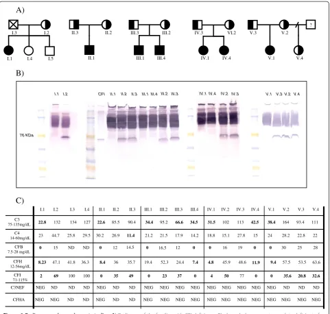
The clinically affected index cases from the five families studied have complete deficiency of CFI protein. Two of the relatives, III.4 and IV.4, brother and sister of III.1 and IV.1 respectively, were also completely CFI deficient, although they had no clinical signs related with this defect by the time the study was performed. All the CFI homozygous deficient had very reduced or no detectable CFI antigen in serum by ELISA and Western Blot (type I CFI deficiency, in which protein production or secretion is below the normal range). Their C3, CFB and CFH levels were also diminished as a consequence of the spontaneous activation and uncontrolled amplification of the complement alternative pathway. In contrast, par-tial CFI deficiency in some patients’relatives was charac-terized by diminished levels of CFI but normal amounts of all other complement proteins analyzed. In all cases, other possible causes of uncontrolled complement con-sumption, like the presence of C3NeF or anti CFH auto antibodies (CFHA), were ruled out. Complement profiles are shown in Figure 1.
A genetic cause of the deficiency was found in all families. The mutations identified in this series (sum-marized in Table 1) behave as recessive traits, as only homozygous carriers develop clinical symptoms.
We found 4 novel mutations causing CFI deficiency. Among them, we describe here the first large gene
dele-tion reported in the CFI locus, which includes exon 2
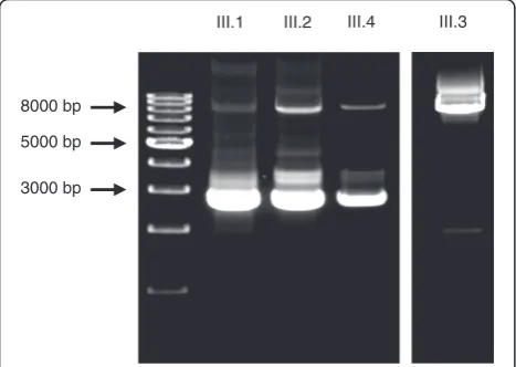
and part of the very large intron 1. This deletion was found in Family III and was characterized by MLPA. The findings were confirmed by long range PCR (XL-PCR), demonstrating a deletion size of approximately 5-6Kb (c.266-?_536 + ?del) in patient III.1, his mother III.2 and his brother III.4. The resulting fragments were resolved in 0.7% ultrapure agarose gel (Figure 2) but the precise break points of this deletion could not be confi-dently determined. In addition to that, patient III.2 car-ries a second, unreported molecular change in exon 4, c.738 A>T; p.N177I. Taking into account that patient
III.2’s serum CFI levels are reduced (23% of normal, see Figure 1) but significantly higher than in the homozy-gous deficient individuals and in her completely deficient offspring (in which CFI is undetectable), the substitution c.738 A>T is probably an unreported polymorphic var-iant. Patient III.2’s two children inherited the exon 2 deletion but not the c.738 A>T substitution, which implies that both changes are located in different alleles. In any case and due to the lack of co-segregation data, additional studies should be performed to confirm or
disprove the polymorphic nature of the c.738 A>T
change.
A novel mutation in exon 4 (c.559 C>T) was identi-fied in families IV and V. The C>T transition converted residue R187 into a premature termination codon. No consanguinity was reported affecting families IV and V, but their close geographic origin hints at a founder effect as a probable cause for the co-occurrence of this muta-tion in these families.
In family IV, a novel AT insertion in exon 13 between nucleotides 1610 and 1611 was also identified. The resultant frameshift change leads to a premature termi-nation codon shortly downstream of the insertion site.
The second mutation found in family V was another unreported mutation. Patient V.I, her mother and half sister, were found to bear an AT deletion (c.80_81delAT) in a region of exon 2 in the N-terminal domain. This deletion converts D27 into an alanine residue and leads to a frame-shifting situation that causes a premature stop codon (p.D27Afs*18).
The remaining mutations found in the patients that we studied had been previously described elsewhere: c.485 G>A [17], c.772 G>A [18], c.1176_1177dupAT [19], c.1420 C>T [20] (Table 1). The second mutation found in patient I.1 (c.485 G>A) is probably ade novo alteration, as both brother and sister of the proband do not carry this change. The father was deceased, so no further investigations could be performed on the segre-gation of this c.485 G>A change.
All the mutations reported here are shown in a sche-matic CFI protein model based on Protein Data Base accession 2XRC (Figure 3).
Discussion
As part of innate immunity, the complement system plays an important role in the defense against pathogens, especially in early childhood. Spontaneous and continu-ous activation of the complement cascade [21] must be tightly regulated by both fluid-phase and membrane-bound inhibitors to prevent uncontrolled amplification of C3 cleavage that may result in a state of acquired severe C3 deficiency.
opsonisation (that enhances microbe uptake into phagocy-tic cells viacomplement receptors), enhancement of anti-gen solubility and immunoanti-genicity, non-inflammatory removal of cell debris and immune complexes by phago-cytes and promotion of the inflammatory environment required for correct immune response [2,5,22]. Deficiency
of Complement Factor I (CFI, OMIM*217030) is a very
rare primary immunodeficiency disease [23], inherited as an autosomal recessive trait. Heterozygous CFI mutations are associated with atypical haemolytic uremic syndrome
(aHUS) [20], a severe disorder characterized by
thrombocytopenia, microangiopathic haemolytic anaemia and acute renal failure.
Defective opsonization in these patients makes them susceptible to recurrent pyogenic infections ( Streptococ-cus pneumoniae, Haemophilus influenzae, Neisseria meningitidis) as well as to aseptic meningitis [12,24]. Furthermore, deregulation of the complement cascade may result in autoimmune disease [22], vasculitis or glo-merulonephritis [25].
Among the CFI deficient individuals reported here, five out of seven homozygotes developed clinical symptoms
A)
B)
C)
I.3 I.2
I.1 I.4 I.5
II.3 II.2
II.1 IV.1
IV.3 VI.2
IV.4
III.3 III.2
III.1 III.4
V.3 V.2
V.1 V.4
? NEG NEG NEG NEG NEG NEG NEG NEG ND ND NEG ND ND NEG NEG CFHA NEG NEG NEG NEG NEG NEG NEG NEG ND ND NEG ND ND ND NEG C3NEF 0 77 50 4 0 37 23 0 49 35 0 100 100 69 2 CFI 71-115% 11.9 48.6 45.9 4.8 7.4 24.4 52.3 19.4 35.7 36 8.4 36.3 41.8 47.1 8.23 CFH 12-56mg/dL 0 19 16 0 0 12 16.5 0 14.5 12 0 ND ND 15 0 CFB 7.5-28 mg/dL 15 27.8 15.1 18.8 14.2 17.9 21.5 21.2 11.4 26.9 30.2 29.5 25.8 44.7 23 C4 14-60mg/dL 42.5 113 102 31.5 34.5 66.6 95.2 34.4 90.4 85.5 22.6 127 134 132 22.8 C3 75-135mg/dL IV.4 IV.3 IV.2 IV.1 III.4 III.3 III.2 III.1 II.3 II.2 II.1 I.4 I.3 I.2 I.1 NEG NEG NEG NEG ND ND ND NEG 32.6 20.8 35.6 0 63.6 53.5 57.5 9.4 28 25 30 0 22 22.8 28.2 24 111 93.4 164 38.4 V.4 V.3 V.2 V.1 I.2
I.1 CFI II.1 II.2 II.3 III.1 III.4 III.2 III.3 IV.1 IV.4 IV.2 IV.3 V.1 V.3 V.2 V.4
76 KDa
characteristic of the disease [16,18,26]. Concerning hetero-zygous carriers, other authors have described symptoms ranging from infections to aHUS [26,27]. To date, none of the relatives with partial CFI deficiency in our series have experienced symptoms.
At the present time, there is no specific therapy for these patients except keeping tight control on their
vac-cination against common bacterial pathogens such asS.
pneumoniae,H. influenzaeandN. meningitidis,and test-ing their correct antibody production. Despite normal polysaccharide antibody production in these patients, antibody weaning is faster than in the common popula-tion [28]. Furthermore, special care has to be taken to prevent associated diseases (using prophylactic antibio-tics if necessary) [3]. The main therapeutic goal is to promote the synthesis of anti-capsular antibodies cap-able of efficiently activating the classical pathway, although plasma infusion may be used in cases of severe infection to ameliorate clinical manifestations.
Familial studies lead us to identify two homozygous children in families III and IV without clinical evidences of CFI deficiency. Their very early diagnoses and the therapeutic precautions taken can most probably explain the lack of clinical manifestations. Even so, we cannot rule out the eventual onset of disease in such patients.
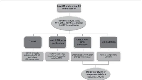
In this study, we aimed to show that in the presence of suggestive pathology and marked, but not absolute, per-manent C3 deficiency, screening of complement regula-tors should be performed. Besides, determination of CFI levels may discard other relatively frequent causes of C3 consumption (like C3NeF or CFH auto antibodies) and thus can guide the clinician in diagnostic tasks. The pre-sence of C3NeF can cause excessive C3 consumption, but clinical signs associated with this autoantibody fit in better with Dense Deposit Disease (DDD) and partial lipodystrophy (PLD) [29,30]. Auto antibodies directed against CFH can also decrease circulating C3 levels while maintaining C4 concentration in its normal range [31-33]. Moreover, functional studies have revealed that autoantibodies provoke the same dysfunction in CFH as mutations in its C-terminal domain, and there seems to be a direct correlation between the extent of CFH dys-function and the amount of CFH-autoantibody com-plexes [34]. Recently, anti-CFI autoantibodies have been described in aHUS, but their relationship with the devel-opment of the disease remains uncertain [35]. We have not tested the presence of anti-CFI antibodies in these 5 families, although this possibility could be explored in cases in which CFI deficiency is suspected.
Complete C3 or CFH deficiency presents clinical out-comes similar to those of CFI deficiency so that bio-chemical and/or functional studies are needed to distinguish among these deficiencies. Identification of the underlying molecular defect can be accomplished following the diagnostic algorithm depicted in Figure 4.
On the other hand, partial defect of C3, CFH or CFI is a susceptibility factor to develop aHUS, one of the most Table 1 Summary of the CFI mutations identified in the five families
Patients Genetic status cDNA ATG +1 Protein with signal peptide Exon Domain References
III.1, III.2, III.4 He c.266-?_536 + ?del - 2 - Not described
V.1, V.2, V.4 He c.80_81delAT p.D27Afs*18 2 N-terminal Not described
I.1 He c.485 G>A p.G162D 4 SRCR Le Quintrec M. et al. 2008 [17]
IV.1, IV.3, IV4, V.1, V.3 He c.559 C>T p.R187X 4 SRCR Not described
II.1 II.2, II.3
Ho He
c.772 G>A p.A258T 5 LDLRA2 Vyse et al. 1996 [18]
I.1, I.2 He c.1176_1177dupAT p.W393Yfs*5 11 SP Baracho et al. 2003 [19]
III.1, III.3, III.4 He c.1420 C>T p.R474X 11 SP Fremaux-Bacchi et al. 2004 [20]
IV.1, IV.2, IV.4 He c.1610_1611insAT p.V537Vfs*2 13 SP Not described
Ho, Homozygote; He, Heterozygote; SRCR, Scavenger Receptor Cysteine Rich domain; LDLRA2, Class A low-density lipoprotein receptor domain 2; SP, Serine Protease domain.
III.3 III.1 III.2 III.4
5000 bp
3000 bp 8000 bp
Figure 3Schematic model of the CFI protein and gene.The protein domains, exon/intron gene structure and the mutations found in our series are depicted.
Molecular study of complement defect (sequencing, MLPA)
C3NeF anti CFH auto antibodies
CFH, CFI or CFB mutations
C3 mutation
L
LoowwCC33ananddnnoorrmmaallCC44 q
quuaannttiiffiiccaattiioonn
C3Nef Haemolytic Assay CFH, CFI and CFB quantification
Anti CFH quantification
Uncontrolled AP activation and C3 consumption C3NeF antibody
stabilizes the AP C3 convertase (C3 consumption)
Anti CFH antibodies neutralize the regulatory
activity of FH.
Lack of complement activation
severe human diseases related to alternative pathway deregulation. When CFI deficiency has been established, family studies are needed in order to predict the conse-quences arising from the segregation of the mutated allele in the family. Multiple concurrent factors, both environmental and genetic, may be necessary in an indi-vidual for disease manifestation of aHUS [36]. Environ-mental factors are thought to initiate endothelium damage, while genetic factors favour disease progression [32]. Moreover, combined deficiencies of two or more complement factors (structural o regulator) have also been described, which adds complexity to these comple-ment deficiencies. Furthermore, a genetic component may also contribute to some typical, infectious HUS cases [33].
Conclusions
Complement Factor I (CFI) is the major complement inhi-bitor that degrades activated complement components C3b and C4b in the presence of specific cofactors. Its complete deficiency results in secondary complement deficiency due to uncontrolled spontaneous alternative pathway activation and causes susceptibility to infections. To date, mutations in several CFI domains have been described worldwide. Here, we report four novel muta-tions and the first large gene deletion in theCFIlocus.
We propose a general screening for this complement immunodeficiency whenever suggestive clinical features are present in the patient. C3 and C4 quantification are available to most laboratories, thus guiding the clinician at an early stage of the study (see Figure 4). Then, if the results are related to an alternative pathway deficiency with low C3 and normal C4, a multistep process may be performed in a more specialized laboratory for the pre-cise characterization of the defect at the molecular level. Such diagnostic process should include complement functional assays, protein quantification and auto anti-body analysis.
Abbreviations
C1INH, C1 inhibitor; CFI, Complement factor I; LDLr, Low-density lipoprotein receptor; FIMAC, FI Membrane attack complex domain; C4BP, C4b-Binding protein; CFH, Complement factor H; CR1 or CD35, Complement receptor 1; MCP or CD46, Membrane cofactor protein; CFB, Complement factor B; DIC, Disseminated intravascular coagulation; CFHA, Anti CFH auto antibodies; C3NeF, C3 nephritic factor; MLPA, Multiplex ligation-dependent probe amplification; XL-PCR, Long range PCR; AHUS, Atypical haemolytic uremic syndrome; DDD, Dense deposit disease; PLD, Partial lipodystrophy.
Competing interest
'The author(s) declare that they have no competing interests'.
Authors’contributions
MA-D carried out the molecular genetic studies, participated in sequence analysis, performed the immunoassays and drafted the manuscript. AL-L carried out the molecular genetic studies, took part in sequence analysis and drafted the manuscript. SG carried out the molecular genetic studies and immunoassays. IGG, JM, PS-P, CC and PN conceived the study and helped to draft the manuscript. ML-T conceived the study, took part in its design and
helped to draft the manuscript. All authors read and approved the final manuscript.
Acknowledgments
This work was supported by grant PS09/00122 (Ministry of Science and Innovation).
Author details 1
Unidad de Inmunología Hospital Universitario La Paz and Hospital La Paz Health Research Institute (IdiPAZ), Madrid, Spain.2Departamento de Pediatría Hospital Universitario 12 de Octubre, Madrid, Spain.3Servicio Inmunología Hospital Infanta Cristina, Badajoz, Spain.4Unidad de Patología Infecciosa e Inmunodeficiencias de Pediatría. Hospital Universitari Vall d’Hebron, Universitat Autònoma de Barcelona, Barcelona, Spain.5Servicio Inmunología Hospital San Pedro de Alcántara, Cáceres, Spain.6Centro de Investigación Biomédica en Red de Enfermedades Raras (CIBERER), Madrid, Spain.
Received: 28 March 2012 Accepted: 11 June 2012 Published: 18 June 2012
References
1. Botto M, Kirschfink M, Macor P, Pickering MC, Würzner R, Tedesco F: Complement in human diseases: Lessons from complement deficiencies.
Mol Immunol2009,46(14):2774–2783.
2. Degn SE, Jensenius JC, Thiel S:Disease-causing mutations in genes of the complement system.Am J Hum Genet2011,88(6):689–705.
3. Sullivan KE, Winkelstein JA:Primary Immunodeficiency Diseases: a molecular and genetic approach. InGenetically Determined Deficiencies of the Complement System. 2nd edition. Edited by Ochs HD, Smith CIE, Puck JM. New York: Oxford University Press; 2007:589–608.
4. Roversi P, Johnson S, Caesar JJ, McLean F, Leath KJ, Tsiftsoglou SA, Morgan BP, Harris CL, Sim RB, Lea SM:Structural basis for complement factor I control and its disease-associated sequence polymorphisms.Proc Natl Acad Sci U S A2011,108(31):12839–12844.
5. Nilsson SC, Sim RB, Lea SM, Fremeaux-Bacchi V, Blom AM:Complement Factor I in health and disease.Mol Immunol2011,48(14):1611–1620. 6. Tsiftsoglou SA, Arnold JN, Roversi P, Crispin MD, Radcliffe C, Lea SM, Dwek
RA, Rudd PM, Sim RB:Human complement factor I glycosylation: structural and functional characterisation of the N-linked oligosaccharides.Biochim Biophys Acta2006,1764(11):1757–1766. 7. Fearon DT:Purification of C3b inactivator and demonstration of its two
polypeptide chain structure.J Immunol1977,119:1248–1252.
8. Goldberger G, Arnaout MA, Aden D, Kay R, Rits M, Colten HR:Biosynthesis and postsynthetic processing of human C3b/C4b inactivator (factor I) in three hepatoma cell lines.J Biol Chem1984,259:6492–6497.
9. Goldberger G, Bruns GA, Rits M, Edge MD, Kwiatkowski DJ:Human complement factor I: analysis of cDNA-derived primary structure and assignment of its gene to chromosome 4.J Biol Chem1987,262 (21):10065–10071.
10. Shiang R, Murray JC, Morton CC, Buetow KH, Wasmuth JJ, Olney AH, Sanger WG, Goldberger G:Mapping of the human complement factor I gene to 4q25.Genomics1989,4(1):82–86.
11. Sadallah S, Gudat F, Laissue JA, Spath PJ, Schifferli JA:Glomerulonephritis in a patient with complement factor I deficiency.Am J Kidney Dis1999, 33(6):x1153–1157.
12. González-Rubio C, Ferreira-Cerdán A, Ponce IM, Arpa J, Fontán G, López-Trascasa M:Complement factor I deficiency associated with recurrent meningitis coinciding with menstruation.Arch Neurol2001,58(11):1923–1928.
13. Abarrategui-Garrido C, Martínez-Barricarte R, López-Trascasa M, de Córdoba SR, Sánchez-Corral P:Characterization of complement factor H-related (CFHR) proteins in plasma reveals novel genetic variations of CFHR1 associated with atypical hemolytic uremic syndrome.Blood2009,114 (19):4261–4271.
14. de Goicoechea Jorge E, Harris CL, Esparza-Gordillo J, Carreras L, Arranz EA, Garrido CA, López-Trascasa M, Sánchez-Corral P, Morgan BP, de Rodríguez Córdoba S:Gain-of-function mutations in complement factor B are associated with atypical hemolytic uremic syndrome.Proc Natl Acad Sci U S A2007,104(1):240–245.
16. Ponce-Castro IM, González-Rubio C, Delgado-Cerviño EM, Abarrategui-Garrido C, Fontán G, Sánchez-Corral P, López-Trascasa M:Molecular characterization of Complement Factor I deficiency in two Spanish families.Mol Immunol2008,45(10):2764–2771.
17. Le Quintrec M, Lionet A, Kamar N, Karras A, Barbier S, Buchler M, Fakhouri F, Provost F, Fridman WH, Thervet E, Legendre C, Zuber J, Frémeaux-Bacchi V: Complement mutation-associated de novo thrombotic microangiopathy following kidney transplantation.Am J Transplant2008,8(8):1694–1701. 18. Vyse TJ, Morley BJ, Bartok I, Theodoridis EL, Davies KA, Webster AD, Walport
MJ:The molecular basis of hereditary complement factor I deficiency.J Clin Invest1996,97(4):925–933.
19. Baracho GV, Nudelman V, Isaac L:Molecular characterization of homozygous hereditary factor I deficiency.Clin Exp Immunol2003,131(2):280–286. 20. Fremeaux-Bacchi V, Dragon-Durey MA, Blouin J, Vigneau C, Kuypers D,
Boudailliez B, Loirat C, Rondeau E, Fridman WH:Complement factor I: a susceptibility gene for atypical haemolytic uraemic syndrome.J Med Genet2004,41(6):e84.
21. Bexborn F, Andersson PO, Chen H, Nilsson B, Ekdahl KN:The tick-over theory revisited: formation and regulation of the soluble alternative complement C3 convertase (C3(H2O)Bb).Mol Immunol2008,45(8):2370–2379. 22. Zipfel PF, Skerka C:Complement regulators and inhibitory proteins.Nat
Rev Immunol2009,9(10):729–740.
23. International Union of Immunological Societies Expert Committee on Primary Immunodeficiencies, Notarangelo LD, Fischer A, Geha RS, Casanova JL, Chapel H, Conley ME, Cunningham-Rundles C, Etzioni A, Hammartröm L, Nonoyama S, Ochs HD, Puck J, Roifman C, Seger R, Wedgwood J:Primary immunodeficiencies: 2009 update.J Allergy Clin Immunol2009,124 (6):1161–1178.
24. Bonnin AJ, Zeitz HJ, Gewurz A:Complement factor I deficiency with recurrent aseptic meningitis.Arch Intern Med1993,153(11):1380–1383. 25. Genel F, Sjöholm AG, Skattum L, Truedsson L:Complement factor I
deficiency associated with recurrent infections, vasculitis and immune complex glomerulonephritis.Scand J Infect Dis2005,37(8):615–618. 26. Nilsson SC, Trouw LA, Renault N, Miteva MA, Genel F, Zelazko M, Marquart
H, Muller K, Sjöholm AG, Truedsson L, Villoutreix BO, Blom AM:Genetic, molecular and functional analyses of complement factor I deficiency.Eur J Immunol2009,39(1):310–323.
27. Grumach AS, Leitão MF, Arruk VG, Kirschfink M, Condino-Neto A: Recurrent infections in partial complement factor I deficiency: evaluation of three generations of a Brazilian family.Clin Exp Immunol
2006,143(2):297–304.
28. Ochs HD, Wedgwood RJ, Frank MM, Heller SR, Hosea SW:The role of complement in the induction of antibody responses.Clin Exp Immunol
1983,53(1):208–216.
29. Licht C, Fremeaux-Bacchi V:Hereditary and acquired complement dysregulation in membranoproliferative glomerulonephritis.Thromb Haemost2009,101(2):271–278.
30. Levy Y, George J, Yona E, Shoenfeld Y:Partial lipodystrophy,
mesangiocapillary glomerulonephritis, and complement dysregulation.
An autoimmune phenomenon. Immunol Res1998,18(1):55–60.
31. Dragon-Durey MA, Loirat C, Cloarec S, Macher MA, Blouin J, Nivet H, Weiss L, Fridman WH, Frémeaux-Bacchi V:Anti-Factor H autoantibodies associated with atypical hemolytic uremic syndrome.J Am Soc Nephrol
2005,16(2):555–563.
32. Sánchez-Corral P, Melgosa M:Advances in understanding the aetiology of atypical Haemolytic Uraemic Syndrome.Br J Haematol2010,150 (5):529–542.
33. Loirat C, Frémeaux-Bacchi V:Atypical hemolytic uremic syndrome.
Orphanet J Rare Dis.2011,6:60.
34. Strobel S, Hoyer PF, Mache CJ, Sulyok E, Liu WS, Richter H, Oppermann M, Zipfel PF, Józsi M:Functional analyses indicate a pathogenic role of factor H autoantibodies in atypical haemolytic uraemic syndrome.Nephrol Dial Transplant2010,25(1):136–144.
35. Kavanagh D, Pappworth IY, Anderson H, Hayes CM, Moore I, Hunze EM, Bennaceur K, Roversi P, Lea S, Strain L, Ward R, Plant N, Nailescu C, Goodship TH, Marchbank KJ:Factor I autoantibodies in patients with
atypical hemolytic uremic syndrome: disease-associated or an epiphenomenon?Clin J Am Soc Nephrol2012,7(3):417–426.
36. Moore I, Strain L, Pappworth I, Kavanagh D, Barlow PN, Herbert AP, Schmidt CQ, Staniforth SJ, Holmes LV, Ward R, Morgan L, Goodship TH, Marchbank KJ:Association of factor H autoantibodies with deletions of CFHR1, CFHR3, CFHR4, and with mutations in CFH, CFI, CD46, and C3 in patients with atypical hemolytic uremic syndrome.Blood2010,115(2):379–387.
doi:10.1186/1750-1172-7-42
Cite this article as:Alba-Domínguezet al.:Complement factor I deficiency: a not so rare immune defect. Characterization of new mutations and the first large gene deletion.Orphanet Journal of Rare Diseases20127:42.
Submit your next manuscript to BioMed Central and take full advantage of:
• Convenient online submission
• Thorough peer review
• No space constraints or color figure charges
• Immediate publication on acceptance
• Inclusion in PubMed, CAS, Scopus and Google Scholar
• Research which is freely available for redistribution