R E S E A R C H
Open Access
Localization of
Helicobacter
spp. in the fundic
mucosa of laboratory Beagle dogs: an
ultrastructural study
Anna Lanzoni
1, Ivo Faustinelli
1, Patrizia Cristofori
2, Mario Luini
3, Kenneth W Simpson
4, Eugenio Scanziani
5,6,
Camilla Recordati
5,6*Abstract
In dogsHelicobacter spp. are found in all gastric regions usually localized in the surface mucus, gastric glands and parietal cells. The aim of this study was to detail the distribution ofHelicobacter spp. in the fundic mucosa of asymptomatic Beagle dogs and their intracellular localization within parietal cells, in order to evaluate species-specific pathogenetic effects on gastric cells. The presence ofHelicobacterspp. was investigated by
immunohistochemistry, TEM, and PCR in the fundic mucosa of six Beagle dogs.Helicobacterspp. were found in all dogs examined, andH. bizzozeronii andH. feliswere identified by PCR and confirmed by TEM. In the lumen of the fundic glands, co-localization was common.H. bizzozeroniiwas present in larger numbers thanH. felisin both intraluminal and intraparietal localization. The amounts ofH. bizzozeroniiwere similar in superficial and basal portions of the glands.H. feliswas predominantly localized in the superficial portions of gastric glands but almost absent from the base. Within parietal cells, mostHelicobacter organisms were intracanalicular, but intact and degenerateHelicobacter organisms were also visualized free in the cytoplasm or in secondary lysosomes. No specific degenerative lesions were found in infected parietal cells.Helicobacterorganisms were also observed within macrophages in the lamina propria. In conclusion, there is a differential distribution ofH. bizzozeroniiand H. felisin the fundic mucosa of Beagle dogs, and their intracellular localization in parietal cells and macrophages suggests novel pathogenic scenarios for the development of immune response and maintenance of chronic gastritis in dogs.
Introduction
Gastric colonization by spiral-shaped bacteria of the
genusHelicobacteris common in dogs and to date
sev-eral species of Helicobacter have been identified in the canine stomach, includingHelicobacter felis, Helicobac-ter bizzozeronii, Helicobacter salomonis, “Candidatus
Helicobacter heilmannii”,Helicobacter bilis,Helicobacter
(Flexispira) rappini, and Helicobacter cynogastricus
[1-6]. Dogs may be infected with more than one species ofHelicobacterthat are indistinguishable on light micro-scopy, appearing as large tightly coiled spiral-shaped bacteria [7]. Different species of canine gastric Helico-bacter can be categorized by their morphological
features (i.e. size, number and degree of tightness of the spirals, presence of periplasmic fibrils, number and posi-tion of flagella) using transmission electron microscopy (TEM). In addition, in the early work of Lockard and Boler (1970) morphologically different organisms were classified as Lockard types 1 to 3 [8,9].
The regional distribution ofHelicobacterspp. through-out the canine stomach (i.e. cardia, fundus, and pylorus) is well studied [7], but knowledge of the distribution of differentHelicobacterspp. within these regions is incom-plete.Helicobacterorganisms in dogs reside in the sur-face mucus, gastric pits, and gastric glands, colonizing both the lumen and the gastric cells. In particular, canine and felineHelicobacterspp. (as“CandidatusH. heilman-nii”in humans) have great predilection for parietal cells of the fundic mucosa, where they are usually described to colonize the intracellular canaliculi and sporadically the
* Correspondence: [email protected]
5Dipartimento di Patologia Animale, Igiene e Sanità Pubblica Veterinaria, Facoltà di Medicina Veterinaria, Università degli Studi di Milano, 20133 Milano, Italy
Full list of author information is available at the end of the article
cytoplasm [7,10-14]. Since the virulence of many patho-genic bacteria is directly related to invasion it is possible that the development ofHelicobacterassociated gastritis in dogs is related to their intracellular localization (intra-canalicular or intracytoplasmic) or possibly invasion of the lamina propria through the basal lamina, as reported forH. pylori, which is now considered as an invasive and facultative intracellular organism [15-17]. The ability of
Helicobacterspp. infection in dogs to establish an intra-cellular niche may also vary by species or even strain, and this may impact on their pathogenicity and ability to evade antibiotics typically employed to treatHelicobacter
gastritis in dogs [18].
The aim of this study was to gain insight into the dis-tribution and localization of distinctHelicobacterspp. in the lumen of gastric glands and within parietal cells in the fundic mucosa, using a limited number of asympto-matic Beagle dogs. This was done by applying a combi-nation of morphological (immunohistochemical and ultrastructural) and molecular methods (species-specific PCR) and allowed to evaluate the presence of distinct colonization niches and possible species-specific patho-genetic effects on gastric cells.
Materials and methods Animals and sampling
Three male (No. 1, 2, 3) and three female (No. 4, 5, 6) Bea-gle dogs (Marshall BeaBea-gles), purchased from Greenhill Italy (Montichiari, Brescia, Italy) were investigated. The dogs were 8-9 months old, and were part of a general toxicology study performed in GlaxoSmithKline S.p.A., where they served as the control group. They were housed singly or in pairs, in standard husbandry and environmental conditions. All the work involving animals was carried out in accor-dance with Italian regulation governing animal welfare and protection (Legislative Decree n. 116/1992), the European
Directive 86/609/EEC, and according to the internal Glax-oSmithKline Committee on Animal Research & Ethics (CARE) review, with related codes of practice.
Dogs were euthanized by intravenous injection of bar-biturate. Prior to necropsy, the animals were deprived of food overnight. The stomach was opened along the inner curvature and macroscopically examined. Three samples were taken from the body (greater curvature) region from each dog: one was snap frozen in liquid nitrogen and stored at -80°C for PCR analysis; one was fixed in 10% buffered formalin for histopathology; one was fixed in 2.5% glutaraldehyde in 0.1 mol/L cacodylate buffer pH 7.2, containing 0.01 mol/L CaCl2 for at least
12 h at 4°C for ultrastructural examination.
DNA Extraction and PCR amplification
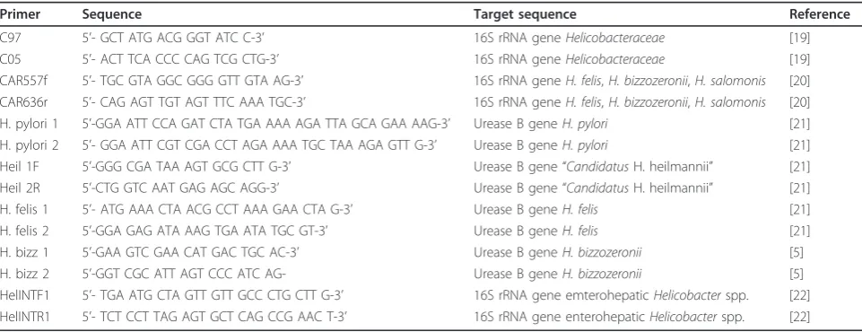
DNA was extracted using DNeasy Tissue kit (QIAGEN, GmbH D-40724, Hilden, Germany) according to the manufacturer’s instructions. Primers used in this study for the amplification ofHelicobacteraceae, canine gastric
Helicobacter spp. (H. bizzozeronii, H. salomonis and
H. felis), canine enterohepatic Helicobacter spp.,
H. pylori, “Candidatus H. heilmannii”, H. felis, and
H. bizzozzeroniiare listed in Table 1.
All PCR reactions were performed in an Eppendorf ther-mocycler (Mastercycler gradient, Eppendorf GA, Hamburg, Germany) in a final volume of 25μL containing 1μL of
extracted DNA, 12.5μL of PCR Master Mix (Promega,
Madison, Wisconsin, USA), and 0.5μM of each primer
(Integrated DNA Technologies, Coralville, IA, USA). Ten microliters of each amplification product were analyzed by gel electrophoresis in 1% agarose gels in Tris-acetate EDTA buffer (TAE). The gel was stained with ethidium bromide (0.5 mg/L) and examined under UV transilluminator for the presence of amplified DNA; the size of the expected fragment was compared to a 100 bp reference marker
Table 1 Oligonucleotide primers used in this study
Primer Sequence Target sequence Reference
(Fermentas Inc, Maryland, USA). Negative controls in which the DNA extract was omitted were included with each reaction.
Helicobacteraceae PCR
The 16S rRNA gene of members of the
Helicobactera-ceae family was amplified by PCR using primers C97/
C05 [19] generating a 1200 bp amplicon. For C97/C05 amplification samples were heated to 94°C for 10 min, followed by 30 amplification cycles of denaturation at 94°C for 1 min, primer annealing at 58°C for 1 min and 30 s, and extension at 72°C for 2 min, with a final extension at 72°C for 10 min.
Gastric Helicobacter spp. PCR
Primers CAR557f and CAR636r were used to amplify a 78 bp fragment of the 16S rRNA gene of the canine gas-tric Helicobacterspecies (H. bizzozeronii, H. salomonis
andH. felis) [20]. For CAR557f/CAR636r amplification samples were heated to 94°C for 9 min, followed by 35 amplification cycles of denaturation at 94°C for 30 s, primer annealing at 60°C for 30 s, and extension at 72°C for 45 s, with a final extension at 72°C for 5 min. Addi-tionally, all gastric samples were investigated using
species-specific primers for H. pylori, “Candidatus
H. heilmannii”, H. felis, andH. bizzozzeronii urease B gene amplification, as previously reported [5,21].
Enterohepatic Helicobacter spp. PCR
Primers HelINTF1 and HelINTR1 were used to amplify a 345 bp fragment of the 16S rRNA gene of enterohepatic
Helicobacterspp. [22]. For HelINTF1/HelINTR1 amplifi-cation samples were heated to 94°C for 2 min, followed by 27 amplification cycles of denaturation at 94°C for 30 s, primer annealing at 60°C for 30 s, and extension at 72°C for 30 s, with a final extension at 72°C for 7 min.
Histopathology
For light microscopy examination, formalin-fixed
sam-ples were embedded in paraffin wax, sectioned at 4 μm
thickness, and stained with hematoxylin and eosin (HE).
For immunohistochemistry (IHC), formalin-fixed, par-affin-embedded 4μm sections were deparaffinized, rehy-drated and treated with 3% hydrogen peroxide in distilled water for 20 min. The sections were labelled by the avidin-biotin-peroxidase (ABC) procedure [23] with a commercial immunoperoxidase kit (Vectastain Stan-dard Elite; Vector Laboratories, Burlingame, CA, USA).
A polyclonal rabbit anti-H. pylori antibody (Dako,
Glostrup, Denmark) was applied at a working dilution of 1:1000 at 4°C overnight. The immunohistochemical
reaction was developed with 3,3’ diaminobenzidine
(DAB) for 1 min and sections were counterstained for 1 min with Mayer’s haematoxylin. Negative controls for each sample were prepared by replacing the primary antibody with PBS containing 10% normal goat serum.
Known positive control sections were included in each immunolabeling assay.
Samples were examined for the presence of inflamma-tion (infiltrates of lymphocytes, plasma cells, and macro-phages) using a semiquantitative scoring system as follows: 0 (absent), +1 (mild), +2 (moderate), +3 (marked).
Immunostained sections were scored for the presence ofHelicobacter antigen within the parietal cells (P) and in the lumen (L) of gastric glands using a semiquantita-tive scoring system as follows: 0 (absence ofHelicobacter
antigen in the parietal cell/lumen of the gastric glands); +1 (presence ofHelicobacterantigen in the parietal cell/ lumen of < 5% of the gastric glands); +2 (presence of
Helicobacterantigen in the parietal cell/lumen of 5-50% of the gastric glands); +3 (presence ofHelicobacter anti-gen in the parietal cell/lumen of > 50% of the gastric
glands); +4 (presence of abundant Helicobacterantigen
in the parietal cell/lumen of > 50% of the gastric glands and extending to the base of the glands).
Transmission electron microscopy (TEM)
Glutaraldehyde fixed samples were transferred into phosphate buffer after 72 h, postfixed for 90 min in 1% osmium tetroxide (wt/vol), and then stained with 2% (wt/vol) uranyl acetate in barbiturate buffer pH 7.3 for 60 min. After dehydration and embedding in Epon
(gly-cide ether 100, Merck, Darmstadt, Germany), 1 μ
m-thick sections were stained with toluidine blue and the areas of interest were selected. Thin sections were stained with uranyl acetate and lead citrate. Electron microscopy was carried out using a Philips Morgagni™ 268D transmission electron microscope (FEI Company, Eindhoven, The Netherlands) operating at 90 kV.
Helicobacter spp. were identified according to their morphological features (size, number of spirals, presence of periplasmic fibrils, number and position of flagella) [2,3,24]. The distribution and number (bacterial load) of
Helicobacterspp. was evaluated in 10-20 gastric glands and in 4-8 parietal cells of the fundic mucosa from 3 dogs (No. 1-3). The superficial (pit/isthmus/neck) and basal portions of the gastric glands were investigated as distinct colonization sites and the number of distinct
morphological types of Helicobacterspp. was evaluated
within parietal cells (including intracanalicular and intracytoplasmic compartments).
Statistical analysis
evaluated using the Wilcoxon signed-rank test. Statisti-cal significance was set atP= 0.001.
Results PCR
Helicobacteraceaeand canine gastricHelicobacter spp. DNA was amplified in all fundic samples from all six dogs. Species-specificHelicobacterPCR detected H. biz-zozeroniiandH. felis in all six dogs. PCR for“
Candida-tus H. heilmannii”, H. pylori and enterohepatic
Helicobacterspp. was negative for all samples.
Gross pathology and histopathology
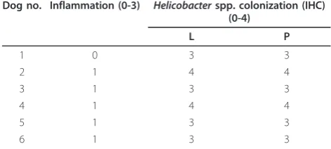
No gross lesions were observed in the stomach of the dogs. Histologically, mild chronic inflammation was pre-sent in the fundic mucosa of 5 of 6 dogs (Table 2). Focal to multifocal infiltrates of small numbers of lym-phocytes, plasma cells, and macrophages were found in the lamina propria, mainly localized in the deepest por-tion of the mucosa, close to themuscularis mucosae. In HE-stained sections, ill-defined elongated bacteria con-sistent with Helicobacterspp. were visible at high power magnification (400×) in the mucus covering the surface epithelium, the gastric pits, lumen of gastric glands, and occasionally in parietal cells. In all dogs, most parietal cells were morphologically normal; however, small num-bers of parietal cells with a large cytoplasmic vacuole containing bacterial organisms were found, localized mainly in the upper third of the gastric gland, around the isthmus region.
The degree of Helicobacterspp. colonization of the
stomach assessed by IHC is summarized in Table 2.
Positive immunostaining of Helicobacter antigen was
characterized by a coarse granular brown precipitate easily detectable against the light blue background.
Moderate to large numbers of Helicobacter spp. were
visible in the fundic mucosa, in particular the Helicobac-terantigen was found in the mucus covering the surface epithelium, the gastric pits, lumen of gastric glands, and
intracellularly within parietal cells, multifocally con-tained in large vacuoles. There was no difference in
Helicobacter colonization density between the lumen and parietal cells. Within parietal cells, theHelicobacter
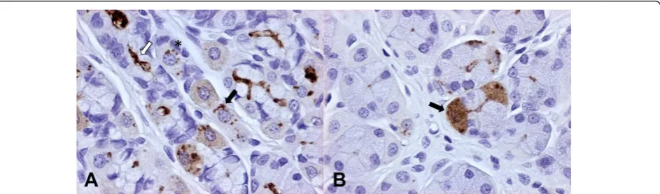
antigen was visible as 1-2μm in diameter, well demar-cated, brown dots or less frequently as a fine brown granular staining diffuse to the whole cytoplasm. Occa-sionally, well defined spiral shaped organisms were visi-ble in parietal cells (Figure 1).
Transmission electron microscopy (TEM)
Bacteria were identified by TEM in the fundic mucosa from all the dogs examined. They appeared as numer-ous, 4-8μm in length × 0.6-0.8 μm in width, tightly to loosely coiled spiral-shaped bacteria with or without periplasmic fibrils, and with bipolar flagellar tufts (com-posed of up to six flagella) consistent with two types of large canine gastric Helicobacterspp. According to the results of PCR and aforementioned ultrastuctural fea-tures they were identified as H. bizzozeronii-like and
H. felis-like (Figures 2A and 2B).
In the superficial mucus layer, low numbers of
free-floating Helicobacter spp. were seen, occasionally
admixed with bacteria with disrupted walls and variably condensed or vacuolated cytoplasm (degenerated bac-teria). In the lumen of the fundic glands, co-localization of the two morphologically distinctHelicobacterspp. was
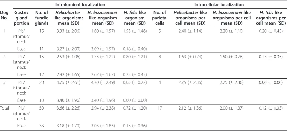
common.Helicobacterspp. load in the lumen of gastric
glands and parietal cells is summarized in Table 3.
Intra-luminal load of total Helicobacter-like organisms and
H. bizzozeronii-like organisms was similar in superficial
and basal portions of the glands (P = 0.4146 andP=
0.4817), while the load ofH. felis-like was significantly higher in the superficial portions compared to the base (P= 0.0007). Both in the superficial and basal portions, there were significantly moreH. bizzozeronii-like organ-isms thanH. felis-like organisms (P< 0.0001). In parietal cells, there were significantly moreH. bizzozeronii-like organisms thanH. felis-like organisms (P= 0.0004).
Intraluminal bacteria were localized close to the lumi-nal surface of gastric epithelial cells and the lumilumi-nal pole of the intracanalicular system of parietal cells. Although H. felis-like organisms were often in contact with parietal cell membranes and microvilli, no adhesion to cellular membranes, pedestal formation, actin con-densation or disruption of intercellular junctions were found. Within parietal cells, electrondense structures morphologically consistent with bacterial organisms were frequently identified. In most cases, bacteria were found in the intracellular portion of the canalicular sys-tem (Figure 2C). Both well preserved spiral-shaped forms, with or without periplasmic fibrils, and rounded bodies (transverse sections of bacteria) were observed (Figure 2C). Bacteria were also present within the Table 2 Gastric inflammation andHelicobacterspp.
colonization of fundic mucosa in 6 laboratory Beagle dogs infected byHelicobacterspp
Dog no. Inflammation (0-3) Helicobacterspp. colonization (IHC) (0-4)
L P
1 0 3 3
2 1 4 4
3 1 3 3
4 1 4 4
5 1 3 3
6 1 3 3
cytoplasm of parietal cells enveloped by a closely apposed membrane, occasionally focally disrupted, or free in the cytoplasm or within secondary lysosomes (Figure 2D). Condensation or lysis of bacterial structures consistent with bacterial degeneration was occasionally visualized within the cytoplasm of parietal cells (Figure 2E). In both colonized and uncolonized parietal cells, secondary lysosomes were often present and degenerat-ing swollen mitochondria were occasionally observed. Macrophages in the lamina propria of fundic mucosa had numerous secondary lysosomes similar to those detected in parietal cells, and, rarely, intracellular Helico-bacter organisms were found in their cytoplasm. In a single case,Helicobacter organisms were in association with remnants of an entire phagocytized cell (Figure 2F).
Discussion
Helicobacter spp. were identified by histology, IHC, TEM, and PCR in the fundic mucosa of all the dogs examined in this study, confirming the high prevalence (100%) of the infection in healthy laboratory Beagle dogs [1,12,25].Helicobacter-like organisms were visualized in the surface mucus, lumen of gastric glands (extending from the gastric pit to the base), and within parietal cells, but not within mucous epithelial cells and chief cells, in agreement with previous studies [12,13,26]. On the basis of species-specific PCR, all of the dogs were found to be coinfected withH. bizzozeroniiandH. felis
and this finding was supported by TEM, which showed
two morphologically distinct Helicobacter types,
H. bizzozeronii-like and H. felis-like, in the fundic
mucosa. Coinfection with H. bizzozeronii-like and
H. felis-like organisms, has been previously observed in laboratory Beagles [12,25].
In depth analysis of TEM images suggests that these
Helicobacter species are adapted for colonization of
distinct niches in the fundic mucosa. In the lumen of fundic glands, co-localization of the two distinct mor-phologic types was common.H. bizzozeronii-like organ-isms colonized the glands from the pit to the base, whileH. felis-like organisms were localized in the more superficial portions of the gastric glands (pit, isthmus and neck regions), but virtually absent from the base. In all dogs and portions of gastric glands evaluated,
H. bizzozeronii-like organisms were significantly more abundant thanH. felis-like organisms. Within parietal
cells,H. bizzozeronnii was also the most common
spe-cies, and this can be simply the result of the lower
abundance of H. felisin the glands of these dogs or a
consequence of the lower affinity of H. felistowards the parietal cells compared to H. bizzozeronii. The latter hypothesis is supported by the presence of higher pro-portion of H. felis-like organisms in the glands versus parietal cells (H. felis in glands/parietal cells = 4.05) compared toH. bizzozeronii-like organisms (H. bizzozer-onii in glands/parietal cells = 1.49). Altogether, these findings may indicate thatH. bizzozeroniicould be more host-adapted thenH. felisin dogs.
Some previous studies have suggested thatH. felis is
more pathogenic thanH. bizzozeronii in dogs and also
experimentally in mice and gerbils, resulting in necrosis and/or apoptosis of parietal cells with subsequent parie-tal cell loss, whereas H. bizzozeroniiappears non patho-genic even in cases of heavy bacterial load [27-30]. In the present study, however, there were no specific and relevant ultrastructural parietal cell degenerative lesions with either of Helicobacterspecies, but the lack of cell damage could also be related to the limited sampling of the fundic mucosa that was performed in this study. Additionally, no evidence of tight attachment to gastric epithelial cells and no intercellular invasion by canine gastric Helicobacterspp. were found and this contrasts Figure 1Dog, fundic mucosa (immunoperoxidase staining, anti-H. pyloriIHC, 400×). Figure 1A - Large amounts ofHelicobacterantigen in the lumen of fundic glands (white arrow) and within parietal cells, whereHelicobacterantigen is detectable as well preserved spiral-shaped organisms (black arrow) or brown round dots (asterisk). In scattered parietal cells, a diffuse light cytoplasmic staining is also visible. Figure 1B
withH. pyloriwhich is commonly found to attach to the cell membrane of epithelial cells by different forms of adhesive structures (i.e. pedestals or membrane fusion), and invade intercellular space with disruption of inter-cellular junctions [15,17,31]. Although there was no attachment to the cell membrane,H. felis-like organisms
were often in direct but loose contact with the plasma cell membranes and microvilli of parietal cells, confirm-ing previous ultrastructural observations performed in dogs and gerbils [27,30].
Canine and felineHelicobacterspp., as well as “ Candi-datusH. heilmannii”in humans, have greater affinity to Figure 2Dog, ultrastructural investigation of the fundic mucosa. Figure 2A. IntraluminalH. bizzozeronii-like organism. A tightly coiled spiral-shaped organism without periplasmic fibrils is free-floating in the superficial mucus (TEM, original magnification 8900×). Figure 2B. Intraluminal
H. felis-like organism. A tightly coiled spiral-shaped bacteria with periplasmic fibrils is visible in the superficial mucus; multifocal contact between
H. felis-like organism and parietal cell microvilli is present (TEM, original magnification 8900×). Figure 2C. Parietal cell containingHelicobacter-like
organisms. Multiple sections ofHelicobacter-like organisms were localized within a canaliculus (black arrow) (TEM, original magnification 4 400×). Inset: other bacterial sections were unclearly delimited by parietal cell membranes and adjacent to secondary lysosomes (TEM, original magnification 28000×). Figure 2D. High magnification of the cytoplasm of a parietal cell containing two electrondense intracytoplasmic
Helicobacter-like organisms (H). In the upper left corner a longitudinal section of anHelicobacter-like organism is surrounded by closely apposed
parietal cells than doesH. pyloriin man, and are usually found in their intracellular canaliculi [7,10-13,27,32,33]. Also in the present study most of the intracellular organisms were intracanalicular, but cross-sections of
Helicobacter organisms or degenerating bacterial frag-ments were found directly free in the cytoplasm of par-ietal cells, in association with secondary lysosome-like structures, or occasionally enveloped by a closely apposed membrane which could be focally interrupted resulting in the partial penetration of the organism into the cytoplasm. These ultrastructural findings are consis-tent with a true intracytoplasmic localization, often asso-ciated with bacterial degradation. It is not clear how
Helicobacterorganisms penetrate into the cytoplasm of parietal cells, and whether the intracytoplasmic organ-isms are associated with parietal cell degeneration, since only sporadic degenerative alterations of organelles were found in both infected and uninfected parietal cells.
By IHC, intracellular Helicobacterantigen within par-ietal cells appeared as well demarcated dots or diffuse cytoplasmic staining of parietal cells. Based on the ultra-structural findings, the immunohistochemical dots are likely the cross-sections of intracellular bacteria or their degrading forms, while the diffuse cytoplasmic staining could be the result of cytoplasmic bacterial degradation and dispersion of bacterial contents (including antigens) throughout the cytoplasm.
Sporadically, Helicobacter spp. and their fragments
were found by TEM, but not by IHC, in the cytoplasm of macrophages in the lamina propria of the fundic
mucosa. The presence of Helicobacterorganisms within
macrophages can be the result of phagocytosis of bac-teria released in the extracellular space after parietal cell
necrosis or phagocytosis of degenerated parietal cells containing intracellular bacteria, as suggested by the presence of intracytoplasmic remnants of an entire phagocytized cell in association withHelicobacter organ-isms within a macrophage found in the lamina propria. Direct invasion of lamina propria with phagocytosis by macrophages seems less likely the mechanism since no evidence of transepithelial invasion was found. To
our knowledge, canineHelicobacterspp. have not been
so far described within macrophages, in contrast to
H. pyloriwhich has been described to invade the lamina propria and translocate via macrophages to gastric lymph nodes, leading to the chronic stimulation of the immune system and thus contributing to the mainte-nance of chronic gastritis in people [15-17,34].
In the fundic mucosa of this small group of Beagle
dogs, we observed high levels of mucosal Helicobacter
colonization but clinical signs of gastritis were absent and only mild chronic lymphoplasmacytic gastritis was
detected. The absence of a correlation between
Helico-bacter colonization density, severity of inflammation, and clinical signs has been reported by several studies [7,13,14,26,35].
Previous studies have demonstrated thatHelicobacter
infection in dogs induces a specific systemic humoral response with reported seroconversion after infection and a Th1-biased local immune response [14,27,35], but the mechanisms of activation of the immune response in association with canineHelicobacterspp. infection have been so far not investigated. The intracytoplasmic locali-zation of canineHelicobacterspp. in parietal cells and macrophages in the lamina propria disclose novel patho-genic scenarios for the development of the cell-mediated Table 3Helicobacterspp. load in gastric glands and parietal cells evaluated by electron microscopy
Intraluminal localization Intracellular localization
Dog No.
Gastric gland portion
No. of fundic glands
Helicobacter -like organisms
mean (SD)
H. bizzozeronii -like organism
mean (SD)
H. felis-like organism mean (SD)
No. of parietal
cells
Helicobacter-like organisms per cell mean (SD)
H. bizzozeronii-like organisms per cell
mean (SD)
H. felis-like organisms per cell mean (SD)
1 Pit/ isthmus/
neck
15 3.33 (± 2.06) 1.80 (± 1.57) 1.53 (± 1.46) 5 2.40 (± 1.14) 2.20 (± 1.10) 0.20 (± 0.45)
Base 11 3.27 (± 2.00) 3.09 (± 1.97) 0.18 (± 0.40)
2 Pit/ isthmus/
neck
15 2.53 (± 1.06) 1.73 (± 1.22) 0.80 (± 1.21) 8 1.63 (± 0.74) 1.50 (± 0.76) 0.13 (± 0.35)
Base 12 2.92 (± 1.65) 2.67 (± 1.67) 0.25 (± 0.45)
3 Pit/ isthmus/
neck
20 4.75 (± 2.61) 4.70 (± 2.49) 0.05 (± 0.22) 4 2.75 (± 2.36) 2.75 (± 2.36) 0.00 (± 0.00)
Base 10 3.40 (± 1.96) 3.40 (± 1.96) 0.00 (± 0.00)
Total Pit/ isthmus/
neck
50 3.66 (± 2.26) 2.94 (± 2.38) 0.72 (± 1.20) 17 2.12 (± 1.36) 2.00 (± 1.37) 0.12 (± 0.33)
and humoral immune response and maintenance of chronic gastritis in dogs. Activation of the immune response might be related to an antigenic stimulation of CD4+ T cells either by direct epithelial cell presentation ofHelicobacterantigens in combination with induced expression of class II MHC and B7 costimulatory mole-cules, or by lamina propria macrophages after phagocyto-sis ofHelicobacterspp. secondary to parietal cell necrosis, with degradation and antigen presentation directly to immunocompetent cells recruited in the lamina propria or after nodal migration, similarly to what was recently suggested for the pathogenesis of chronic gastritis elicited byH. pyloriin humans [17,34,36].
In conclusion, we found that morphologically different
Helicobacterspecies tend to differ in their abundance and regional distribution in the fundic mucosa. Free
mixing of Helicobacterspp. was observed all along the
length of gastric glands. The amounts ofH. bizzozeronii
were similar in superficial and base portions of the
glands. OverallH. bizzozeroniiwas more abundant than
H. felis in both intraluminal and intraparietal localiza-tion.H. feliswas predominantly localized in the superfi-cial portions of the gastric glands and was almost absent from the base. We also showed a true intracytoplasmic localization of both viable and degenerate Helicobacter
organisms in parietal cells (within lysosomes and free in the cytoplasm), and rarely in macrophages in the lamina propria. The intraparietal and intramacrophagic pre-sence ofHelicobacterspp. are of interest in elucidating the mechanisms involved in the modulation of the
immune response against gastric Helicobacter spp. in
affected dogs. However, despite the high levels of coloni-zation and ultrastructural findings, no clinical signs and only mild histological gastritis were observed, suggesting that immune tolerance might be involved in canine
Helicobacterspp. infection.
Acknowledgements
We thank Dr Robert J Munn (Center for Comparative Medicine, University of California, Davis) for his precious advice on TEM and F Davis (Department of Clinical Sciences, College of Veterinary Medicine, Cornell University, Ithaca) for technical support.
Author details
1Pathology Department, GlaxoSmithKline S.p.A. Medicine Research Centre, 37135 Verona, Italy.2Pathology Department, GlaxoSmithKline R&D, Park Road SG120DP, Ware, Herts, UK.3Istituto Zooprofilattico Sperimentale della Lombardia e dell’Emilia Romagna, Sezione di Lodi, 26900 Lodi, Italy. 4
Department of Clinical Sciences, College of Veterinary Medicine, Cornell University, Ithaca, NY 14853, USA.5Dipartimento di Patologia Animale, Igiene e Sanità Pubblica Veterinaria, Facoltà di Medicina Veterinaria, Università degli Studi di Milano, 20133 Milano, Italy.6Mouse and Animal Pathology Laboratory, Fondazione Filarete, 20139 Milano, Italy.
Authors’contributions
AL carried out the ultrastructural studies, and drafted the manuscript. IF participated in the ultrastructural studies. PC coordinated the ultrastructural studies and contributed to the interpretation of the ultrastructural data. ML
performed part of the molecular genetic studies. KS performed part of the molecular genetic studies and the statistical analysis. ES participated in the design of the study and critically revised the manuscript. CR participated in the design of the study, carried out the histopathological studies and co-drafted the manuscript. All authors read and approved the final manuscript.
Competing interests
The authors declare that they have no competing interests.
Received: 10 November 2010 Accepted: 2 March 2011 Published: 2 March 2011
References
1. Eaton KA, Dewhirst FE, Paster B, Tzellas JN, Coleman BE, Paola J, Sherding R:
Prevalence and varieties ofHelicobacterspecies in dogs from random sources and pet dogs: animal and public health implications.J Clin
Microbiol1996,34:3165-3170.
2. Hänninen ML, Happonen I, Saari S, Jalava K:Culture and characteristics of
Helicobacter bizzozeronii, a new canine gastricHelicobactersp.Int J Syst
Bacteriol1996,46:160-166.
3. Jalava K, Kaartinen M, Utriainen M, Happonen I, Hänninen ML:Helicobacter salomonissp. nov., a canine gastricHelicobactersp. related to
Helicobacter felisandHelicobacter bizzozeronii.Int J Syst Bacteriol1997,
47:975-982.
4. Jalava K, On SL, Vandamme PA, Happonen I, Sukura A, Hänninen ML:
Isolation and identification ofHelicobacterspp. from canine and feline gastric mucosa.Appl Environ Microbiol1998,64:3998-4006.
5. Priestnall SL, Wiinberg B, Spohr A, Neuhaus B, Kuffer M, Wiedmann M, Simpson KW:Evaluation of“Helicobacter heilmannii“subtypes in the gastric mucosas of cats and dogs.J Clin Microbiol2004,42:2144-2151. 6. Van den Bulck K, Decostere A, Baele M, Vandamme P, Mast J, Ducatelle R,
Haesebrouck F:Helicobacter cynogastricusssp. nov., isolated from the canine gastric mucosa.Int J Syst Evol Microbiol2006,56:1559-1564. 7. Happonen I, Saari S, Castren L, Tyni O, Hänninen ML, Westermarck E:
Occurrence and topographical mapping of gastricHelicobacter-like organisms and their association with histological changes in apparently healthy dogs and cats.Zentralbl Veterinarmed A1996,43:305-315. 8. Haesebrouck F, Pasmans F, Flahou B, Chiers K, Maele M, Meyns T,
Decostere A, Ducatelle R:GastricHelicobactersin domestic animals and nonhuman primates and their significance for human health.Clin
Microbiol Rev2009,22:202-223.
9. Lockard VG, Boler RK:Ultrastructure of a spiraled microorganism in the gastric mucosa of dogs.Am J Vet Res1970,31:1453-1462.
10. Salomon H:Ueber das Spirillum des Saugetiermagens und sein Verhalten zu den Belegzellen.Zentbl Bakteriol1896,19:433-442. 11. Weber AF, Hasa O, Sautter JH:Some observations concerning the
presence of spirilla in the fundic glands of dogs and cats.Am J Vet Res 1958,19:677-680.
12. Henry GA, Long PH, Burns JL, Charbonneau DL:Gastric spirillosis in Beagles.Am J Vet Res1987,48:831-836.
13. Hermanns W, Kregel K, Breuer W, Lechner J:Helicobacter-like organisms: histopathological examination of gastric biopsies from dogs and cats.
J Comp Pathol1995,112:307-318.
14. Wiinberg B, Spohr A, Dietz HH, Egelund T, Greiter-Wilke A, McDonough SP, Olsen J, Priestnall S, Chang YF, Simpson KW:
Quantitative analysis of inflammatory and immune responses in dogs with gastritis and their relationship toHelicobacterspp. infection.J Vet
Intern Med2005,19:4-14.
15. Petersen AM, Krogfelt KA:Helicobacter pylori: an invading microorganism? A review.FEMS Immunol Med Microbiol2003,36:117-126.
16. Dubois A, Borén T:Helicobacter pyloriis invasive and it may be a facultative intracellular organism.Cell Microbiol2007,9:1108-1116. 17. Necchi V, Candusso ME, Tava F, Luinetti O, Ventura U, Fiocca R, Ricci V,
Solcia E:Intracellular, intercellular, and stromal invasion of gastric mucosa, preneoplastic lesions, and cancer byHelicobacter pylori.
Gastroenterology2007,132:1009-1023.
18. Leib MS, Duncan RB, Ward DL:Triple antimicrobial therapy and acid suppression in dogs with chronic vomiting and gastricHelicobacterspp.
J Vet Intern Med2007,21:1185-1192.
in bile and gallbladder tissue from Chileans with chronic cholecystitis.
Gastroenterology1998,114:755-763.
20. De Groote D, Haesebrouck F, van Doorn LJ, Vandamme P, Ducatelle R:
Evaluation of a group-specific 16S ribosomal DNA-based PCR for detection ofHelicobacter bizzozeronii,Helicobacter felis, andHelicobacter
salomonisin fresh and paraffin-embedded gastric biopsy specimens.J
Clin Microbiol2001,39:1197-1199.
21. Neiger R, Dieterich C, Burnens A, Waldvogel A, Corthésy-Theulaz I, Halter F, Lauterburg B, Schmassmann A:Detection and prevalence ofHelicobacter infection in pet cats.J Clin Microbiol1998,36:634-637.
22. Recordati C, Gualdi V, Craven M, Sala L, Luini M, Lanzoni A, Rishniw M, Simpson KW, Scanziani E:Spatial distribution ofHelicobacterspp. in the gastrointestinal tract of dogs.Helicobacter2009,14:180-191.
23. Hsu SM, Raine L, Fanger H:Use of avidin-biotin-peroxidase complex (ABC) in immunoperoxidase techniques: a comparison between ABC and unlabeled antibody (PAP) procedures.J Histochem Cytochem1981,
29:577-580.
24. Lee A, Hazell SL, O’Rourke J, Kouprach S:Isolation of a spiral-shaped bacterium from the cat stomach.Infect Immun1988,56:2843-2850. 25. Simpson KW, Strauss-Ayali D, McDonough PL, Chang YF, Valentine BA:
Gastric function in dogs with naturally acquired gastric Helicobacter spp. infection.J Vet Intern Med1999,13:507-515.
26. Happonen I, Linden J, Saari S, Karjalainen M, Hänninen ML, Jalava K, Westermarck E:Detection and effects of helicobacters in healthy dogs and dogs with signs of gastritis.J Am Vet Med Assoc1998,213:1767-1774. 27. Lee A, Krakowka S, Fox JG, Otto G, Eaton KA, Murphy JC:Role of
Helicobacter felisin chronic canine gastritis.Vet Pathol1992,29:487-494. 28. De Bock M, Decostere A, Van den Bulck K, Baele M, Duchateau L,
Haesebrouck F, Ducatelle R:The inflammatory response in the mouse stomach toHelicobacter bizzozeronii,Helicobacter salomonisand two
Helicobacter felisStrains.J Comp Pathol2005,133:83-91. 29. De Bock M, D’Herde K, Duchateau L, Hellemans A, Decostere A,
Haesebrouck F, Ducatelle R:The effect ofHelicobacter felisand
Helicobacter bizzozeroniion the gastric mucosa in Mongolian gerbils: a sequential pathological study.J Comp Pathol2006,135:226-236. 30. De Bock M, Decostere A, Hellemans A, Haesebrouck F, Ducatelle R:
Helicobacter felisandHelicobacter bizzozeroniiinduce gastric parietal cell loss in Mongolian gerbils.Microbes Infect2006,8:503-510.
31. Noach LA, Rolf TM, Tytgat GN:Electron microscopic study of association betweenHelicobacter pyloriand gastric and duodenal mucosa.J Clin
Pathol1994,47:699-704.
32. Heilmann KL, Borchard F:Gastritis due to spiral shaped bacteria other thanHelicobacter pylori: clinical, histological, and ultrastructural findings. Gut1991,32:137-140.
33. Scanziani E, Simpson KW, Monestiroli S, Soldati S, Strauss-Ayali D, Del Piero F:Histological and immunohistochemical detection of different
Helicobacterspecie in the gastric mucosa of cats.J Vet Diagn Invest2001,
13:3-12.
34. Ito T, Kobayashi D, Uchida K, Takemura T, Nagaoka S, Kobayashi I, Yokoyama T, Ishige I, Ishige Y, Ishida N, Furukawa A, Muraoka H, Ikeda S, Sekine M, Ando N, Suzuki Y, Yamada T, Suzuki T, Eishi Y:Helicobacter pylori
invades the gastric mucosa and translocates to the gastric lymph nodes.
Lab Invest2008,88:664-681.
35. Simpson KW, McDonough PL, Strauss-Ayali D, Chang YF, Harpending P, Valentine BA:Helicobacter felisinfection in dogs: effect on gastric structure and function.Vet Pathol1999,36:237-248.
36. Ye G, Barrera C, Fan X, Gourley WK, Crowe SE, Ernst PB, Reyes VE:
Expression of B7-1 and B7-2 costimulatory molecules by human gastric epithelial cells. Potential role in CD4+ T cell activation during
Helicobacter pyloriinfection.J Clin Invest1997,99:1628-1636.
doi:10.1186/1297-9716-42-42
Cite this article as:Lanzoniet al.:Localization ofHelicobacterspp. in the fundic mucosa of laboratory Beagle dogs: an ultrastructural study. Veterinary Research201142:42.
Submit your next manuscript to BioMed Central and take full advantage of:
• Convenient online submission
• Thorough peer review
• No space constraints or color figure charges
• Immediate publication on acceptance
• Inclusion in PubMed, CAS, Scopus and Google Scholar
• Research which is freely available for redistribution