K
etoacidosis and hyperosmolarhyperglycemia are the two most serious acute metabolic compli-cations of diabetes, even if managed properly. These disorders can occur in both type 1 and type 2 diabetes. The mortality rate in patients with diabetic ketoacidosis (DKA) is <5% in experi-enced centers, whereas the mortality rate of patients with hyperosmolar hyper-glycemic state (HHS) still remains high at ~15%. The prognosis of both condi-tions is substantially worsened at the extremes of age and in the presence of coma and hypotension.1–10This position statement will outline precipitating factors and recommenda-tions for the diagnosis, treatment, and prevention of DKA and HHS. It is based on the technical review,11which should be consulted for further information.
PATHOGENESIS
Although the pathogenesis of DKA is better understood than that of HHS, the basic underlying mechanism for both disorders is a reduction in the net effec-tive action of circulating insulin cou-pled with a concomitant elevation of counterregulatory hormones, such as glucagon, catecholamines, cortisol, and growth hormone. These hormonal alter-ations in DKA and HHS lead to increased hepatic and renal glucose production and impaired glucose
uti-lization in peripheral tissues, which result in hyperglycemia and parallel changes in osmolality of the extracellu-lar space.12,13The combination of insulin deficiency and increased coun-terregulatory hormones in DKA also leads to release of free fatty acids into the circulation from adipose tissue (lipolysis) and to unrestrained hepatic fatty acid oxidation to ketone bodies (-hydroxybutyrate [-OHB] and ace-toacetate), with resulting ketonemia and metabolic acidosis. HHS on the other hand may be due to plasma insulin con-centration inadequate to facilitate glu-cose utilization by insulin-sensitive tis-sues but adequate (as determined by residual C-peptide) to prevent lipolysis
and subsequent ketogenesis, although the evidence for this is weak.14Both DKA and HHS are associated with gly-cosuria, leading to osmotic diuresis with loss of water, sodium, potassium, and other electrolytes.3,15–20The labora-tory and clinical characteristics of DKA and HHS are summarized in Tables 1 and 2. As can be seen, DKA and HHS differ in magnitude of dehydration and degree of ketosis (and acidosis).
PRECIPITATING FACTORS
The most common precipitating factor in the development of DKA or HHS is infection. Other precipitating factors include cerebrovascular accident, alco-hol abuse, pancreatitis, myocardial
DKA
Mild Moderate Severe HHS
Plasma glucose (mg/dl) >250 >250 >250 >600
Arterial pH 7.25–7.30 7.00–7.24 <7.00 >7.30
Serum bicarbonate (mEq/l) 15–18 10–<15 <10 >15
Urine ketones* Positive Positive Positive Small
Serum ketones* Positive Positive Positive Small
Effective serum osmolality Variable Variable Variable >320 (mOsm/kg)†
Anion gap‡ >10 >12 >12 <12
Alteration in sensorium Alert Alert/drowsy Stupor/coma Stupor/coma or mental obtundation
*Nitroprusside reaction method; †calculation: 2[measured Na (mEq/l)] + glucose (mg/dl)/18; ‡calculation: (Na+) - (Cl–
+ HCO3
–
) (mEq/l). See text for details.
Table 1. Diagnostic Criteria for DKA and HHS
Hyperglycemic Crises in Patients
With Diabetes Mellitus
AMERICANDIABETESASSOCIATION
The recommendations in this paper are based on the evidence reviewed in the following publication: Management of hyperglycemic crises in patients with diabetes (Technical Review). Diabetes Care 24:131–153, 2001. The initial draft of this position statement was prepared by Abbas E. Kitabchi, PhD, MD, Guillermo E. Umpierrez, MD, Mary Beth Murphy, RN, MS, CDE, MBA, Eugene J. Bar-rett, MD, PhD, Robert A. Kreisberg, MD, John I. Malone, MD, and Barry M. Wall, MD. The paper was peer-reviewed, modified, and approved by the Professional Practice Committee and the Executive Committee, October 2000.
betes may be present for several days, the metabolic alterations typical of ketoacidosis usually evolve within a short time frame (typically <24 h). Occasionally, the entire symptomatic presentation may evolve or develop more acutely, and the patient may pres-ent in DKA with no prior clues or symp-toms. For both DKA and HHS, the clas-sical clinical picture includes a history of polyuria, polydipsia, polyphagia, weight loss, vomiting, abdominal pain (only in DKA), dehydration, weakness, clouding of sensorium, and finally coma. Physical findings may include poor skin turgor, Kussmaul respirations (in DKA), tachycardia, hypotension, alteration in mental status, shock, and ultimately coma (more frequent in HHS). Up to 25% of DKA patients have emesis, which may be coffee-ground in appear-ance and guaiac positive. Endoscopy has related this finding to the presence of hemorrhagic gastritis. Mental status can vary from full alertness to profound lethargy or coma, with the latter more frequent in HHS. Although infection is a common precipitating factor for both DKA and HHS, patients can be nor-mothermic or even hypothermic primari-ly because of peripheral vasodilation. Hypothermia, if present, is a poor prog-nostic sign.21Caution needs to be taken with patients who complain of abdomi-nal pain on presentation, because the symptoms could be either a result or a cause (particularly in younger patients) of DKA. Further evaluation is necessary if this complaint does not resolve with resolution of dehydration and metabolic acidosis.
Laboratory findings
The initial laboratory evaluation of patients with suspected DKA or HHS should include determination of plasma glucose, blood urea nitrogen/creatinine, serum ketones, electrolytes (with calcu-lated anion gap), osmolality, urinalysis, urine ketones by dipstick, as well as ini-tial arterial blood gases, complete blood count with differential, and electrocar-diogram. Bacterial cultures of urine,
P O S I T I O N S T A T E M E N T
infarction, trauma, and drugs. In addi-tion, newly onset type 1 diabetes or dis-continuation of or inadequate insulin in established type 1 diabetes commonly leads to the development of DKA. Elderly individuals with newly onset diabetes (particularly residents of chron-ic care facilities) or individuals with known diabetes who become hyper-glycemic and are unaware of it or are unable to take fluids when necessary are at risk for HHS.6
Drugs that affect carbohydrate metabolism, such as corticosteroids, thi-azides, and sympathomimetic agents (e.g., dobutamine and terbutaline), may precipitate the development of HHS or DKA. In young patients with type 1 dia-betes, psychological problems compli-cated by eating disorders may be a con-tributing factor in 20% of recurrent ketoacidosis. Factors that may lead to insulin omission in younger patients include fear of weight gain with improved metabolic control, fear of hypoglycemia, rebellion from authority, and stress of chronic disease.13
DIAGNOSIS
History and physical examination
The process of HHS usually evolves over several days to weeks, whereas the evolution of the acute DKA episode in type 1 diabetes or even in type 2 dia-betes tends to be much shorter. Although the symptoms of poorly controlled
dia-blood, and throat, etc., should be obtained and appropriate antibiotics given if infection is suspected. HbA1c may be useful in determining whether this acute episode is the culmination of an evolutionary process in previously undiagnosed or poorly controlled dia-betes or a truly acute episode in an oth-erwise well-controlled patient. A chest X ray should also be obtained if indicat-ed. Tables 1 and 2 depict typical labora-tory findings in patients with DKA or HHS.
The majority of patients with hyper-glycemic emergencies present with leukocytosis proportional to blood ketone body concentration. Serum sodi-um concentration is usually decreased because of the osmotic flux of water from the intracellular to the extracellular space in the presence of hyperglycemia, and less commonly, serum sodium con-centration may be falsely lowered by severe hypertriglyceridemia. Serum potassium concentration may be elevat-ed because of an extracellular shift of potassium caused by insulin deficiency, hypertonicity, and acidemia. Patients with low-normal or low serum potassi-um concentration on admission have severe total-body potassium deficiency and require very careful cardiac moni-toring and more vigorous potassium replacement, because treatment lowers potassium further and can provoke car-diac dysrhythmia. The occurrence of stupor or coma in diabetic patients in the absence of definitive elevation of effec-tive osmolality (≥320 mOsm/kg) demands immediate consideration of other causes of mental status change. Effective osmolality may be calculated by the following formula: 2[measured Na (mEq/l)] + glucose (mg/dl)/18. Amylase levels are elevated in the majority of patients with DKA, but this may be due to nonpancreatic sources, such as the parotid gland. A serum lipase determination may be beneficial in the differential diagnosis of pancreati-tis. However, lipase could also be elevat-ed in DKA. Abdominal pain and eleva-tion of serum amylase and liver
DKA HHS
Total water (liters) 6 9
Water (ml/kg)* 100 100–200
Na+
(mEq/kg) 7–10 5–13
Cl–
(mEq/kg) 3–5 5–15
K+
(mEq/kg) 3–5 4–6
PO4(mmol/kg) 5–7 3–7
Mg2+(mEq/kg) 1–2 1–2
Ca2+(mEq/kg) 1–2 1–2
*Per kilogram of body weight.
‡From Ennis et al.15and Kreisberg.8
drugs such as salicylate, methanol, eth-ylene glycol, and paraldehyde, and chronic renal failure (which is more typ-ically hyperchloremic acidosis rather than high–anion gap acidosis). Clinical history of previous drug intoxications or metformin use should be sought. Measurement of blood lactate, serum salicylate, and blood methanol level can be helpful in these situations. Ethylene glycol (antifreeze) is suggested by the presence of calcium oxalate and hippu-rate crystals in the urine. Paraldehyde ingestion is indicated by its characteris-tic strong odor on the breath. Because these intoxicants are low–molecular weight organic compounds, they can produce an osmolar gap in addition to enzymes are noted more commonly in
DKA than in HHS.
Differential diagnosis
Not all patients with ketoacidosis have DKA. Starvation ketosis and alcoholic ketoacidosis (AKA) are distinguished by clinical history and by plasma glu-cose concentrations that range from mildly elevated (rarely >250 mg/dl) to hypoglycemia. In addition, although AKA can result in profound acidosis, the serum bicarbonate concentration in starvation ketosis is usually not lower than 18 mEq/l. DKA must also be dis-tinguished from other causes of high–anion gap metabolic acidosis, including lactic acidosis, ingestion of
the anion gap acidosis.14–16
TREATMENT
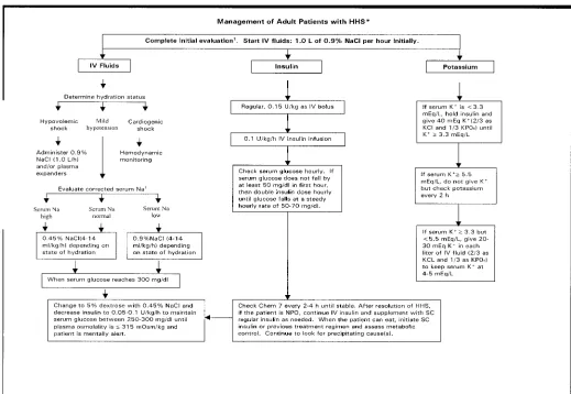
Successful treatment of DKA and HHS requires correction of dehydration, hyperglycemia, and electrolyte imbal-ances; identification of comorbid precip-itating events; and above all, frequent patient monitoring. Guidelines for the management of patients with DKA and HHS follow and are summarized in Figs. 1, 2, and 3. Table 3 includes a summary of major recommendations and evidence gradings.
Fluid therapy
corrected serum sodium is low. Once renal function is assured, the infusion should include 20–30 mEq/l potassium (2/3 KCl and 1/3 KPO4) until the patient is stable and can tolerate oral supplementation. Successful progress with fluid replacement is judged by hemodynamic monitoring (improve-ment in blood pressure), measure(improve-ment of fluid input/output, and clinical exam-ination. Fluid replacement should cor-rect estimated deficits within the first 24 h. The induced change in serum osmo-lality should not exceed 3 mOsm .kg-1
P O S I T I O N S T A T E M E N T
intravascular and extravascular volume and restoration of renal perfusion. In the absence of cardiac compromise, isoton-ic saline (0.9% NaCl) is infused at a rate of 15–20 ml .kg-1
body wt .h-1 or greater during the 1st hour (~1–1.5 liters in the average adult). Subsequent choice for fluid replacement depends on the state of hydration, serum electrolyte levels, and urinary output. In general, 0.45% NaCl infused at 4–14 ml .kg-1. h-1
is appropriate if the corrected serum sodium is normal or elevated; 0.9% NaCl at a similar rate is appropriate if
H2O .h-1.14–20,22In patients with renal or cardiac compromise, monitoring of serum osmolality and frequent assess-ment of cardiac, renal, and assess-mental sta-tus must be performed during fluid resuscitation to avoid iatrogenic fluid overload.14–20,22
Pediatric patients (<20 years of age). Initial fluid therapy is directed toward expansion of the intravascular and extravascular volume and restoration of renal profusion. The need for vascular volume expansion must be offset by the risk of cerebral edema associated with
(2/3 KCl or potassium-acetate and 1/3 KPO4). Once serum glucose reaches 250 mg/dl, fluid should be changed to 5% dextrose and 0.45–0.75% NaCl, with potassium as described above. Therapy should include monitoring mental status to rapidly identify changes that might indicate iatrogenic fluid overload, which can lead to symptomatic cerebral edema.23–25
Insulin therapy
Unless the episode of DKA is mild (Table 1), regular insulin by continuous intravenous infusion is the treatment of choice. Once hypokalemia (K+
<3.3 mEq/l) is excluded, an intravenous bolus rapid fluid administration. The 1st hour
of fluids should be isotonic saline (0.9% NaCl) at the rate of 10–20 ml .kg-1.
h-1 . In a severely dehydrated patient, this may need to be repeated, but the initial reexpansion should not exceed 50 ml/kg over the first 4 h of therapy. Continued fluid therapy is calculated to replace the fluid deficit evenly over 48 h. In general, 0.9% NaCl infused at a rate of 1.5 times the 24-h maintenance requirements (~5 ml .kg-1.
h-1
) will accomplish a smooth rehydration, with a decrease in osmolali-ty not exceeding 3 mOsm .kg-1
H2O .h-1. Once renal function is assured and serum potassium is known, the infusion should include 20–40 mEq/l potassium
of regular insulin at 0.15 U/kg body wt, followed by a continuous infusion of regular insulin at a dose of 0.1 U .kg-1. h-1
(5–7 U/h in adults), should be admin-istered. This low dose of insulin usually decreases plasma glucose concentration at a rate of 50–75 mg .dl-1.
h-1 , similar to a higher dose insulin regimen.26If plasma glucose does not fall by 50 mg/dl from the initial value in the 1st hour, check hydration status; if accept-able, the insulin infusion may be dou-bled every hour until a steady glucose decline between 50 and 75 mg/h is achieved. When the plasma glucose reaches 250 mg/dl in DKA or 300 mg/dl in HHS, it may be possible to decrease
Figure 3. Protocol for the management of pediatric patients (<20 years) with DKA or HHS. *DKA diagnostic criteria: blood glu-cose >250 mg/dl, venous pH <7.3, bicarbonate <15 mEq/l, and moderate ketonuria or ketonemia. †HHS diagnostic criteria: blood glucose >600 mg/dl, venous pH >7.3, bicarbonate >15 mEq/l, and altered mental status or severe dehydration. ‡After the initial history and physical examination, obtain blood glucose, venous blood gases, electrolytes, blood urea nitrogen, creatinine,
calci-um, phosphorous, and urine analysis STAT. §Usually 1.5 times the 24-h maintenance requirements (~5 ml .kg-1.
h-1
) will accom-plish a smooth rehydration; do not exceed two times the maintenance requirement. ||The potassium in solution should be 1/3
response to therapy. During therapy for DKA or HHS, blood should be drawn every 2–4 h for determination of serum electrolytes, glucose, blood urea nitro-gen, creatinine, osmolality, and venous pH (for DKA). Generally, repeat arterial blood gases are unnecessary; venous pH (which is usually 0.03 U lower than arterial pH) and anion gap can be fol-lowed to monitor resolution of acidosis. With mild DKA, regular insulin given either subcutaneously or intramuscular-ly every hour is as effective as intra-venous administration in lowering blood glucose and ketone bodies.27Patients with mild DKA should first receive a “priming” dose of regular insulin of 0.4–0.6 U/kg body wt, half as an intra-venous bolus and half as a subcuta-neous or intramuscular injection.22 Thereafter, 0.1 U .kg-1.
h-1of regular insulin should be given subcutaneously or intramuscularly.
P O S I T I O N S T A T E M E N T
the insulin infusion rate to 0.05–0.1 U . kg-1.
h-1
(3–6 U/h), and dextrose (5–10%) may be added to the intra-venous fluids. Thereafter, the rate of insulin administration or the concentra-tion of dextrose may need to be adjusted to maintain the above glucose values until acidosis in DKA or mental obtun-dation and hyperosmolarity in HHS are resolved.
Ketonemia typically takes longer to clear than hyperglycemia. The nitro-prusside method only measures ace-toacetic acid and acetone. However, -OHB, the strongest and most prevalent acid in DKA, is not measured by the nitroprusside method. During therapy,
-OHB is converted to acetoacetic acid, which may lead the clinician to believe that ketosis has worsened. Therefore, assessments of urinary or serum ketone levels by the nitroprusside method should not be used as an indicator of
After resolution of DKA (glucose <200 mg/dl, serum bicarbonate ≥18 mEq/l, venous pH >7.3, anion gap <12 mEq/l) and when patients are able to take fluids orally, a multidose regimen may be initiated based on history of previous treatment. However, for newly diagnosed patients, a total insulin dose of 0.6–0.7 U .kg-1.
day-1
may be initi-ated as a multidose regimen of short-and intermediate-/long-acting insulin, with subsequent modification based on glucose testing. Finally, some type 2 diabetic patients may be discharged on oral agents and dietary therapy.
Potassium
Despite total-body potassium depletion, mild to moderate hyperkalemia is not uncommon in patients with hyper-glycemic crises. Insulin therapy, correc-tion of acidosis, and volume expansion decrease serum potassium concentration. To prevent hypokalemia, potassium replacement is initiated after serum lev-els fall below 5.5 mEq/l, assuming the presence of adequate urine output. Generally, 20–30 mEq potassium (2/3 KCl and 1/3 KPO4) in each liter of infu-sion fluid is sufficient to maintain a serum potassium concentration within the normal range of 4–5 mEq/l. Rarely, DKA patients may present with signifi-cant hypokalemia. In such cases, potas-sium replacement should begin with fluid therapy, and insulin treatment should be delayed until potassium con-centration is restored to >3.3 mEq/l to avoid arrhythmias or cardiac arrest and respiratory muscle weakness.
Bicarbonate
Bicarbonate use in DKA remains con-troversial.28At a pH >7.0, reestablishing insulin activity blocks lipolysis and resolves ketoacidosis without any added bicarbonate. Prospective randomized studies have failed to show either benefi-cial or deleterious changes in morbidity or mortality with bicarbonate therapy in DKA patients with pH between 6.9 and 7.1.29No prospective randomized stud-ies concerning the use of bicarbonate in Table 3. Summary of Major Recommendations
Recommendations Grading
Initiate insulin therapy according to recommendations in position statement. A Unless the episode of DKA is mild, regular insulin by continuous intravenous B
infusion is preferred.
Assess need for bicarbonate therapy, and if necessary, follow treatment C recommendations in position statement: Bicarbonate may be beneficial in patients with a pH <6.9; not necessary if pH is >7.0.
Studies have failed to show any beneficial effect of phosphate replacement on the A clinical outcome in DKA. However, to avoid cardiac and skeletal muscle weakness and respiratory depression due to hypophosphatemia, careful phosphate
replacement may sometimes be indicated in patients with cardiac dysfunction, anemia, or respiratory depression and in those with serum phosphate concentration <1.0 mg/dl.
Studies of cerebral edema in DKA are limited in number. Therefore, to avoid the C occurrence of cerebral edema, follow the recommendations in the position statement regarding a gradual correction of glucose and osmolality as well as the judicious use of isotonic or hypotonic saline, depending on serum sodium and the hemodynamic status of the patient.
Initiate fluid replacement therapy based on recommendations in position statement. A
the initial hour of hydration, it seems prudent to administer 1–2 mEq/kg um bicarbonate over an hour. This sodi-um bicarbonate can be added to 0.45 NaCl, with any required potassium, and this solution can be used as the rehydra-tion solurehydra-tion for that hour. No bicarbon-ate therapy is required if pH is ≥7.0.30,31 Insulin, as well as bicarbonate thera-py, lowers serum potassium; therefore, potassium supplementation should be maintained in intravenous fluid as described above and carefully moni-tored. (See Fig. 1 for guidelines.) Thereafter, venous pH should be DKA with pH values <6.9 have been
reported. Given that severe acidosis may lead to a myriad of adverse vascular effects, it seems prudent that for adult patients with a pH <6.9, 100 mmol sodi-um bicarbonate be added to 400 ml ster-ile water and given at a rate of 200 ml/h. In patients with a pH of 6.9–7.0, 50 mmol sodium bicarbonate is diluted in 200 ml sterile water and infused at a rate of 200 ml/h. No bicarbonate is neces-sary if pH is >7.0.
In the pediatric patient, there are no randomized studies in patients with pH <6.9. If the pH remains below 7.0 after
assessed every 2 h until the pH rises to 7.0, and treatment should be repeated every 2 h if necessary. (See Kitabchi et al.11for a complete description of stud-ies done to date on the use of bicarbon-ate in DKA.)
Phosphate
Despite whole-body phosphate deficits in DKA that average ~1.0 mmol/kg body wt, serum phosphate is often nor-mal or increased at presentation. Phosphate concentration decreases with insulin therapy. Prospective randomized studies have failed to show any benefi-cial effect of phosphate replacement on the clinical outcome in DKA,32and overzealous phosphate therapy can cause severe hypocalcemia with no evi-dence of tetany.17,32However, to avoid cardiac and skeletal muscle weakness and respiratory depression due to hypophosphatemia, careful phosphate replacement may sometimes be indicat-ed in patients with cardiac dysfunction, anemia, or respiratory depression and in those with serum phosphate concentra-tion <1.0 mg/dl. When needed, 20–30 mEq/l potassium phosphate can be added to replacement fluids. No studies are available on the use of phosphate in the treatment of HHS.
Continuous monitoring using a flow-sheet (Fig. 4) aids in the organization of recovery parameters and treatment inter-ventions.
COMPLICATIONS
The most common complications of DKA and HHS include hypoglycemia due to overzealous treatment with insulin, hypokalemia due to insulin administration and treatment of acidosis with bicarbonate, and hyperglycemia secondary to interruption/discontinuance of intravenous insulin therapy after recovery without subsequent coverage with subcutaneous insulin. Commonly, patients recovering from DKA develop hyperchloremia caused by the use of excessive saline for fluid and electrolyte replacement and transient non–anion gap metabolic acidosis as chloride from
the patient becomes clinically stable.35 Hypoxemia and, rarely, noncardio-genic pulmonary edema may complicate the treatment of DKA. Hypoxemia is attributed to a reduction in colloid osmotic pressure that results in increased lung water content and decreased lung compliance. Patients with DKA who have a widened alveolo-arteriolar oxygen gradient noted on ini-tial blood gas measurement or with pul-monary rales on physical examination appear to be at higher risk for the devel-opment of pulmonary edema.
PREVENTION
Many cases of DKA and HHS can be prevented by better access to medical care, proper education, and effective communication with a health care provider during an intercurrent illness. The observation that stopping insulin for economic reasons is a common precipi-tant of DKA in urban
African-Americans36,37is disturbing and under-scores the need for our health care deliv-ery systems to address this problem, which is costly and clinically serious.
Sick-day management should be reviewed periodically with all patients. It should include specific information on 1) when to contact the health care provider, 2) blood glucose goals and use of supplemental short-acting insulin dur-ing illness, 3) means to suppress fever and treat infection, and 4) initiation of an easily digestible liquid diet contain-ing carbohydrates and salt. Most impor-tantly, the patient should be advised never to discontinue insulin and to seek professional advice early in the course of the illness. Successful sick-day man-agement depends on involvement by the patient and/or a family member. The patient/family member must be able to accurately measure and record blood glucose, urine ketone determination when blood glucose is >300 mg/dl, insulin administered, temperature, respi-ratory and pulse rate, and body weight and must be able to communicate this to a health care professional. Adequate supervision and help from staff or
fami-P O S I T I O N S T A T E M E N T
intravenous fluids replaces ketoanions lost as sodium and potassium salts dur-ing osmotic diuresis. These biochemical abnormalities are transient and are not clinically significant except in cases of acute renal failure or extreme oliguria.
Cerebral edema is a rare but fre-quently fatal complication of DKA, occurring in 0.7–1.0% of children with DKA. It is most common in children with newly diagnosed diabetes, but has been reported in children with known diabetes and in young people in their twenties.33,34Fatal cases of cerebral edema have also been reported with HHS. Clinically, cerebral edema is char-acterized by a deterioration in the level of consciousness, with lethargy, decrease in arousal, and headache. Neurological deterioration may be rapid, with seizures, incontinence, pupillary changes, bradycardia, and respiratory arrest. These symptoms progress as brain stem herniation occurs. The pro-gression may be so rapid that papillede-ma is not found. Once the clinical symp-toms other than lethargy and behavioral changes occur, mortality is high (>70%), with only 7–14% of patients recovering without permanent morbidity. Although the mechanism of cerebral edema is not known, it likely results from osmotically driven movement of water into the cen-tral nervous system when plasma osmo-lality declines too rapidly with the treat-ment of DKA or HHS. There is a lack of information on the morbidity associ-ated with cerebral edema in adult patients; therefore, any recommenda-tions for adult patients are clinical judgements, rather than scientific evi-dence. Prevention measures that might decrease the risk of cerebral edema in high-risk patients are gradual replace-ment of sodium and water deficits in patients who are hyperosmolar (maxi-mal reduction in osmolality 3 mOsm . kg-1
H2O .h-1) and the addition of dex-trose to the hydrating solution once blood glucose reaches 250 mg/dl. In HHS, a glucose level of 250–300 mg/dl should be maintained until hyperosmo-larity and mental status improves and
ly may prevent many of the admissions for HHS due to dehydration among eld-erly individuals who are unable to rec-ognize or treat this evolving condition. Better education of care givers as well as patients regarding signs and symp-toms of new-onset diabetes; conditions, procedures, and medications that worsen diabetes control; and the use of glucose monitoring could potentially decrease the incidence and severity of HHS.
The annual incidence rate for DKA from population-based studies ranges from 4.6 to 8 episodes per 1,000 patients with diabetes, with a trend toward an increased hospitalization rate in the past two decades.38The incidence of HHS accounts for <1% of all primary diabetic admissions. Significant resources are spent on the cost of hospitalization. Based on an annual average of ~100,000 hospitalizations for DKA in the U.S., with an average cost of $13,000 per patient, the annual hospital cost for patients with DKA may exceed $1 bil-lion per year. Many of these hospitaliza-tions could be avoided by devoting ade-quate resources to apply the measures described above.
Because repeated admissions for DKA are estimated to drain approxi-mately one out of every two health care dollars spent on adult patients with type 1 diabetes, resources need to be redirect-ed toward prevention by funding better access to care and educational programs tailored to individual needs, including ethnic and personal health care beliefs. In addition, resources should be directed toward the education of primary care providers and school personnel so that they can identify signs and symptoms of uncontrolled diabetes and newly onset diabetes can be diagnosed at an earlier time. This has been shown to decrease the incidence of DKA at the onset of diabetes.30,39
REFERENCES
1McGarry JD, Woeltje KF, Kuwajima M,
Foster DW: Regulation of ketogenesis and the renaissance of carnitine palmitoyl transferase.
Diabetes Metab Rev 5:271–284, 1989
27Fisher JN, Shahshahani MN, Kitabchi AE:
Diabetic ketoacidosis: low dose insulin therapy by various routes. N Engl J Med 297:238–247, 1977
28Barnes HV, Cohen RD, Kitabchi AE,
Murphy MB: When is bicarbonate appropriate in treating metabolic acidosis including diabetic ketoacidosis? In Debates in Medicine. Gitnick G, Barnes HV, Duffy TP, et al., Eds. Chicago, Yearbook, 1990, p. 172
29Morris LR, Murphy MB, Kitabchi AE:
Bicarbonate therapy in severe diabetic ketoacido-sis. Ann Int Med 105:836–840, 1986
30Vanelli M, Chiari G, Ghizzoni L, Costi G,
Giacalone T, Chiarelli F: Effectiveness of a pre-vention program for diabetic ketoacidosis in chil-dren. Diabetes Care 22:7–9, 1999
31Viallon A, Zeni F, Lafond P, Venet C, Tardy
B, Page Y, Bertrand JC: Does bicarbonate thera-py improve the management of severe diabetic ketoacidosis? Critical Care Medicine 27, December 1999
32Fisher JN, Kitabchi AE: A randomized
study of phosphate therapy in the treatment of diabetic ketoacidosis. J Clin Endocrinol Metab 57:177–180, 1983
33Rosenbloom AL: Intracerebral crises during
treatment of diabetic ketoacidosis. Diabetes Care 13:22–33, 1990
34Duck SC, Wyatt DT: Factors associated
with brain herniation in the treatment of diabetic ketoacidosis. J Pediatr 113:10-14, 1988
35Holsclaw DS Jr, Torcato B: Acute
pul-monary edema in juvenile diabetic ketoacidosis.
Pediatr Pulmonology 24:438–443, 1997
36Musey VC, Lee JK, Crawford R, Klatka
MA, McAdams D, Phillips LS: Diabetes in urban African Americans. I. Cessation of insulin thera-py is the major precipitating cause of diabetic ketoacidosis. Diabetes Care 18:483–489, 1995
37Umpierrez GE, Kelly JP, Navarrete JE,
Casals MMC, Kitabchi AE: Hyperglycemic crises in urban blacks. Arch Int Med 157: 669–675, 1997
38Fishbein HA, Palumbo PJ: Acute metabolic
complications in diabetes. In Diabetes in
America. National Diabetes Data Group, Ed.
Bethesda, MD, National Institutes of Health, 1995, p. 283–291 (NIH pub. no. 95-1468)
39Kaufman FR, Halvorsen M: The treatment
and prevention of diabetic ketoacidosis in chil-dren and adolescents with type 1 diabetes melli-tus. Pediatr Annals 28:576–582, 1999
Joslin’s Diabetes Mellitus. 13th ed. Kahn CR,
Weir GC, Eds. Philadelphia, Lea & Febiger, 1994, p. 738–770
15Ennis ED, Stahl EJVB, Kreisberg RA: The
hyperosmolar hyperglycemic syndrome. Diabetes
Rev 2:115–126, 1994
16Marshall SM, Walker M, Alberti KGMM:
Diabetic ketoacidosis and hyperglycaemic non-ketotic coma. In International Textbook of
Diabetes Mellitus. 2nd ed. Alberti KGMM,
Zimmet P, DeFronzo RA, Eds. New York, John Wiley, p. 1215–1229, 1997
17Carroll P, Matz R: Uncontrolled diabetes
mellitus in adults: experience in treating diabetic ketoacidosis and hyperosmolar coma with low-dose insulin and uniform treatment regimen.
Diabetes Care 6:579–585, 1983
18Ennis ED, Stahl EJ, Kreisberg RA: Diabetic
ketoacidosis. In Diabetes Mellitus: Theory and
Practice. 5th ed. Porte D Jr, Sherwin RS, Eds.
Amsterdam, Elsevier, 1997, p. 827–844
19Hillman K: Fluid resuscitation in diabetic
emergencies: a reappraisal. Intensive Care Med 13:4–8, 1987
20Fein IA, Rackow EC, Sprung CL, Grodman
R: Relation of colloid osmotic pressure to arterial hypoxemia and cerebral edema during crystalloid volume loading of patients with diabetic ketoaci-dosis. Ann Intern Med 96:570–575, 1982
21Matz R: Hypothermia in diabetic acidosis.
Hormones 3:36–41, 1972
22Kitabchi AE, Sacks HS, Young RT, Morris
L: Diabetic ketoacidosis: reappraisal of therapeu-tic approach. Ann Rev Med 30:339–357, 1979
23Mahoney CP, Vleck BW, DelAguila M:
Risk factors for developing brain herniation dur-ing diabetic ketoacidosis. Pediatr Neurology 21: 721–727, 1999
24Finberg L: Why do patients with diabetic
ketoacidosis have cerebral swelling, and why does treatment sometimes make it worse? Pediatr
Adolescent Med 150:785–786, 1996
25Duck SC, Wyatt DT: Factors associated
with brain herniation in the treatment of diabetic ketoacidosis. J Pediatr 113:10–14, 1988
26Kitabchi AE, Ayyagari V, Guerra SMO,
Medical House Staff: The efficacy of low dose versus conventional therapy of insulin for treat-ment of diabetic ketoacidosis. Ann Int Med 84:633–638, 1976
Diabetic ketoacidosis: a combined metabolic-nephrologic approach to therapy. Diabetes Rev 2:209–238, 1994
3Atchley DW, Loeb RF, Richards DW,
Benedict EM, Driscoll ME: A detailed study of electrolyte balances following withdrawal and reestablishment of insulin therapy. J Clin Invest 12:297–326, 1933
4Halperin ML, Cheema-Dhadli S: Renal and
hepatic aspects of ketoacidosis: a quantitative analysis based on energy turnover. Diabetes
Metab Rev 5:321–336, 1989
5Malone ML, Gennis V, Goodwin JS:
Characteristics of diabetic ketoacidosis in older versus younger adults. J Am Geriatr Soc 40: 1100–1104, 1992
6Matz R: Hyperosmolar nonacidotic diabetes
(HNAD). In Diabetes Mellitus: Theory and
Practice. 5th ed. Porte D Jr, Sherwin RS, Eds.
Amsterdam, Elsevier, 1997, p. 845–860
7Morris LE, Kitabchi AE: Coma in the
dia-betic. In Diabetes Mellitus: Problems in
Management. Schnatz JD, Ed. Menlo Park, CA,
Addison-Wesley, 1982, p. 234–251
8Kreisberg RA: Diabetic ketoacidosis: new
concepts and trends in pathogenesis and treat-ment. Ann Int Med 88:681–695, 1978
9Klekamp J, Churchwell KB: Diabetic
ketoacidosis in children: initial clinical assess-ment and treatassess-ment. Pediatric Annals 25: 387–393, 1996
10Glaser NS, Kupperman N, Yee CK,
Schwartz DL, Styne DM: Variation in the man-agement of pediatric diabetic ketoacidosis by specialty training. Arch Pediatr Adolescent Med 151:1125–1132, 1997
11Kitabchi AE, Umpierrez GE, Murphy MB,
Barrett EJ, Kreisberg RA, Malone JI, Wall BM: Management of hyperglycemic crises in patients with diabetes mellitus (Technical Review).
Diabetes Care 24:131–153, 2001
12Beigelman PM: Severe diabetic
ketoacido-sis (diabetic coma): 482 episodes in 257 patients: experience of three years. Diabetes 20:490–500, 1971
13Polonsky WH, Anderson BJ, Lohrer PA,
Aponte JE, Jacobson AM, Cole CF: Insulin omission in women with IDDM. Diabetes Care 17:1178–1185, 1994
14Kitabchi AE, Fisher JN, Murphy MB,