Consequences of Getting the Head Covered During Sleep in Infancy
Britt T. Skadberg, MD and Trond Markestad, MD
ABSTRACT. Objective. To study the consequences of getting the head covered by bedding (fiber quilt) on carbon dioxide (CO2) accumulation around the face, be-havior, and physiologic responses during prone and su-pine sleep in infants to add understanding to why vic-tims of sudden infant death syndrome are often found under the bedding.
Methodology. Of 33 healthy term, usually nonprone sleeping infants, behavior and computerized polysom-nography were successfully recorded for 30 during prone and supine sleep at 21⁄2months and for 23 prone and 25 supine at 5 months.
Results. For both ages and body positions, covering the head resulted in significant CO2 accumulation around the face, fewer apneas (3 to 10 seconds), shorter duration of apneas after sighs, higher heart and respira-tory rates, and peripheral skin temperature. Differences were generally greater at 21⁄2 than at 5 months. While covered, the prone position was associated with higher CO2levels close to the face, slightly higher transcutane-ous PCO2, and higher heart rates and peripheral skin temperatures than the supine position. In the supine position 23% were able to remove the cover from the head at 21⁄2and 60% at 5 months, whereas only 1 infant of 5 months managed to remove the cover when prone.
Conclusions. The observed responses are consistent with a potential for distress when the head is covered, particularly when placed prone. Probably most impor-tant with respect to sudden infant death syndrome is the infants’ inability to remove the bedding from the head
upon awakening from prone sleep. Pediatrics
1997;100(2). URL: http://www.pediatrics.org/cgi/content/ full/100/2/e6; behavior, CO2 rebreathing, hyperthermia,
SIDS, sleeping position.
ABBREVIATIONS. SIDS, sudden infant death syndrome; CO2, carbon dioxide; ALTE, apparent life-threatening event; REM, rapid eye movement; EEG, electroencephalogram; EOG, elec-trooculogram; EMG, electromyogram; ECG, electrocardiogram; Po
2, partial pressure of oxygen; Pco2, partial pressure of carbon dioxide; O2, oxygen.
I
n 1944, Abramson
1discussed accidental
mechan-ical suffocation as a cause of sudden death in
infants sleeping prone, but the next year Wolley
et al
2rejected suffocation and rebreathing into the
bedding as the sole mechanisms for sudden infant
death. Recently, rebreathing has reappeared as a
pos-sible explanation for sudden infant death syndrome
(SIDS).
3Although the concentration of carbon
diox-ide (CO
2) in the atmosphere is .03% to .04%, the
exhaled air of infants contains 4% to 5% CO
2,
4and it
has been speculated that under certain conditions,
such as sleeping face down on soft bedding,
exten-sive CO
2accumulation around the infant’s face may
result in rebreathing, asphyxia, and suffocation.
3,5,6The possibility of CO
2rebreathing has mainly been
studied in experimental in vitro and animal
mod-els,
3,5–10whereas studies in infants are limited.
11–13We have previously shown that a significant
num-ber of SIDS victims are found with their head
cov-ered by bedding and usually in the prone position.
14The purpose of the present investigation was to
study the effect of getting the head covered by
bed-ding on CO
2accumulation around the face, behavior,
and physiologic responses in sleeping infants, and to
compare outcome measures for the prone and supine
sleeping positions.
SUBJECTS AND METHODS Subjects
Thirty-three infants (17 girls and 16 boys) were recruited for an overnight sleep study both at 21⁄2(mean, 74 days; range, 71 to 79 days) and 5 (mean, 152 days; range, 147 to 158 days) months of age. The ages were chosen to correspond with the age of highest risk for SIDS (21⁄2months) and a period of significantly lower risk (5 months) in Norway.15Only term infants ($ 37-weeks-postcon-ceptual age) without prenatal or perinatal complications were included. No infants with a history of family diseases, apparent life-threatening events (ALTE), or SIDS in siblings were allowed. Mean birth weight was 3631 g (range, 2870 g to 4840 g) and all were healthy at the time of study.
A questionnaire containing information on infant care prac-tices, such as feeding, preferred sleeping position, sleep environ-ment, weight gain, and psychomotor development in relation to the ability to move, was completed by the parents at the time of each study.
Informed written consent was obtained from the parents of all the infants, and the study was approved by the Regional Com-mittee on Medical Research Ethics.
Study Conditions
The infants were admitted early in the evening for a 12-hour polysomnographic recording of sleep-related behavior. Their nor-mal feeding schedule was followed during the evening, and they were further fed in the middle of the night before the sleeping position was changed. After the evening feed the room was dark-ened and the infants were placed to sleep in the supine position. The mother was allowed to stay with her infant until it fell asleep if this was the routine at home. The study conditions were iden-tical at 21⁄2and 5 months of age.
The infants were studied in an infant bed (bars as side walls) with a firm foam rubber mattress (normal hospital cot mattress; polyurethane-30N, 9.5 cm thick) lined with a thin blanket. All were dressed in a diaper and two shirts, and covered with a duvet (polyester fiber quilt; 1453115 cm with a cotton quilt cover, total weight 1300 g; tog value 13).16The same fiber quilt and identical cotton quilt covers were used in all the studies. Duvets are the From the Department of Pediatrics, University Hospital of Bergen, Bergen,
Norway.
Received for publication Sep 11, 1996; accepted Feb 11, 1997.
Reprint requests to (B.T.S.) Department of Pediatrics, University of Bergen, N-5021 Bergen, Norway.
covers most commonly used in Norway, and both the fiber quilt and the cotton quilt covers used in the present study were similar to those used at home for infants at this age. A light clothing (2.5 tog)16was chosen to ease the nursing of the infant and the super-vision of the sensors, and the room temperature was kept at 23°C (6.5°C) to obtain an environmental temperature close to the cor-responding thermoneutral range.17The duvet covered the lower part of the body (up to midthorax).
The overnight sleep was divided into two equal periods of prone and supine sleep. The initial position was chosen at random using the sealed envelope method. Because most infants were usually placed supine to sleep, and none slept prone at home all were allowed to fall asleep while supine. During the first non-REM (quiet sleep) interval, one-half of the infants were turned to the prone position according to the randomization. All were moved to the opposite position midway through the study after being fed. The study of covering the infants’ head in addition to the body with the duvet was performed once during each sleep period, and was started during the third sleep cycle after the last feed. Ten minutes after a change of sleep state into non-REM was documented, the duvet was gently draped over the infant’s head.
Polysomnography
A computer-aided multichannel system with the operating CARDAS software (Computer Aided Record Display and Analy-sis Systems) and the Oxcams/Pi Logic IMS-2000 multi-channel monitor (Oxcams/Pi Logic Ltd., Dyfed, United Kingdom) were used for the polysomnographic recordings. For each infant the sleep variables recorded included two electroencephalograph der-ivations (C3-A2, C4-A1; EEG), two electrooculographic deriva-tions (ROC/A1, LOC/A2; EOG), and one chin electromyogram (EMG). Continuously recorded physiologic parameters were: elec-trocardiogram (ECG, three leads placed on the upper part of the trunk), thoracic and abdominal respiratory movements (strain gauge, Sensorband, Mediplus AB, Malmø, Sweden), peripheral skin temperature (left foot, Skin Surface Temp xHH-10005-x, Ellab as, Denmark), oxygen saturation (right foot, oxygen trend curve, pulse rate, and pulse waveform, 50 Hz fast response mode, data average time 3 seconds, update interval .375 seconds, Ohmeda Biox-3700, Ohmeda Medical System Division, Louisville, CO), intermittent transcutaneous pressure of oxygen (Po2) and CO2
(Pco
2) (limited to the last 11 infants at 21⁄2months and the last 13 infants at 5 months of age, left or right side of abdomen depending on sleep position; Microgas 7640, Kontron Instruments Ltd, United Kingdom), body movements (pressure sensitive pad placed under the blanket on the mattress; Pad-HF, B8208, Pi Logic Ltd., Dyfed, United Kingdom), and sound (microphone in front of the face during prone and supine face to side position, on the neckband of the shirt in the supine face up position; Sound, B8202, Pi Logic Ltd., Dyfed, United Kingdom). Attempts to estimate core temperature were abandoned because a method for central skin temperature measurement (Thermistor Zeal, Pi Logic Ltd., Dyfed, United Kingdom) did not correlate sufficiently with rectal tem-perature, and rectal monitoring was unsuccessful in a pilot project because it interfered with sleep behavior. It has been shown that peripheral skin and rectal temperatures change simultaneously, and that changes in skin temperature fluctuate less throughout the night than core temperature.18Furthermore, upon warming, pe-ripheral vasodilatation and subsequent increases in pepe-ripheral body temperature occur to promote heat loss.19 We therefore found it acceptable to record peripheral skin temperature alone to fulfill the purpose of this study. Attempts to measure air flow by thermistors were abandoned mainly because sleep behavior was disturbed, especially in the prone position, but also because of low sensor sensitivity in the younger infants (EdenTech infant airflow sensor, EdenTech Corp, Eden Prairie, MN).
CO2Measurement
The CO2accumulation around the infant’s face was measured intermittently with a CO2gas analyzer (infrared, real time moni-tor, accuracy of CO2measurement .1%, sampling period 60 sec-onds, Binos 11/GAV 100, Leybold-Haraeus AG, West Germany). The flow of gas through the analyzer necessary for measurement was 100 mL/min. The gas analyzer was calibrated with standard gases before each measurement. To keep the gas volume around the baby’s head as constant as possible, the gas volume used for CO2analysis was recirculated by a pump (peristaltic pump;
Mil-lipore model 80 002 30; MilMil-lipore Corp, Bedford, MA). A special quality tube with low oxygen permeability (Norprene food grade A-60F, 3.1 mm inside diameter; Cole Parmer) was used to connect the different parts of the system. The two openings of the tube were placed 3 and 4 cm in front of and 3 cm cranial to the nostrils when the head was in the face to the side position. If the infant accepted the test situation under the duvet, the CO2concentration was measured 1, 5, 15, 30, 45, and 60 minutes after covering of the head. The CO2concentration was also measured before the head was covered, and repeated after the duvet was removed.
Recorded Responses
Sleep states were defined according to the behavioral, EEG, EOG, and EMG criteria recommended by other investigators.20 –22 Each minute of the recording was coded as non-REM, REM (ac-tive), indeterminate sleep, or awakening. Non-REM sleep was defined as eyes closed with no eye movements, no body move-ments except occasional generalized startles, high voltage and slow-wave pattern or sleep spindles on EEG, resting muscle tone on EMG and decreased heart and respiratory rates. REM sleep was defined as visible eye movements under the eye lids inde-pendent of facial or gross body movements, frequent small move-ments of head, face, and limbs, low voltage and fast desynchro-nized pattern on EEG, muscle atonia on EMG, and rapid and irregular heart and respiratory rates. Periods of sleep not meeting these criteria were classified as indeterminate sleep, and periods with open eyes, body movements and vocalization were classified as awake. When the head was covered, eye movements were interpreted on the basis of EOG. Only technically unblemished data were used for the analyses of physiologic responses, ie, if arousals or artefact time exceeded 15 seconds the sequence was excluded from the analysis as were intervals of sleep in which the heart rate from the ECG tracing differed with more than an average of 3 beats/min from the pulse rate recorded by the pulse oximeter. Behavior was charted separately, but plotted along with the polysomnography.
Behavioral and physiologic parameters before the head was covered refer to the mean values obtained from periods of non-REM and non-REM sleep of similar length as after covering the head for both prone and supine sleep.
As all parameters were continuously stored on a hard disk either as trend curves or as digitalized signals, the values of physiologic parameters corresponding to the CO2measurements in the air close to the face were available. Each parameter was determined as the mean of the last 10 seconds of recording before each CO2measurement. If the quality of the recorded signals were unsatisfactory, the mean value of the 10-second interval closest to, but within 20 seconds of the CO2measurements, was chosen.
A movement of the head or extremities was defined by artefacts in a limited number of channels and an increase in the amplitude of the activity-channel whereas arousals caused artefacts in most channels. Arousals were further subdivided into short arousals, ie, body movements lasting between 3 and 15 seconds without changes in EEG, and longer arousals lasting longer than 15 sec-onds, often associated with changes in EEG and subsequent awak-ening. A sigh was defined as an isolated and sudden change in the amplitude of either the thoracic or abdominal respiratory channel. The duration of an apnea was measured from the end of inspiration in the last breath before to the end of inspiration in the first breath after the apnea. Apneas lasting 3 seconds or longer were recorded and further divided into 3 through 10, and 11 seconds and longer. Periodic breathing was defined as the succes-sion of more than two apneas longer than 3 seconds separated from each other by less than 20 seconds of breathing movements. The ECG was stored continuously both as single heart beats and as a trend curve. The heart rate could therefore be counted manually despite artefacts from arousals and body movements. Respiratory rate was determined from the thoracic and/or ab-dominal amplitude in the polysomnography.
Transcutaneous Pco2 and Po2 were measured intermittently
throughout the night to prevent skin damage (electrode temper-ature144°C). The sensor was applied to the skin during non-REM sleep in the second sleep cycle. The Microgas was calibrated with standard gases and adjusted to the actual atmospheric pressure before each application. Transcutaneous Pco
As the signals of the pulse oximeter are very sensitive to body movements, it was often difficult to interpret the oxygen satura-tion during and immediately after arousals and body movements. Only intervals with distinct pulse waves were used for further analysis.
The cover was removed from the head if awakening with continuous head and body movements beyond 60 seconds oc-curred, if the infant managed to remove the duvet from the head, or after a maximum of 60 minutes under the duvet. The cover was also removed if a permanent increase in heart rate beyond 20% and/or respiratory rate beyond 40% of pretest values occurred, if the oxygen saturation dropped to 90% or less, or if peripheral skin temperature increased beyond 38°C.
The data were analyzed with SPSS for MS windows release 6. The paired t test and the Wilcoxon signed rank test for dependent samples were used for statistical analyses where appropriate. Analysis of differences in response between supine and prone sleep (paired t test) was limited to those who accepted both sleeping positions at each particular age. Analysis of variation with age (paired t test) was limited to infants who accepted each particular position after being covered at 21⁄2and 5 months. Before analysis of differences between mean number of events during non-REM and REM sleep before and after covering the head (sleep behavior), the number (n) was adjusted to a rate of events per 20 minutes.
RESULTS
Of the 33 infants enrolled in the study 30 were
successfully tested in both the prone and supine
sleeping positions at 2
1⁄
2months. At 5 months, 26
infants were tested; 22 in both positions, one only in
the prone, and 3 only in the supine position. One
infant was excluded at 2
1⁄
2months attributable to
maternal illness, and one at 5 months attributable to
possible seizures. Three infants were excluded from
the analysis attributable to technical problems with
the CO
2gas analyzer (one infant at 2
1⁄
2and two
infants at 5 months). The others were not tested
because they were withdrawn from the study by the
parents (two infants at 5 months) or because they
woke up immediately when the head was covered in
either or both sleeping positions (one infant at 2
1⁄
2and six at 5 months).
The characteristics of the infants tested are listed in
Table 1, and did not differ from those who were not
tested. The mean weight was at the 50th percentile
for Norwegian infants. Supine was the dominating
sleeping position and none slept prone, but all
in-fants were regularly put in the prone position when
awake. They were regularly covered with a duvet
(fiber quilt) and none used a pillow during sleep.
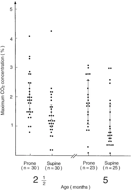
CO2Accumulation
The CO
2concentration close to the infants’ face
never exceeded .1% before the head was covered or
after removal of the duvet. There was a significant
CO
2accumulation around the face under the duvet
at 2
1⁄
2and 5 months of age, and although there was
a considerable overlap between body positions, the
maximum median CO
2concentration was
signifi-cantly higher in the prone than in the supine position
at both ages (Fig 1). The time to attain maximum CO
2concentration did not differ significantly for the
prone and supine positions at 2
1⁄
2(12.5 min
6
12.6
min vs 12.6 min
6
16.0 min; P
5
1.0) and 5 months
(9.0 min
6
10.9 min vs 6.9 min
6
7.1 min; P
5
.3).
There was no significant difference in median CO
2concentration in either position with increasing age.
At 2
1⁄
2months, the highest CO
2
concentration was
recorded during non-REM sleep in the majority of
prone (87%) and supine infants (60%). At 5 months
there were no significant differences according to
sleep state (76%, both positions).
Subsequent to body movements or arousals
dur-ing sleep, the duvet was regularly lifted from the
mattress. When such movements of the duvet
coin-cided with CO
2measurements, a sudden drop in CO
2was observed. Visible temporary or permanent air
channels were commonly created after such
move-ments and resulted in low and stable CO
2levels
throughout the rest of the study.
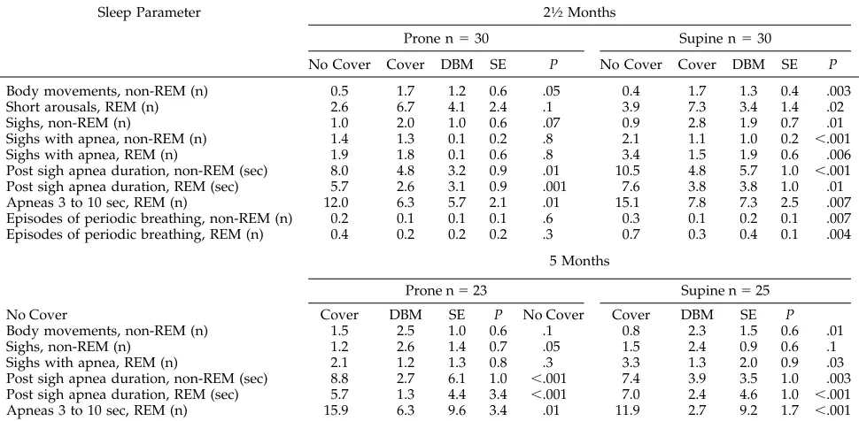
Sleep Behavior
Indeterminate sleep represented less than 2% of
total sleep under the duvet, and was excluded from
TABLE 1. Characteristics of the Infants Tested at 21⁄2 and 5 Months of Age
21⁄2Months (n530)
5 Months (n526)
Gender, female/male 15/15 14/12 Breastfeeding 24 (80%) 19 (73%) Weight in g (mean61 SD) 58216696 74686831 Sleeping in bed with parents 0 1 Supine preferred position 23 26 Side or supine preferred position 3 0 Side preferred position 4 0
the analysis. The mean time tolerated under the
du-vet varied from 19 to 33 minutes depending on age
and body position (range, 68 seconds to 60 min). For
both positions and for either or both non-REM or
REM sleep, covering the head at 2
1⁄
2months resulted
in more frequent body movements, significantly
shorter duration of apneas after sighs, and fewer
apneas (3 to 10 seconds) (Table 2). In the supine
position covering was also associated with more
fre-quent short arousals and isolated sighs, and fewer
sighs with apneas, and episodes of periodic
breath-ing (Table 2). At 2
1⁄
2months covering the head did
not cause changes in number of short arousals or
apneas during non-REM sleep, or in number of body
movements or apneas exceeding 10 seconds during
REM sleep. Comparing prone and supine while
cov-ered at 2
1⁄
2months, the infants spent relatively more
time in REM sleep (P
5
.04), and had later arousals
(P
5
.05) and change of sleep state (P
5
.001) when
prone. For both positions at 5 months covering
re-sulted in fewer apneas (3 to 10 seconds) and shorter
duration of apneas after sighs (Table 2). Sleep
behav-ior did not differ with position after covering at 5
months.
Independent of body position and age,
spontane-ous awakening terminated the test in most infants
(22 of 30 prone and 23 of 30 supine infants at 2
1⁄
2months, and 20 of 23 prone and 22 of 25 supine
infants at 5 months). In the supine position 7 (23%)
infants at 2
1⁄
2months and 15 (60%) at 5 months
managed to remove the duvet from the face by using
all four extremities. In the prone position one (4%)
infant at 5 months managed to remove the cover by
moving the head up and down. The rate of removal
was significantly higher for the supine position at
both ages (P
,
.01 and P
,
.001). For the infants who
awoke spontaneously but did not remove the duvet,
head and body movements continued until the cover
was removed by the attendant. Although the
major-ity of infants cried upon awakening under the duvet,
the noise was markedly muffled by the cover.
Ac-cording to the criteria given in the study protocol,
four tests were actively terminated during prone
sleep at 2
1⁄
2and one at 5 months attributable to
increases in heart and respiratory rates, and a
simul-taneous drop in oxygen saturation and a rise in body
temperature in one (89% and 38°C). No tests were
terminated because of such responses in the supine
position (P
5
.07 and P
5
.3) for 2
1⁄
2and 5 months,
respectively.
Physiologic Responses
Covering the head was associated with significant
increases in heart and respiratory rates, and
periph-eral skin temperature in both body positions and at
both ages except for heart rate at 5 months when
sleeping supine (Table 3). There was a minor, but
significant increase in transcutaneous P
co
2
after
cov-ering during prone sleep at both ages, and
transcu-taneous P
o
2
was higher after covering in both
posi-tions at 2
1⁄
2months (Table 3). Comparing prone and
supine sleep when the head was covered, prone was
associated with a higher mean heart rate at both
ages, and a higher mean peripheral skin temperature
and transcutaneous P
o
2at 2
1⁄
2months, but there
were no significant differences in transcutaneous
P
co
2or oxygen saturation (Table 3). With increasing
age, heart and respiratory rates decreased
signifi-cantly for both positions before and after covering,
and peripheral skin temperature was significantly
lower during prone sleep after covering at 5 months.
There were no differences with age for other
param-eters. The skin on the trunk and face of all but four
infants was warm upon touch when the cover was
TABLE 2. Comparison of Sleep Behavior Before and After Covering the Head With the Duvet During Prone and Supine Sleep
Sleep Parameter 21⁄2Months
Prone n530 Supine n530
No Cover Cover DBM SE P No Cover Cover DBM SE P
Body movements, non-REM (n) 0.5 1.7 1.2 0.6 .05 0.4 1.7 1.3 0.4 .003 Short arousals, REM (n) 2.6 6.7 4.1 2.4 .1 3.9 7.3 3.4 1.4 .02
Sighs, non-REM (n) 1.0 2.0 1.0 0.6 .07 0.9 2.8 1.9 0.7 .01
Sighs with apnea, non-REM (n) 1.4 1.3 0.1 0.2 .8 2.1 1.1 1.0 0.2 ,.001 Sighs with apnea, REM (n) 1.9 1.8 0.1 0.6 .8 3.4 1.5 1.9 0.6 .006 Post sigh apnea duration, non-REM (sec) 8.0 4.8 3.2 0.9 .01 10.5 4.8 5.7 1.0 ,.001 Post sigh apnea duration, REM (sec) 5.7 2.6 3.1 0.9 .001 7.6 3.8 3.8 1.0 .01 Apneas 3 to 10 sec, REM (n) 12.0 6.3 5.7 2.1 .01 15.1 7.8 7.3 2.5 .007 Episodes of periodic breathing, non-REM (n) 0.2 0.1 0.1 0.1 .6 0.3 0.1 0.2 0.1 .007 Episodes of periodic breathing, REM (n) 0.4 0.2 0.2 0.2 .3 0.7 0.3 0.4 0.1 .004
5 Months
Prone n523 Supine n525
No Cover Cover DBM SE P No Cover Cover DBM SE P
Body movements, non-REM (n) 1.5 2.5 1.0 0.6 .1 0.8 2.3 1.5 0.6 .01
Sighs, non-REM (n) 1.2 2.6 1.4 0.7 .05 1.5 2.4 0.9 0.6 .1
Sighs with apnea, REM (n) 2.1 1.2 1.3 0.8 .3 3.3 1.3 2.0 0.9 .03 Post sigh apnea duration, non-REM (sec) 8.8 2.7 6.1 1.0 ,.001 7.4 3.9 3.5 1.0 .003 Post sigh apnea duration, REM (sec) 5.7 1.3 4.4 3.4 ,.001 7.0 2.4 4.6 1.0 ,.001 Apneas 3 to 10 sec, REM (n) 15.9 6.3 9.6 3.4 .01 11.9 2.7 9.2 1.7 ,.001
Abbreviations: n, mean number of events per 20 min; DBM, differences between means; SE, standard error of differences between means;
removed from the head. In one half—and relatively
more pronounced the longer the head was covered
with the duvet—the skin was wet with additional
flushing of the face whereas the limbs felt cool. Four
infants were generally wet and cool, and eight were
pale instead of flushed. These findings did not differ
with position or age.
DISCUSSION
In the present study covering the head with
bed-ding resulted in accumulation of CO
2around the
face, significantly less apneas, and shorter duration
of apneas after sighs, higher heart and respiratory
rates, and higher peripheral skin temperature at 2
1⁄
2and 5 months of age. At 2
1⁄
2months the infants also
had more frequent body movements after being
cov-ered. While covered, the prone position was
associ-ated with higher CO
2levels close to the face, higher
heart rates, peripheral skin temperatures, and
slightly higher transcutaneous P
co
2than the supine
position, and the infants were less able to remove the
cover when prone.
The infants in the present study slept on a firm
mattress with the face to the side, and the duvet
(fiber quilt) used to cover the head is the cover in
common use in Norway. The study condition of
totally covering the head was chosen to mimic the
circumstances in which SIDS victims are commonly
found.
14The observed CO
2
concentrations at the face
while the head was covered were similar to those
observed by Chiodini et al
11in uncovered infants
sleeping face down on soft bedding, but greater than
the levels of .3 to 2.0% observed by Malcolm et al
12in
prone infants whose heads were covered with a baby
blanket. The higher CO
2levels in the present
com-pared with the latter study probably reflect a higher
resistance to transport of gases through the duvet
than a blanket, and possibly a greater difficulty in
creating air channels to the surrounding air
attribut-able to the heavier weight of the duvet. The range of
CO
2accumulation measured around the face during
the first 15 minutes of the study was comparable to
the values obtained during the first 5 minutes in an
experimental CO
2rebreathing model.
10Lack of
fur-ther rise probably reflected the establishment of
channels from spontaneous lifting of the duvet with
subsequent exchange of air to the surroundings.
Hy-percapnia is known to be a strong respiratory and
arousal stimulant in normal infants.
23In this study,
the immediate increases in respiratory rate, number
of body movements and arousals, and fewer apneas
and shorter duration of apneas after sighs after
cov-ering the head, are consistent with such an effect of
CO
2. Although not always significant, the rise in
oxygen saturation and transcutaneous P
o
2
, and only
a minimal rise in transcutaneous P
co
2further
indi-cate that normal infants are able to eliminate CO
2immediately by hyperventilation.
An increase in CO
2in the respiratory air is
accom-panied by a decrease in O
2. Although most infants
will respond to both hypoxia and hypercarbia with
hyperventilation, around two-thirds of normal
in-fants may have an inappropriate response to mild
and moderate hypoxia
24and 2% may have a
dimin-TABLE 3. Physiological Parameters Before and After Covering the Head With the Duvet 2
1⁄2
ished response to hypercarbia.
25Impaired alveolar
ventilation and significantly higher arousal
thresh-olds to high CO
2have also been found in some
infants with ALTE.
26Ventilatory and arousal
thresh-olds to hypercapnia and hypoxia are also increased
during sleep and especially during REM sleep.
23,27In
adults, the common response to a single exposure of
CO
2seems to be an enhanced rather than a decreased
sensitivity to CO
2,
28but this condition has not been
studied in infants. However, in adult divers, the
ventilatory response to an increased CO
2in
inspira-tory gases is commonly decreased, and it has been
suggested that the blunted response may be caused
by adaptation.
29We speculate that a subgroup of
normal infants may respond inappropriately to
hyp-oxia and hypercarbia if repeatedly exposed to
re-breathing which may occur with consistent
unto-ward care-giving practices such as prone sleeping on
soft bedding.
The mean peripheral skin temperature in the
present study was higher in the prone than the
su-pine position both before and after covering the
head. The prone position is associated with reduced
heat loss partly because a higher fraction of the body
surface is in direct contact with the mattress, and
partly because air exchange around the body is
de-creased,
19,30,31(B.T. Skadberg and T. Markestad,
per-sonal observation). The additional rise in peripheral
skin temperature observed after covering the head
may reflect a combined effect from the cover itself
because the head is the main route of heat loss in
young infants,
32–36and the increase in metabolic rate
attributable to hyperventilation from CO
2rebreath-ing. This is further supported by the circulatory
re-sponse with a rise in heart rate. Most of the infants
were sweating and the skin often felt cool. Therefore,
the moderate rise in peripheral skin temperature
may not fully reflect the increased thermal stress
because sweating and evaporation of sweat may
have limited the rise in temperature at the surface of
the skin.
Subsequent to a change in body position or
move-ments during sleep, infants may happen to slip
un-der the bedding,
13,14,37,38and a situation for CO
2
re-breathing and risk of overheating may occur.
10,13,19Although most infants in the present study woke up
when covered, there was a striking difference in
subsequent behavior depending on the infants’ body
position. Only one prone infant of 5 months
man-aged to remove the duvet from the head compared
with 23% of the supine infants at 2
1⁄
2months and
60% at 5 months. The inability of prone sleepers to
remove the duvet may be explained by the age
de-pendent neuromotor development. Coordination of
head and body movements is still very limited at 5
months,
39,40and the present study shows that the
complex series of movements which is necessary to
remove the bedding when prone, may be virtually
impossible.
Victims of SIDS apparently die in close
relation-ship to sleep. Infants’ early crawling movements are
reciprocal which may tend to bring them under the
bedding while prone. It has also been shown that
infants sleeping on the side may be able to pull the
cover over the head,
13and that they may roll from
the unstable side position to prone.
13,14,37,38The
inabil-ity to remove the cover while prone and subsequent
awakening and struggling may increase the
likeli-hood of overheating, exhaustion, and a face down
position with CO
2rebreathing, all possible
contribu-tors to SIDS.
ACKNOWLEDGMENTS
This research was supported by the Norwegian Research Coun-cil for Science and the Humanities, the Norwegian SIDS Society, and the local SIDS society.
REFERENCES
1. Abramson H. Accidental mechanical suffocation in infants. J Pediatr. 1944;25:404 – 413
2. Wolley JR. Mechanical suffocation during infancy. J Pediatr. 1945;26: 72–75
3. Kemp JS, Thach BT. Sudden death in infants sleeping an polystyrene-filled cushions. N Engl J Med. 1991;324:1858 –1864
4. Barrow GM. Physical Chemistry. 3rd ed. Tokyo, Japan: McGraw-Hill; 1973:741
5. Bolton DPG, Taylor BJ, Campell AJ, Galland BC, Cresswell C. Rebreath-ing expired gases from beddRebreath-ing. Arch Dis Child. 1993;69:187–190 6. Kemp JS, Thach BT. A sleep position-dependent mechanism for infant
death on sheepskins. Am J Dis Child. 1993;147:642– 646
7. Bolton DPG, Cross KW, McKettrick AC. Are babies in carry cots at risk from CO2rebreathing? Br Med J. 1972;4:80 – 81
8. Ryan EL. Distribution of expired air in carry cots—a possible explana-tion for some sudden infant deaths? Australas Phys Eng Sci Med. 1991; 14:112–118
9. Corbyn JA. Atmospheric carbon dioxide levels in the vicinity of sleep-ing infants and sudden infant death. Trans Inst Eng Aust Multidiscipl. 1991;15:9 –13
10. Skadberg BT, Oterhals A, Finborud K, Markestad T. CO2rebreathing: a possible contributory factor to some cases of sudden infant death? Acta
Paediatr. 1995;84:988 –995
11. Chiodini BA, Thach BT. Impaired ventilation in infants sleeping facedown: Potential significance for sudden infant death syndrome.
J Pediatr. 1993;5:686 – 691
12. Malcolm G, Cohen G, Henderson-Smart D. Carbon dioxide concentra-tions in the environment of sleeping infants. J Paediatr Child Health. 1994;30:45– 49
13. Waters KA, Gonzales A, Jean C, Marielli A, Brouillette RT. Face-straight down and face-near-straight-down positions in healthy, prone-sleeping infants. J Pediatr. 1996;128:616 – 625
14. Markestad T, Skadberg B, Hordvik E, Morild I, Irgens L. Sleeping position and sudden infant death syndrome (SIDS): effect of an inter-vention programme to avoid prone sleeping. Acta Paediatr. 1995;84: 375–378
15. Irgens LM, Skjærven R, Lie RT. Secular trends of sudden infant death syndrome and other causes of post perinatal mortality in Norwegian births cohorts 1967–1984. Acta Paediatr Scand. 1989;78:228 –232 16. Culow EE. Thermal insulating properties of fabrics. Textiles. 1978;1:
47–52
17. Wigfield RE, Fleming PJ, Azaz YEZ, et al. How much wrapping do babies need at night? Arch Dis Child. 1993;69:181–186
18. Wailoo MP, Petersen SA, Whittaker H, Goodenough P. Sleeping body temperatures in 3- to 4-month-old infants. Arch Dis Child. 1989;64: 596 –599
19. Nelson EAS, Taylor BJ, Weatherall IL. Sleeping position and infant bedding predispose to hyperthermia and the sudden infant death syn-drome. Lancet. 1989;28:199 –201
20. Anders T, Embde R, Parmelee A. A Manual of Standardised Terminology,
Techniques and Criteria for Scoring of States of Sleep and Wakefulness in Newborne Infants. Los Angeles, CA: Brain Information Service/Brain
Research Institute, University of California; 1970
21. Hoppenbrowers T, Hodgeman JE, Kazuko A, Geidel SA, Sterman MB. Sleep and waking states in infancy: normative studies. Sleep. 1988;60: 387– 401
22. Prechtl HFR. The behavioral states of the newborn infant (a review).
Brain Res. 1974;76:185–212
in Infants and Children. Baltimore, MD: Williams & Wilkins; 1992:
112–124
24. Davidson Ward SL, Bautista DB, Keens TG. Hypoxic arousal response in normal infants. Pediatrics. 1992;89:860 – 864
25. Bolton DPG. The prevalence of immature respiratory control in a neo-natal population. N Z Med J. 1990;103:89 –92
26. McCulloch K, Brouillette RT, Guzzetta JA, Hunt CE. Arousal response in near-miss sudden infant death syndrome and normal infants. J
Pedi-atr. 1982;101:911–917
27. Fewell JE, Taylor BJ, Kondo CS, Dasclau V, Filyk SC. Influence of carotid denervation on the arousal and cardiopulmonary responses to upper airway obstruction in lambs. Pediatr Res. 1990;28:374 –378 28. Florio JT, Mackenzie DAR. Ventilatory response to carbon dioxide
following simulated saturation diving in man. J Physiol. 1985;362:10 29. Schaefer KE, Carey CR, Dougherty JH, Morgan C, Messier A. Effect of
intermittent exposure to 3% CO2on respiration, acid-base balance and calcium-phosphorus metabolism. Undersea Biomed Res. 1979;(suppl): 115–134
30. Douthitt TC, Brackbill Y. Differences in sleep, waking and motor activ-ity as a function of prone or supine resting position in the human neonate. Psychophysiology. 1972;9:99 –100
31. Kahn A, Grosswasser J, Sottiaux M, Rebuffat E, Franco E, Dramaix M. Prone or supine position and sleep characteristics in infants. Pediatrics. 1993;6:1112–1115
32. Azaz Y, Fleming PJ, Levine M, McCabe R, Stewart A, Johnson P. The relationship between environmental temperature, metabolic rate, sleep
state, and evaporative water loss in infants from birth to three months.
Pediatr Res. 1992;4:417– 423
33. Stothers JK. Head insulation and heat loss in the newborn. Arch Dis
Child. 1981;56:530 –534
34. Mestyan J, Jarai I, Bata G, Fekete M. The significance of facial temper-ature in the chemical heat regulation of premtemper-ature infants. Biol Neonate. 1964;7:243–254
35. Bruck K. Temperature regulation in the newborn infant. Biol Neonate. 1961;3:65–119
36. North RG, Petersen SA, Wailoo MP. Lower body temperature in sleep-ing supine infants. Arch Dis Child. 1995;72:340 –342
37. Willinger M, Hoffman MA, Hartford RB. Infant sleep position and risk for sudden infant death syndrome: Report of meeting held January 13 and 14, 1994, National Institutes of Health, Bethesda, Maryland.
Pedi-atrics. 1994;5:814 – 820
38. Fleming PJ, Blair PS, Bacon C, et al. Environment of infants during sleep and risk of the sudden death syndrome: results of 1993–1995 case-control study for confidental inquiry into stillbirths and deaths in infancy. Br Med J. 1996;313:191–195
39. Vaughan VC, Litt IF. Developmental pediatrics. In: Behrman RE, Vaughan VC, eds. Nelson Textbook of Pediatrics. 13th ed. Philadelphia, PA: WB Saunders; 1987:33–35
40. Frankenberg WK, Dodds JB, Fandal AW, Kazuk E, Cohrs M. Denver
Developmental Screening Test. Reference Manual. Denver, CO: University