UDC 616-006+616.4+615.3
The role of reacTive oxygen species in Tumor
cells apopTosis induced by landomycin a
L. V. Lehka1, R. R. PaNchUk1, W. BeRgeR2, Ju. RohR3, R. S. StoIka1
1Institute of cell Biology, National academy of Sciences of Ukraine, Lviv; 2Institute of cancer Research, Medical University of Vienna, austria; 3Department of Pharmaceutical Sciences, University of kentucky, USa;
e-mail: lilyalehka@gmail.com
Landomycin a (La) is a new antitumor antibiotic of angucycline group, possessing high antitumor activity against cancer cells of different origin, which induces early apoptosis in target cells. It was shown that under La action the level of reactive oxygen species (RoS) in human t-leukemia cells had increased 5.6 times in comparison to control already at the 1st hour after the addition of studied antibiotic to the culture medium. at the 6th hour after incubation of cells with La the nucleosomal DNa cleavage, chromatin condensation and nucleus fragmentation were observed, indicating apoptotic cell death. catalase (scavenger of hydrogen peroxide), mannitol (scavenger of hydroxyl radicals) and superoxide dismutase (scavenger of superoxide radicals) reduced the level of RoS production under La, suggesting the generation of h2o2, oh• and o2 -radicals, respectively. It was revealed that catalase and mannitol effectively inhibited La-mediated tumor cell death, increasing 2.5 times the percentage of alive cells in comparison to La. however, superoxide dismutase had no significant inhibitory effect on cytotoxic activity of LA, indicating the minor role of superoxide anions in the implementation of antitumor activity of this antibiotic. combination of catalase, mannitol and superoxide dismutase with La increased 4-fold the percentage of alive cells in comparison to the action of La. Dynamics of ROS formation confirms that the increase of ROS is a very rapid process, but at the same time it is not a direct consequence of apoptosis triggering, mediated by mitochondria.
k e y w o r d s: landomycins, apoptosis, reactive oxygen species (RoS), malignant cells.
C
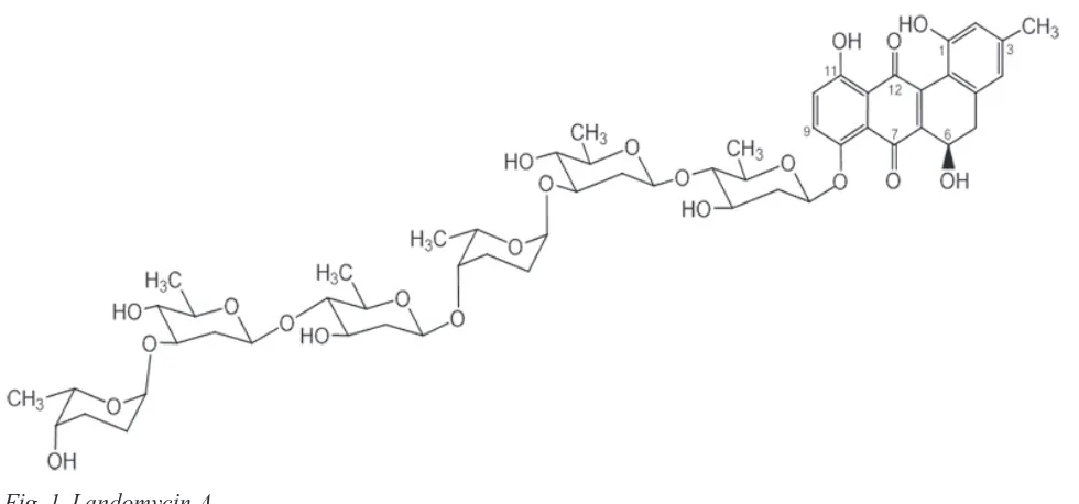
hemotherapy, targeted on the level of reac-tive oxygen species modulating (ROS) in tumor cells, is considered the most promi-sing and effective strategy in the fight against can -cer [1-3]. It is known that normal cells are charac-terized by a powerful antioxidant defense system that effectively eliminates free radicals, which are the byproducts of oxidative phosphorylation in the mitochondria. Due to this, healthy cells can tolera-te high level of exogenous ROS emerging under ex-treme conditions. But antioxidant enzyme system of the tumor cells is unbalanced and the intensity of metabolic processes is considerably higher, resulting in a persistent oxidative stress [4]. As a consequence, above threshold ROS concentrations are not toxic to malignant cells, however, a further sudden increase in the level of free radicals in tumor cells by exog-nous ROS-induced agents causes their rapid death. This special feature of malignant cells makes them promising target for anticancer drugs which induce oxidative stress in tumor cells [2].Landomycin A is a new antitumor angucycline antibiotic consisting of benzoantharacene tetracyclic aglycone and hexasaccharide chain that contains two repetitive trisaccharide subunits (α-L-rhodinose-(1-3)-β-D-olivose-(1-4)-β-D-olivose) joined by O-gly -coside bond (Fig. 1). Landomycin A possesses high cytotoxic activity toward cancer cells as well as ability to overcome acquired multidrug resistance of cancer cells to chemotherapy treatment [5]. Previous studies showed that landomycin A acts as a prooxi-dant, so the aim of this study was to clarify the role of ROS in apoptotic death of tumor cells induced by this antibiotic.
materials and methods
Landomycin A (99% purity according to thin-layer chromatography) was obtained in the labora-tory of professor Jurgen Rohr (Department of Phar-maceutical Sciences, University of Kentucky, USA). cells and cell culture. Jurkat cell line (human T-cell leukemia), HL-60 cell line (human acute
myelocytic leukemia) and MG-63 cell line (human osteosarcoma) were used in the study.
Cells were grown in RPMI-1640 medium (Sig-ma, USA) supplemented with 10% fetal bovine se-rum (Sigma, USA) and 50 µg/ml gentamicin (Sigma, USA) in 5% CO2-containing humidified atmosphere at 37 ºC. Cells were passaged every two days at the rate of 5×10⁵ cell/ml of culture medium for the sus-pension cultures and 2.5x10⁵ cell/ml of culture me-dium for the substrate-dependent lines [6].
Study of cytotoxic activity of La. Cells were seeded in 24-tissue culture plates (Greiner Bio One, Germany) in culture medium at a concentration of 1×10⁶ cell/ml (suspension cell) and 1×10⁵ cell/ml (substrate-dependent cells). The studied compound was added to the cells at various concentrations. Af-ter 24 h incubation the number of cells was calcula-ted in hemocytometer chamber by estimation of the number of dead cells using 0.1% Trypan Blue dye. This dye stains dead cells with damaged membrane in blue, whereas alive cells remain unstained [7].
Study of DNa fragmentation using agarose gel electrophoresis. Cells were plated in Petri dishes at 1×10⁶ cell/ml concentration with the addition of the studied compound in the medium. After 6 h incuba-tion, cells were washed with 1-x phosphate-buffered saline (PBS) and centrifuged at 300 g for 6-7 min. The cell pellet was gently resuspended in a tube on ice with lysis buffer (50 mM Tris-HCl, pH 7.5, 20 mM EDTA-Na, 1% NP-40 detergent) at the rate of 10 µl buffer per 1×10⁶ cells. The cells were then Fig. 1. Landomycin a
centrifuged at 4090 g for 5-10 min, the supernatant was collected, and sodium dodecyl sulfate (SDS) to a final concentration of 1% and RNase A (Invi-trogen, USA) to a final concentration of 5 mg/ml were added. The mixture was incubated for 2 h at 56 °C. Thereafter proteinase K (Invitrogen, USA) was added to a final concentration of 2.5 mg/ml and incubated for 2 h at 37 °C. After the incubation com-pletion, 10 M ammonium acetate (½ of volume) and chilled isopropanol (2 volumes) were added to the mixture and then centrifuged at 14000 g for 15-20 min. The precipitate was washed with ethanol and re-centrifuged. Ethanol was then pipetted. The pre-cipitate was dried at room temperature. DNA was dissolved in TBE buffer (Tris-borate-EDTA); 5 µl buffer per 1×10⁶ cells. Before the electrophoresis, a loading buffer (final concentration 7-10% sucrose) and bromophenol blue were added to samples. The samples were loaded at the rate of 1×10⁶ cells per well. Electrophoresis was performed in 1-x TBE buffer, in 1% agarose at 35 V using a BioRad ap-paratus (Sweden).
Measurement of reactive oxygen species pro-duction. To detect ROS, the specific fluorescent dye
and 60 mM, respectively, and incubated for other 30 min. Next, landomycin A was added to the cells and incubated for a certain time. The fluorescence inten -sity was measured by flow cytometry using FACS Calibur (Becton Dickinson, Palo Alto, USA).
Double cell staining with fluorescent dyes Hoe -chst 33258 and propidium iodide. Cells were seeded in 6-well plate (105 cells per well). After 24 h, the studied compounds were added and the cells were incubated for 24 h. Thereafter, Hoechst 33258 (1 mg/ ml) 1 µl/ml and propidium iodide (2.5 mg/ml) 1 µl/ ml were added with further incubation for 1 h. At the end of the experiment, digital images of the cells were produced using fluorescence microscope digital camera Nikon Eclipse 9000.
Measurement of mitochondrial transmembrane potential (ΔΨm). Mitochondrial transmembrane po-tential (ΔΨm) was determined semi-quantitatively with JC-1 dye (5,5′-tetrachloro-1,1′,3,3′-tetraethylben-zimidazolocarbocyanine iodide, Sigma, USA) using flow cytometry. Cells (1×10⁶) were incubated for a certain time with the studied compound. Cells were washed with 1-x PBS and incubated for 10 min at 37 °C with a freshly prepared solution of JC-1 (final concentration 10 µg/ml). JC-1 remains were removed by washing the cells with PBS. Fluorescence inten-sity was evaluated by flow cytometry using FACS Calibur (Becton Dickinson, Palo Alto, USA).
Statistical data analysis. The experiments were performed in triplicate for each variant. Each point of graphs in the figures and the column ordinate on the diagram corresponds to the mean value M, cal-culated on the results of the three measurements in one of several similar experiments. Standard error «m» was calculated using the mean square deviation «σ». Significant differences between the statistical groups were determined by Student’s test. The data with P < 0.05 were considered statistically signifi -cant. The percentage of apoptotic cells (determined by the cell staining with Hoechst 33258) was counted using Image-Pro Plus 7.0 software. A computer based on AMD’s with windows 7 (Microsoft, USA) was used in the study. Statistical data processing was performed using Microsoft Excel software (Micro-soft Office, 2007).
results and discussion
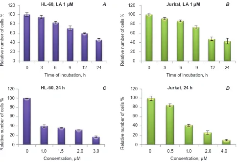
To estimate landomycin A (LA) cytotoxic ac-tivity on Jurkat cell line (human T-cell leukemia) and HL-60 cell line (human promyelocytic leuke-mia), Trypan Blue assay was used. The data shown
in Fig. 2 (a, B) demonstrate that the number of alive cells in both studied lines decreases depending on the duration of incubation with LA. A negative cor-relation between the cell number and LA concen-tration was observed (Fig. 2, c, D). LC50 and LC75 values (the concentrations of compounds which cause the death of 50% or 75% of the cells compared to the control) after incubation for 24 h were 1 and 2 µM for Jurkat cells and 0.8 and 2.4 µM for HL-60 cells, respectively.
For analysis of the cytomorphological features of alterations in the target cells under LA action, MG-63 human osteosarcoma cells were stained with fluorescent dye Hoechst 33258 (Fig. 3). Owing to changes in the plasma membrane permeability, apo-ptotic cells accumulated this dye significantly faster in comparison to intact cells. Therefore, intact cells usually have a weak fluorescence in the nucleus, whereas apoptotic cells are characterized by bright fluorescence of condensed chromatin and fragmenta -tion of nucleus [8, 9]. On the microscopy images we can see condensed chromatin sites (marked by red arrows) in cells treated with the studied antibiotic, indicating the induction of apoptosis, the percentage of it was about 53% for 1 µM LA and 79% for 2 µM LA.
Internucleosomal DNA fragmentation is con-sidered as one of the most important biochemical features of apoptosis [10, 11]. To detect the DNA fragmentation in tumor cells of HL-60 line after exposure to LA, agarose gel electrophoresis was performed. Already at the 6th hour after incubation the cells with 1 µM of LA the DNA-ladder could be observed confirming the cell death by apoptosis (Fig. 4).
Fig. 2. cytotoxic effect of landomycin a (La) on Jurkat cell line (human t-cell leukemia) and hL-60 line (hu-man promyelocytic leukemia) (see the text for explanations)
0 3 6 9 12 24 80
HL-60, LA 1 μM
0 60 40 20 R e lat iv e n u m b e r o f c e lls % 100 120
Time of incubation, h
A
0 3 6 9 12 24
Time of incubation, h 80 0 60 40 20 R e lat iv e n u m b e r o f c e lls % 100
120 Jurkat, LA 1 μM B
0 1.0 1.5 2.0 3.0 80
HL-60, 24 h
0 60 40 20 R e lat iv e n u m b e r o f c e lls % 100 120 Concentration, μM C 80 0 60 40 20 R e lat iv e n u m b e r o f c e lls % 100
120 Jurkat, 24 h D
0 0.5 1.0 2.0 4.0 Concentration, μM
duration with studied antibiotic the level of ROS decreased but it still remained higher than in control.
ROS are oxygen-containing reactive molecules including O2-, H
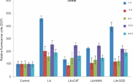
2O2 and OH˙ radicals. In our work a series of ROS scavengers such as superoxide dis-mutase, catalase and mannitol was used to study their influence on ROS production in tumor cells under landomycin A action (Fig. 6). It was found that catalase already at early time points (1, 3 h) significantly reduced ROS level (almost 3.5 fold in comparison to the amount of ROS produced under LA action). This indicates an increase in the genera-tion of hydrogen peroxide in the presence of studied antibiotic. Mannitol and superoxide dismutase also decreased the level of ROS, but to much less extent (2- and 1.3-fold, respectively). It confirmes the gene-ration of OH˙ and O2- radicals under LA action. The similar tendency was observed at late time points (6, 12, 24 h) after LA addition to the culture medium, however the inhibitory effect of ROS scavengers, produced in the presence of LA, was significantly
lower compared to values obtained at early time points.
It is known that the antitumor activity of most anticancer drugs used in chemotherapy (alkylating factors, plant alkaloids, antimetabolites, taxanes, platinum compounds, camptothecin, etoposide, an-thracyclines etc.) is achieved, among other things, through the generation of ROS. Free radicals formed during the oxidative stress influence on different cellular targets, causing cell cycle arrest, damage of DNA, lipids, proteins and cell membranes. Lipid peroxidation in mitochondrial membrane leads to membrane permeability, which causes disorder of the electron transport chain, release of cytochrome c into cytosol, activating processes of programmed cell death [13-15].
Fig. 3. cytomorphological analysis of the chromatin state in human osteosarcoma Mg-63 cells (staining with hoechst 33258, 24 h): 1 – control; 2 – landomycin a (1 µM); 3 – landomycin a (2 µM). the arrows point to condensed chromatin regions
Hoechst 33258 Differential-interference contrast
It was found that catalase (scavenger of H2O2) and mannitol (scavenger of hydroxyl radicals) effec-tively inhibited LA-induced cell death, increasing 2.5-fold the percentage of alive cells in comparison to studied antibiotic (Fig. 7). SOD did not exhibit significant inhibitory effect on the cytotoxic activity of LA that indicates a minor role of superoxide ani-ons (compared to hydrogen peroxide and hydroxyl radicals) in the implementation of antitumor activi-ty of this antibiotic. The combined use of catalase,
superoxide dismutase and mannitol had the highest inhibitory effect on the cytotoxic effect of LA, and the percenta ge of the alive cells increased by almost 4-fold compared to the effect of studied antibiotic.
Fig. 4. DNa fragmentation in the cells of hL-60 line (human acute promyelocytic leukemia) under lando mycin a action, 6 h: 1 – control; 2 – landomy-cin a, 1 µM
1 2
characterized by intensive condensation and frag-mentation of nuclear chromatin that indicates apo-ptosis in these cells. Preliminary incubation of cells with mannitol (before the addition of LA) led to a partial decrease of the above-mentioned changes in the nuclear chromatin structure that confirms the in -volvement of hydroxyl radicals in the process of cell death in the presence of this antibiotic. In turn, the addition of catalase to the culture medium almost completely eliminated the chromatin condensation induced by LA. The obtained data agreed with the results of the cytotoxic activity of studied antibiotic as well as the results of analysis of ROS modulators influence on free radical level in tumor cells in the presence of LA. It was found that catalase possessed the highest inhibitory effect on anti-tumor activi-ty as well as ROS production by tumor cells under studied compound. These results indicate that ROS, in particular hydrogen peroxide, hydroxyl and partly superoxide radicals are mediators of cell death and involved in LA-induced apoptosis.
The decreasing of mitochondrial membrane potential (MMP) is one of the earliest apoptosis markers [15]. To find the connection between the inducing of cell death and oxidative stress in tumor cells under LA action, Jurkat cells were stained with
a fluorescent dye JC-1. At normal mitochondrial transmembrane potential, the dye enters the orga-nelles, accumulates there and forms J-aggregates, which fluoresce in the red spectrum. As MMP de -creases, JC-1 cannot accumulate in organelles and remains in the cytoplasm in monomeric form, emit-ting green fluorescence that can be detected by flow cytometry [16]. It was observed that after 6th h of cells incubation with LA the percentage of depolari-zed mitochondria statistically significant (P < 0.05) increased and was 22% with further increasing (Fig. 9). Thus, the apoptosis-associated alterations in mitochondria were observed at the 6th hour after LA addition to the culture medium.
Antitumor agents, widely used in chemothera-py, which induce high level of ROS in cancer cells, include platinum compounds (cisplatin, oxyplatin), etoposide, camptothecins (topotecan) as well as an-thracyclines (doxorubicin, epirubicin) [17, 18]. It was established that most of these anticancer drugs may affect the mitochondria, causing the elevation of O2- radical level, due to either blocking the electron transport chain or capacity to accept electrons from NADPH-dependent dehydrogenases and transfer them to molecular oxygen with no inhibitory effect on the respiratory chain. Thereafter a superoxide radical can undergo spontaneous or enzymatic dis-mutations to form hydrogen peroxide, which in turn is able to transform in a hydroxyl radical [15, 17, 19, 20]. Our results suggest that a key role in antitumor activity of studied antibiotic rather belongs to hydro-gen peroxi de than to superoxide-radicals. Dynamics of ROS production in cancer cells in the presence of LA illustrates that the elevation of H2O2 level is a very fast process (the first hour after LA addition to the culture medium) however it is not a direct conse-quence of mitochondria damage or electron transport chain blocking. This distinguishes the mechanisms of ROS generation in tumor cells under studied an-tibiotic from the effect of above-mentioned antican-cer drugs. Further investigations of ROS generation mechanism and molecular targets of landomycin A are in progress.
Fig. 6. effect of catalase (cat), mannitol (MaN) and superoxide dismutase (SoD) on RoS production of hu-man t-leukemia cells of Jurkat line under the action of landomycin a (La), 1 µM
Fig. 5. Influence of landomycin A (LA) on ROS production in human T-leukemia cells of Jurkat line 0 1 3 6 12 24
Jurkat
0
R
el
at
iv
e fl
uo
re
sc
en
ce u
ni
ts (
D
C
F)
100 200
Time, h 300
400 500 600 700
La 1 μM La 2 μM
Control LA LA+CAT LA+MAN LA+SOD 0
R
el
at
iv
e fl
uo
re
sc
en
ce u
ni
ts (
D
C
F)
100 200 300 400 500 600
1 h
3 h
6 h
12 h
24 h
Jurkat
Thus, we have shown that landomycin A pos-sesses significant anti-tumor activity against leuke -mia and carcinoma cells and induces early apoptosis in target cells. Thus, already at the 6th h after cells in-cubation with studied antibiotic it the
Fig. 7. Inhibitory effect of RoS-scavengers on the induction of cell death under the action of landomycin a, 1 µM: 1 – control (c); 2 – c+cat; 3 – c+MaN; 4 – c+SoD; 5 – La; 6 – La+cat; 7 –La+MaN; 8 – La+SoD; 9 – La+cat+MaN+SoD
Fig. 8. cytomorphological analysis of chromatin state in human osteosarcoma Mg-63 cells after incubation with landomycin a (1 µM), catalase (cat) or mannitol (MaN), 24 h
Control
LA
LA+MAN
LA+CAT
C+MAN
Hoechst 33258 PI
C+CAT
1 2 3 4 5 6 7 8 9
80
0 60
40
20
R
e
lat
iv
e n
u
m
b
e
r o
f c
e
lls % 100
120 Jurkat, 24 h
role of ROS, especially H2O2, in this process. It has been shown that the ROS generation by tumor cells in the presence of LA is a very rapid process, but is not a consequence of apoptosis triggering, mediated by mitochondria.
acknowledgements. This work was partially
Fig. 9. Influence of landomycin A (1 µM) on the level of mitochondrial transmembrane potential in human Jurkat t-leukemia cells
Роль активних фоРм кисню в апоптозі пухлинних
клітин, індукованому ландоміцином а
Л. В. Легка1, Р. Р. Панчук1, В. Бергер2, Ю. Рор3, Р. С. Стойка1
1інститут біології клітини нан України, львів;
2інститут ракових досліджень, медичний
університет Відня, австрія;
3Університет кентуккі, відділ
фармацевтичних наук, сШа;
e-mail: lilyalehka@gmail.com
ландоміцин а (ла) – новий протипухлин -ний антибіотик ангуциклінового ряду, який виявляє виражену протипухлинну активність щодо ракових клітин різного походження та індукує ранній апоптоз у клітинах-мішенях. по -казано, що рівень активних форм кисню (аФк) у клітинах лінії Jurkat т-лейкозу людини зростав у 5,6 раза порівняно з контролем уже на 1-шу годину після додавання ла в культуральне се -редовище. на 6-ту годину інкубації клітин із ла спостерігали міжнуклеосомне розщеплен -ня Днк, що разом із конденсацією хроматину
та фрагментацією ядра вказувало на апоптичну загибель клітин. каталаза (скевенджер перок -сиду водню), манітол (скевенджер гідроксил-радикалів) і супероксиддисмутаза (скевенджер супероксидних радикалів) знижували рівень продукції аФк під впливом ла, вказуючи на генерацію н2о2, он˙ і о2- радикалів відповідно. Встановлено, що каталаза та манітол ефективно пригнічували ла-опосередковану загибель пух -линних клітин і збільшували в 2,5 раза відсоток живих клітин порівняно з ла. проте супероксид -дисмутаза не виявляла значного інгібувального ефекту на цитотоксичну активність ла, що свідчить про мінорну роль супероксид-аніонів у реалізації протипухлинної дії останнього. Ви -користання каталази, манітолу і супероксиддис -мутази разом із ла збільшувало відсоток живих клітин у 4 рази порівняно з дією досліджуваного антибіотика. результати утворення аФк підтверджують, що збільшення їхньої кількості є дуже швидким процесом, але, разом з тим, не є прямим наслідком запуску апоптозу, опосеред -кованого мітохондріями.
к л ю ч о в і с л о в а: ландоміцини, апоптоз, активні форми кисню, злоякісні клітини.
1 3 6 12 24 80
0 60
40
20 100
Time, h 90
70
30
10 50
intact mitochondria
Роль активных фоРм кислоРода в апоптозе опухолевых клеток, индуциРованном ландомицином а
Л. В. Легка1, Г. Г. Панчук1, В. Бергер2, Ю. Рор3, Р. С. Стойка1
1институт биологии клетки нан Украины, львов;
2институт раковых исследований,
медицинский университет Вены, австрия;
3Университет кентукки, отдел
фармацевтических наук, сШа;
e-mail: lilyalehka@gmail.com
ландомицин а (ла) – новый противоопу -холевый антибиотик ангуциклинового ряда c выраженной противоопухолевой активностью в отношении раковых клеток различного про -исхождения и способностью вызывать ранний апоптоз в клетках-мишенях. показано, что уро -вень активных форм кислорода (аФк) в клетках линии Jurkat т-лейкоза человека повышался в 5,6 раза по сравнению с контролем уже в течение 1-го часа после добавления ла в культуральную среду. на 6-й час инкубации клеток с ла наблю -далось межнуклеосомное расщепление Днк, что вместе с конденсацией хроматина и фраг -ментацией ядра указывало на апоптическую гибель клеток. каталаза (скэвенджер пероксида водорода), маннитол (скэвенджер гидроксил-ра -дикалов) и супероксиддисмутаза (скэвенджер супероксидных радикалов) снижали уровень продукции аФк под влиянием ла, указывая на генерацию н2о2, он˙ и о2- радикалов соответ -ственно. Установлено, что каталаза и маннитол эффективно подав ляли ла-опосредованную ги -бель опухолевых клеток, увеличивая в 2,5 раза процент живых клеток по сравнению с действи -ем ла. однако супероксиддисмутаза не вызвала значительного ингибирующего эффекта на ци -тотоксическую активность ла, что свидетель -ствует о минорной роли супероксид-анионов в реализации противоопухолевого действия по -следнего. использование каталазы, маннитола и супероксиддисмутазы в сочетании c ла увели -чивало процент живых клеток в 4 раза по срав -нению с действием исследуемого антибиотика. результаты образования аФк подтверждают, что увеличение их количества является очень быстрым процессом, но, вместе с тем, не являет
-ся прямым следствием запуска апоптоза, опос -редованного митохондриями.
к л ю ч е в ы е с л о в а: ландомицины, апоптоз, активные формы кислорода, злокаче-ственные клетки.
references
1. Kong Q., Beel J. A., Lillehei K. O. A threshold concept for cancer therapy. Med. hypotheses. 2000;55(1):29-35.
2. Trachootham D., Alexandre J., Huang P. Targeting cancer cells by ROS-mediated mechanisms: a radical therapeutic approach? Nat. Rev. Drug Discov. 2009;8(7):579-591.
3. Gorrini C., Harris I. S., Mak T. w. Modulation of oxidative stress as an anticancer strategy. Nat. Rev. Drug Discov. 2013;12(12):931-947.
4. Manuel M., Mario D. C. Oxidative Stress and Diseases. InTech, 2012. 519 p.
5. Ostash B., Korynevska A., Stoika R., Fedoren-ko V. Chemistry and biology of landomycins, an expanding family of polyketide natural products. Mini Rev Med chem. 2009;9(9):1040-51.
6. Adams R., Laboratory techniques in bioche-mistry and molecular biology. Elsevier. 1990. P. 16-94.
7. Freshney R. I. Culture of animal cells: a manual of basic technique and specialized applications, 6th edition. Wiley-Backwell, 2010. 768 p.
8. Ziegler U., Groscurth P. Morphological Features of Cell Death. News Physiol. Sci. 2004;(19):124-128.
9. Saraste A., Pulkki K. Morphologic and biochemical hallmarks of apoptosis. cardiovasc. Res. 2000;45(3):528-37.
10. Nagata S. Apoptotic DNA Fragmentation. exp. cell Res. 2000;256(1):12-18.
11. Matassov D., Kagan T., Leblanc J., Sikorska M., Zakeri Z. Measurement of apoptosis by DNA fragmentation. Methods Mol. Biol. 2004;282:1-17.
12. Noriko N., wakasugi H. Cancer and Oxidative Stress. J. Japan Med. assoc. 2000;124(11):1571-1574.
13. Manda G., Nechifor M., Neagu T-M. Reactive Oxygen Species, Cancer and Anti-Cancer Therapies. curr. chem. Biol. 2009;3(1):22-46. 14. Conklin K. A. Cancer chemotherapy and
15. Deavall D. G., Martin E. A., Horner J. M., Roberts R. Drug-induced oxidative stress and toxicity. J. toxicol. 2012;2012:645460.
16. Nuydens R., Novalbos J., Dispersyn G., weber C., Borgers M., Geerts H. A rapid method for the evaluation of compounds with mitochondria-protective properties. J. Neurosci. Methods. 1999 Oct 15;92(1-2):153-9.
17. Conklin K. A. Chemotherapy-associated oxi-dative stress: impact on chemotherapeutic effecti-veness. Integr. cancer ther. 2004;3(4):294-300. 18. Angsutararux P., Luanpitpong S., Issaragrisil S.
Chemotherapy-Induced Cardiotoxicity: Over-view of the Roles of Oxidative Stress. oxidative Medicine and cellular Longevity. 2015. P. 1-13.
19. Minotti G., Menna P., Salvatorelly E., Cairo G., Gianti L. Anthracyclines: Molecular Advances and Pharmacologic Developments in Antitumor Activity and Cardiotoxicity. Pharmacol. Rev.
2004;56(2):185-229.
20. Turrens J. F. Mitochondrial formation of reactive oxygen species. J. Physiol. 2003;552(Pt 2):335-344.
21. Kirshner J. R., He S., Balasubramanyam V., Kepros J., Yang C. Y., Zhang M., Du Z., Barsoum J., Bertin J. Elesclomol induces cancer cell apoptosis through oxidative stress. Mol cancer ther. 2008;7(8):2319-2327.