e x p e r i m e n t a l w o r k s
e x p e r i m e n t a l w o r k s
© 2019 Guzyk M. M. et al. This is an open-access article distributed under the terms of the Creative Commons Attribution License, which permits unrestricted use, distribution, and reproduction in any medium, provided the original author and source are credited. UDC 577.151.6:612.04:616.8
altered sirtuins 1 and 2 expression
in the brain of rats induced by experimental
diabetes and the ways of its correction
M. M. GUzyk1, T. M. TykhoNeNko1, k. o. DyakUN1, L. V. yaNITSka2, I. B. PryVroTSka3, T. M. kUchMeroVSka1 1Palladin Institute of Biochemistry, National academy of Sciences of Ukraine, kyiv;
2Bogomolets National Medical University, kyiv, Ukraine; 3I. horbachevsky Ternopil State Medical University, Ukraine;
e-mail: tkuchmerovska@gmail.com
received: 03 August 2018; accepted: 13 December 2018
The molecular pathogenesis of diabetic encephalopathy (De), one of the serious complications of dia-betes mellitus, is complex. In this study, we examined whether expression levels of SIrT1 and SIrT2 were the
key for the development of brain dysfunctions and whether PARP-1 inhibitors could affect the expression of
these proteins for prevention the development of De in rats with type 1 diabetes. after 10 weeks of the strep-tozotocin-induced diabetes mellitus (70 mg/kg), Wistar male rats were treated by i.p. injection with ParP-1 inhibitors, 1.5-isoquinolinediol (ISo) or nicotinamide (Nam) (3 or 100 mg/kg/daily i.p., respectively) for 2 weeks. The rats with blood glucose levels over 19.7 ± 2.1 mmol/l were taken into experiments. Western blots
were performed to evaluate effects of PAPR-1 inhibitors on the levels of sirtuins, SIRT1 and SIRT2 expres
-sion. Diabetes induced significant reduction of SIRT1 expression and SIRT2 overexpression in brain nuclear
extracts of diabetic rats compared to non-diabetic control. In brain, Nam attenuated SIrT2 overexpression
in nuclear extracts of diabetic rats and slightly elevated SIRT1 expression, while ISO didn’t affect expres -sion of both sirtuins in diabetic rats. Furthermore, it was observed that in brain of diabetic rats, the ratio of free NaD/NaDh couples decreased 3.1-fold compared to non-diabetic control. The administration of ISo increased only slightly the ratio of free NaD/NaDh couples in the brain of diabetic rats while Nam increased this parameter 1.7-fold compared to diabetic rats. Therefore, we concluded that alterations in the expression of SIrT1 and SIrT2 in brain cell nuclei of diabetic rats can lead to the development of brain dysfunctions. one of the neuroprotective mechanisms of Nam action can also be realized through inhibition of SIrT2 expression in brain cell nuclei that down-regulate progression of diabetes-induced alterations and can be a therapeutic option for treatment of brain dysfunctions.
k e y w o r d s: diabetes mellitus, expression of sirtuins, SIrT1, SIrT2, ParP-1 inhibitors, 1.5-isoquinolinediol (ISo), nicotinamide (Nam).
T
here are increasing evidences that diabe-tes mellitus (DM) predisposes to cognitive dysfunctions in animal models and humans with both Type 1 (T1DM) and Type 2 (T2DM) dia-betes [1-3]. Type 1 diabetic encephalopathy (DE) is likely to increase due to the global increase in theprocesses which can be potential targets and search-ing of new therapeutic strate gies. Development of diabetic encephalopathy is associated with a com-plex interplay between neurons, glia and vascular components of the brain. However, the precise mo-lecular mechanisms, underlying development of dia-betes-induced brain dysfunctions are not completely elucidated. Numerous evidences including our inves-tigations suggest that activation and/or overexpres-sion of poly(ADP-Ribose)polymerase-1 (PARP-1) in the brain and the retina of diabetic rats in response to excessive DNA damage induce cell death [6, 12]. Moreover, under apoptosis, PARP-1 is cleaved by activated caspase-3 and such limited proteolysis of PARP-1 through this split lead to the enzyme inacti-vation that further facilitates apoptotic cell death [7]. It is strongly believed that PARP-1 inhibitors to be important factors leading to protection of pathologi-cal events in diabetic neuropathy [8, 9]. Indeed, we
have shown that PARP-1 inhibitors reduce inflam -mation and production of reactive oxygen species (ROS) that activate an apoptotic death program and may contribute to neurodegeneration [10]. Moreover, an additional hallmark of astroglial reactivity is
en-hanced expression of glial fibrillary acidic protein
(GFAP), a major constituent of astrocyte cytoskele-ton. GFAP is responsible for morpholo gy support of hypertrophic astrocytes and involved in other glial functional alterations aimed to protect neurons
against harmful effects [11].
Thus, possible non-invasive treatment strate-gy of DE would be aimed to improve expression of NAD-dependent proteins such as special proteins, endogenous deacetylases (SIRT, Silent Information Regulators). Almost in all organisms including bac-teria, plants and animals are present NAD-depen-dent sirtuins [16]. For specialized cells the functio-ning of the genes that express the proteins necessary
for the specific functions of one or another tissue is
inherent. Silencing of genes provide sirtuins which belong to the class III histone deacetylases. Note-worthy that mammalian sirtuins are highly regulated at the posttranscriptional level, but at the same time they are also transcriptionally regulated [15]. It is believed that SIRT1 could be a vital mediator provi-ding reduction of calorie intake without malnutrition that is very important for diabetic patients [17-22]. In the nucleus SIRT1 modulates chromatin structure by
deacetylating specific lysine residues in histones H1,
H3 and H4 [23]. Interestingly enough, that in mam-mals all seven sirtuins, which are homologs to yeast
Sir2, have not only a highly conserved NAD-bin ding site, but also common catalytic core domain and may act as a mono-ADP-ribosyl transferases and/ or NAD-dependent deacetylases. Despite of SIRT2 was originally described as a cytosolic sirtuin recent
findings reveal that it also localized in the nucleus
where it regulates cell cycle [28, 29]. SIRT1 func-tions more investigated in liver, heart, white adipose tissue and skeletal muscle, where it inhibits glyco-lysis [30].
PARP-1 inhibitors are thought to be ones of the promising approaches to improve brain dysfunc-tions, because their target is PARP-1 and possibly can be sirtuins. That is why it was important to
as-sess possible positive effects of PARP-1 inhibitors
on level sirtuins expression in diabetes mellitus that has not been yet completely investigated. Previously, we have revealed that the amide form of vitamin B3 (nicotinamide, NAm) and its derivative, N-methylni-cotinamide exert pronounced neuroprotective action and ameliorates diabetes-induced diabetic encepha-lopathy [12, 13]. Our observations, as well as those of other authors, indicate that 1, 5-isoquinolinediol (ISO), potent inhibitor of inducible nitric oxide syn-thase (iNOS; NOS II) in mouse macrophages and
PARP, has the ability to prevent intensification of
angiogenesis in some types of retinopathies [40, 44]. Sirtuins have been involved in key cellular pro-cesses, such as cell survival and cell cycle regula-tion, apoptosis, gene expression control, DNA repair, genome stability stress response, and autophagy [14]. Nevertheless at present very little it is known about role of all sirtuins in the nervous system.
The purpose of present study was to examine
the effects of PARP-1 inhibitors, 1.5-isoquinolinediol
(ISO) and nicotinamide (NAm), on the expression of SIRT1 and SIRT2 proteins in brain of diabetic rats.
materials and methods
chemicals and experimental design. All chemi-cals used were of analytical reagent grade quality and purchased from Sigma Chemical Co. (USA),
except for those specified in the text. All procedures fulfilled in accordance with international guidelines
by a single intraperitoneal (i.p.) injection of freshly prepared solution of streptozotocin (STZ) in citrate
buffer, pH 4.5 at a dose 70 mg/kg b.w. Rats of the
control group of the same weight, gender and age were intraperitoneally injected only with 0.5 ml of
citrate buffer. The animals were maintained on 12-h
light/dark cycle and randomly divided into the fol-lowing groups (n = 5 in each group): control rats (Control); diabetic rats (Diabetes, D); diabetic rats after 10 weeks of diabetes were treated by i.p. in-jection with 1,5-isoquinolinediol (D + ISO) or with nicotinamide (D + NAm) at doses 3 and 100 mg/kg b. w., respectively, daily for 2 weeks. The dose levels of these compounds and treatment period were
cho-sen on the basis of abcho-sence their toxic effects. The
rats with blood glucose levels over 19.7 ± 2.1 mmol/l were taken into experiments. During the study pe-riod body weight and blood glucose were recorded weekly and then in the end of experiments. After 12 weeks experimental rats after a fasting (12 hours)
were sacrificed via cervical dislocation under mild
diethyl ether narcosis and blood was collected from the retrobulbar venous sinus of the eye. Blood glu-cose levels were determined using Precision Xtra Plus (MediSense UK Ltd., Great Britain).
Fractioning and separation of brain nuclear proteins. Whole brain tissue immediately was frozen
in liquid nitrogen and сrushed. Then to 100 mg of the сrushed brain tissue was added 0.5% solution of
NP40-PBS containing a mixture of protease
inhibi-tors and phosphatase (Thermo Scientific, USA) for
lysis of the cell membranes. The homogenate was stored at 4 °C for 20 min [37]. The nuclear extracts of brain tissue were obtained as we described
al-ready [10]. A 5×Laemmli sample buffer was added
to the interphase (nuclear fraction of proteins) and boiled for 5 min. The samples were stored at –80 °C before analysis.
electrophoresis of proteins in polyacrylamide gel (PaaG). Electrophoresis of proteins was
per-formed during 3-4 h in PAAG (8-10%) in the presen-ce of 0.1% SDS, using a chamber for electrophoresis,
Mini-PROTEAN II (BIO-RAD, Sweden) [38] for subsequent immunoblotting analysis. The molecu-lar mass of proteins in electrophoregrams was de-termined using standards of proteins obtained from
Thermo Scientific (USA) and Fermentas (Lithuania).
Immunoblotting of proteins. Blotting of pro-teins from PAAG on a nitrocellulose membrane (GE Healthcare, Great Britain) was performed on a Mini Trans-Blot Cell device (BIO-RAD, Sweden) at a voltage of 100 V during 90 min. At the end of the
process, the membrane was stained during 5-10 min
with 1% solution of the Ponceau S dye, prepared on a 3% solution of trichloroacetic acid. Then, free
binding sites on the membrane were blocked during
60 min by a 5% solution of skimmed milk powder (APEX Research, USA) in PBS buffer with the ad
-dition of 0.1% Tween-20 (PBSt). Subsequently, the
membrane was incubated with primary antibodies
in the buffer for blocking overnight at 4 °C; this was followed by washing off with PBSt (three times
for 5 min). As secondary antibodies, anti-mouse or anti-rabbit IgGs, conjugated with horseradish peroxidase, in dilutions 1:10 000 and 1:1000 in the
blocking buffer , respectively, were used. Incuba -tion with secon dary antibodies was carried out for 60 min at room temperature, and then the
mem-brane was washed off with PBSt three times for
5 min. The following antibodies were used in the study: anti-Poly (ADP-ribose) (Trevigen, USA),
anti-β-Actin-Peroxidase (Sigma, USA), anti-SIRT1
(Cell Signaling Technolo gy, USA), anti-SIRT2 (Cell Signa ling Technology, USA), anti-mouse IgG (Sig-ma, USA) and anti-rabbit IgG (Sig(Sig-ma, USA). Im-munoreactive zones were detected by measuring the intensity of chemiluminescence [16]. The den-sitometric analysis was performed using TotalLab TL120 (Nonlinear Inc., USA) software. The protein contents are shown below in arbitrary units (a.u.).
The values of free NAD/NADH couples ratios were calculated from the concentrations of the me-tabolites (lactate, pyruvate), taking into account the equilibrium constants of the corresponding dehydro-genase systems [39].
Measurement of the proteins content in the samples. Proteins content in the samples was esti-mated by the Bradford technique [41].
Statistical analysis. Protein contents evalua-ted by Western blots are expressed in arbitrary units (a.u.) and presented in histograms as mean ± standard error of the mean (SEM). Quantitative re-sults were analyzed by one-way analysis of variance (ANOVA) followed by Bonferroni post-hoc tests. P values < 0.05 were considered to indicate statistical
significance. Each determination was performed at
least in triplicate.
results and discussion
vs. 353 ± 32 g, P < 0.05) than that of control group while blood glucose level was elevated (19.7 ± 2.1 vs. 5.2 ± 0.4 mmol/l, P < 0.05), this is mean that single STZ injections induced the development of strong uncompensated hyperglycemia. Chronic
treatment of diabetic rats with ISO or NAm affected
neither body weight nor blood glucose level as com-pared with untreated diabetic animals. Western blot
analysis demonstrated significant down-regulation
of SIRT1 expression in brain nuclei of diabetic rats compared to non-diabetic group (Fig. 1).
The reduction in the level of SIRT1 expression in brain of diabetic rats may indicate a violation of
its regulatory effect on posttranslational modifica -tion of histone and non-gistone proteins in response to cellular stress induced by T1DM, as well as on
the process of neuronal precursor cells differentia -tion. According to literature data activation of SIRT1
expression can have benefitial effect on brain neu -rons because its activation protects neu-rons against
β-amyloid-induced toxicity via inhibition of NF-κB
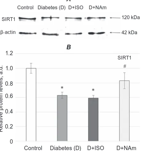
Fig. 1. Effects of ISO or NAm on SIRT1 expression
in brain cell nuclei. SIrT1 expression was lower in diabetic rats, while treatment with ISo does not
influen ce its expression, NAm had slight normalizing effect on SIRT1 expression restoration. All values
are expressed as mean ± SeM of three experiments (n = 3) in duplicate. A – Blottogram; B – results of densitometry. *P < 0.05 vs. control; #P < 0.05 vs.
diabetes
1.2
R
e
lat
iv
e p
rot
e
in l
e
v
e
ls
, a
.u
.
Control Diabetes (D) D+ISO D+NAm 1.0
0.8
0.6
0.4
0.2
0
SIRT1
B
Control Diabetes (D) D+ISO D+NAm
SIRT1
β-actin
120 kDa
42 kDa
A
signalling in microglia [42]. However administration of PARP-1 inhibitors to diabetic rats demonstrated
that while ISO had virtually no effect on the expres -sion of SIRT1, NAm slightly increased its expres-sion
(Fig. 1). Nevertheless, slight influence of NAm on in
-creasing SIRT1 expression can have protective effect
on metabolic processes in brain, altered by T1DM, resulting in a life prolongation [43]. Noteworthy that
such slight effect of NAm on SIRT1 expression in
the nuclei extracts of brain tissue can be the result of the fact that SIRT1 is not only localized in the nucleus, where it deacetylates histones thereby
in-fluencing genes expression [26], but SIRT1 can also
shuttle to cytoplasm and through this
nucleocyto-plasmic mechanism may participate in differentia -tion and in inhibi-tion of cell death [27]. Moreover,
sirtuins are structurally different with regard to their
N- and C-ends, their sub-cellular localization, and
their different substrates [24, 25]. It is not excluded
that even slight elevation of SIR1expression in turn
can lead to activation of 5′-AMP-activated protein
kinase which controls energy intake under physio-logical and pathophysio-logical conditions [45]. According to literature data SIRT1 activation can be involved in mitochondrial metabolism in conditions of PARP-1 inhibition [36]. So, the data concerning overexpres-sing SIRT1 in the brain of rats may provide valuable clues as to how SIRT1 realizes its neuroprotective action in vivo. It is known that SIRT1 in the heart increases ischemic tolerance via an activation of eNOS [31] and it also protects against cardiac
hyper-trophy through PPARα activation [32]. These data
suggest that SIRT1 alterations expression in brain in-duced by diabetes might impact on encephalopathy develop ment.
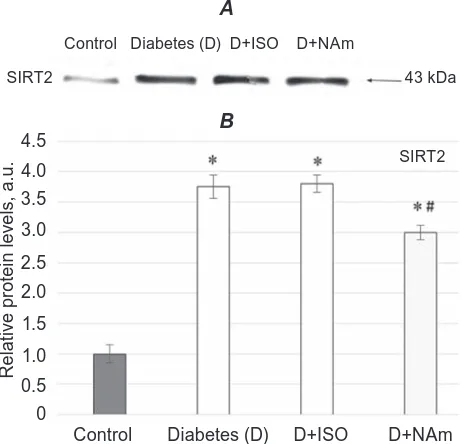
perception [34]. However it is still considerable work to be done to determine the precise role of SIRT2 in the initiation and progression of brain dysfunctions induced by diabetes in vivo. Diabetic rats treated by ISO had no changes in SIRT2 expression in brain cell nuclei as compared to untreated animals (Fig. 2), whereas after NAm administration to diabetic rats its expression was slightly decreased, P < 0.05.
Therefore, it is not excluded, that exist the con-nection between the regulatory process under diabe-tes and level of sirtuins expression in nuclei brain. So, it is mean that sirtuins can become
pharmaceu-tical targets for their modulation and modification. These findings concerning SIRT1 up-regulation and
SIRT2 down-regulation are complementary to our previous study showing that PARP-1 inhibition in rats by ISO and NAm increased nicotinamide ade-nine dinucleotide (NAD+) content and improved
brain functions [10]. However, the molecu lar role of sirtuins in diabetes still remains completely unclear. To better understand of sirtuin functions we have to determine the complete range of their biochemical and enzymatic activities. It should be noted that
de-spite of the fact that ISO did not influence on SITR1
and SIRT2 expression of nuclei brain the protective mechanism of its action on dia betes-induced brain
dysfunction might be realized through its influence
on other molecular prosesses in brain. As example ISO can inhibit lipid peroxidation, improve the ace-tylcholinesterase functio ning and partially normalize the levels of GABA and glutamate in the hippocam-pus [54]. Since PARP-1, SIRT1 and SIRT2 are NAD-dependent, it is quite obvious that they can compete for the availability of NAD+, reducing not only its
content, but also the ratio of free NAD/NADH cou-ples in cells. That is why it was reasonable to evalu-ate the ratio of free NAD/NADH couples in the brain
because it reflects the metabolic state of oxidized and
reduced substrates, that is related to two aspects of cellular metabolism: determination the direction of reverse reactions and assessment of alterations of free ener gy in extracellular oxidoreduction (for
exam ple, transport of electrons from NADH to fla -voproteins in the electron transport chain [55]. As it is known NAD+ plays important role in cellular me-tabolism and realized its action involving in different
a key processes including redox reactions which are regulators of cells functioning [46, 47]. Noteworthy,
even insignificant changes in metabolism are accom -panied by changes in the free NAD/NADH couples
ratio, which in turn causes a change in the flow of
carbon to the glycolysis or gluconeogenesis at the
Fig. 2. Western blotting of SIrT2 (43 kDa) in the nu-clear extracts of rat brain tissues. A – representa-tive Western blot and B – densitometric analysis of SIrT2 protein content. The control values are taken
as 1. Statistically significant differences are ex -pressed as mean ± SeM of three experiments (n = 3) in duplicate. *P < 0.05 vs. control; #P < 0.05 vs.
diabetes
Control Diabetes (D) D+ISO D+NAm
SIRT2 43 kDa
A
4.5
R
e
lat
iv
e p
rot
e
in l
e
v
e
ls
, a
.u
.
Control Diabetes (D) D+ISO D+NAm 3.0
2.0
1.5
1.0
0.5
0
SIRT2
B
4.0
3.5
2.5
level of the glyceral dehyde-3-phosphate dehydro-genase reaction [48]. Results obtained in our previ-ously published data revealed that the NAD+ content
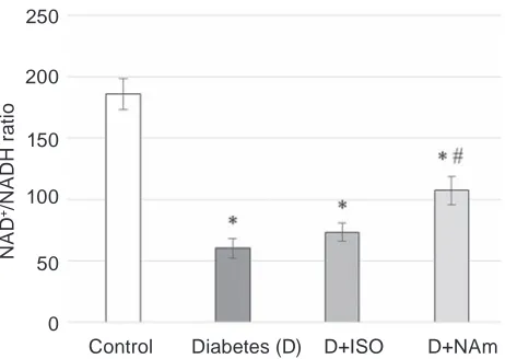
in brain of diabetic rats was reduced [10]. That is why it is not excluded that the ratio of free NAD/ NADH couples also will be changed whereas its maintenance is very important. Indeed, we observed the alterations of metabolites equilibrium concen-trations which lead to the reduction of the ratio of free NAD/NADH couples in the brain of diabetic rats to 60.1 versus 186.2 in the control (Fig. 3). Such changes in the oxidation-reduction state can lead to disturbances in the dynamic equilibrium between the oxidation of glucose in NAD-dependent glyco-lysis and the NADPH-dependent pentose phosphate pathway and enhance DE develop ment.
It was shown that administration of ISO or NAm to diabetic rats lead to slight increase the ra-tio of free NAD/NADH couples in the brain to 73.4 in case of ISO and more profound for NAm up to 107.5, that may result in the enhanced fuel
oxida-tion and amplificaoxida-tion of NAD+-dependent SIRT-1
be noted that our findings are consistent with results
concerning Parkinson’s disease that does not exclude the common mechanisms of neurodegenerative pro-cesses development in the nervous system [52].
In conclusion, it is becoming clear that both SIRT1 and SIRT2 play an important role in brain functioning and alterations of their expression in-duced by diabetes lead to brain dysfunctions which can be accompanied by cognitive impairment develop ment. However, the results of the present study have shown that among investigated PAPR-1 inhibitors only NAm can partially ameliorate sirtuins expression and improve redox state in brain of
dia-betic rats. Unlike ISO, anti-diadia-betic efficacy of NAm
may also be associated with formation of 1-methyl-nicotinamide, one of the endogenous products of NAm, due to its ability to attenuate deleterious ef-fects of diabetes in the CNS as we observed erlier [13]. We also emphasize here that the inhibition of PAPR-1 may be linked with improvement of SIRT1 and SIRT2 functioning during hyperglycemia,
pos-sibly due to influence of NAm on the sirtuins expres -sion. More importantly, our results indicate the need
to develop more specific modulators of these sirtuins that would not cause any side effects. Therefore these
sirtuins can be promising and potential therapeutic targets for treatment of diabetic encephalopathy.
Conflict of interest. Authors have completed
the Unified Conf licts of Interest form at http:// ukrbio chemjournal.org/wp-content/uploads/2018/12/ coi_disclosure.pdf and declare no conf lict of interest .
Fig. 3. The ratio of free NaD/NaDh couples in
the rat brain, mean ± SEM of five of experiments.
*P < 0.05 compared to control group, #P < 0.05
compared to diabetic group
250
NA
D
+/N
A
D
H r
at
io
Control Diabetes (D) D+ISO D+NAm 150
100
50
0 200
Зміна експресії сИртуїнів 1 і 2 в моЗку щурів З індукованИм експерИментальнИм
діабетом та шляхИ її корекції
M. M. Гузик1, T. M. Тихоненко1, К. О. Дякун1,
Л. В. Яніцька2, I. Б. Привроцька3,
T. M. Кучмеровська1
1Інститут біохімії ім. О. В. Палладіна
НАН України, Київ;
2Національний медичний університет імені
О. О. Богомольця МОЗ України, Київ;
3ДВНЗ «Тернопільский державний медичний
університет імені І. Я. Горбачевського», Україна;
e-mail: tkuchmerovska@gmail.com
Молекулярні механізми патогенезу діабетичної енцефалопатії (ДЕ), одного із серйоз
-них ускладнень цукрового діабету, є складними. У цій роботі досліджували експресію SIRT1 та SIRT2 як ключову в розвитку дисфункції мозку та можливість інгібіторів PARP-1 впливати на експресію цих протеїнів з метою попереджен
-ня розвитку ДЕ в щурів із діабетом 1-го типу. Через 10 тижнів розвитку цукрового діабету, індукованого стрептозотоцином (70 мг/кг), щурам-самцям лінії Wistar вводили інгібітори PARP-1, 1,5-ізохіноліндіол (ISO) або нікотинамід (NAm) (3 або 100 мг/кг/добу відповідно) про
можуть призводити до розвитку дисфункцій мозку. Один з нейропротекторних механізмів дії NAm також може бути реалізований шляхом гальмування експресії SIRT2 в ядрах клітин го
-ловного мозку, що знижує рівень прогресування індукованих діабетом змін і може бути терапев
-тичним засобом для лікування дисфункцій мозку.
К л ю ч о в і с л о в а: цукровий діабет, експресія сиртуїнів, SIRT1, SIRT2, інгібітори PARP-1, 1,5-ізохіноліндіол, нікотинамід.
references
1. Grünblatt E, Bartl J, Riederer P. The link between iron, metabolic syndrome, and Alzheimer's disease. J Neural Transm (Vienna). 2011; 118(3): 371-379.
2. Monette MC, Baird A, Jackson DL. A meta-analysis of cognitive functioning in nondemented adults with type 2 diabetes mellitus. can J Diabetes. 2014; 38(6): 401-408.
3. Wong RH, Scholey A, Howe PR. Assessing premorbid cognitive ability in adults with type 2 diabetes mellitus – a review with implications for future intervention studies. curr Diab rep. 2014; 14(11): 547.
4. Sima AA. Encephalopathies: the emerging diabetic complications. acta Diabetol. 2010; 47(4): 279-293.
5. Wayhs CA, Mescka CP, Guerreiro G, Moraes TB, Jacques CE, Rosa AP, Ferri MK, Nin MS, Dutra-Filho CS, Barros HM, Vargas CR. Diabetic encephalopathy-related depression: experimental evidence that insulin and clonazepam restore antioxidant status in rat brain. cell Biochem Funct. 2014; 32(8): 711-719. 6. Ray Chaudhuri A, Nussenzweig A. The
multifaceted roles of PARP1 in DNA repair and chromatin remodelling. Nat rev Mol cell Biol. 2017; 18(10): 610-621.
7. Chaitanya GV, Steven AJ, Babu PP. PARP-1 cleavage fragments: signatures of cell-death proteases in neurodegeneration. cell commun Signal. 2010; 8: 31.
8. Drel VR, Pacher P, Stavniichuk R, Xu W, Zhang J, Kuchmerovska TM, Slusher B, Obrosova IG. Poly(ADP-ribose)polymerase inhibition counteracts renal hypertrophy and multiple manifestations of peripheral neuropathy in diabetic Akita mice. Int J Mol Med. 2011; 28(4): 629-635.
9. Virág L, Szabó C. The therapeutic potential of poly(ADP-ribose) polymerase inhibitors. Pharmacol rev. 2002; 54(3): 375-429.
10. Guzyk MM, Dyakun KO, Yanytska LV, Pryvrotska IB, Krynytska IYa, Pishel’ IM, Kuchmerovska TM. Inhibitors of poly(ADP-ribose)polymerase-1 as agents providing correction of brain dysfunctions induced by experimental diabetes. Neurophysiology. 2017; 49(3): 183-193.
11. Sugaya-Fukasawa M, Watanabe T, Tamura M,
Egashira S, Hisatomi H. Glial fibrillary acidic
protein is one of the key factors underlying neuron-like elongation in PC12 cells. exp Ther Med. 2011; 2(1): 85-87.
12. Kuchmerovska T, Shymanskyy I, Donchenko G, Kuchmerovskyy M, Pakirbaieva L, Klimenko A. Poly(ADP-ribosyl)ation enhancement in brain cell nuclei is associated with diabetic neuropathy. J Diabetes complications. 2004; 18(4): 198-204. 13. Kuchmerovska T, Shymanskyy I, Chlopicki S,
Klimenko A. 1-methylnicotinamide (MNA) in prevention of diabetes-associated brain disorders. Neurochem Int. 2010; 56(2): 221-228. 14. Verdin E, Hirschey MD, Finley LW, Haigis MC.
Sirtuin regulation of mitochondria: energy production, apoptosis, and signaling. Trends Biochem Sci. 2010; 35(12): 669-675.
15. Flick F, Lüscher B. Regulation of sirtuin function
by posttranslational modifications. Front Pharmacol. 2012; 3: 29.
16. Haigis MC, Sinclair DA. Mammalian sirtuins: biological insights and disease relevance. annu rev Pathol. 2010; 5: 253-295.
17. Chalkiadaki A, Guarente L. Sirtuins mediate mammalian metabolic responses to nutrient availability. Nat rev endocrinol. 2012; 8(5): 287-296.
18. Austad SN. Ageing: Mixed results for dieting monkeys. Nature. 2012; 489(7415): 210-211. 19. Bishop NA, Guarente L. Genetic links between
diet and lifespan: shared mechanisms from yeast to humans. Nat rev Genet. 2007; 8(11): 835-844. 20. Jiang H, Medintz I, Zhang B, Michels CA.
Metabolic signals trigger glucose-induced inactivation of maltose permease in Saccharo-myces. J Bacteriol. 2000; 182(3): 647-654. 21. Mair W, Goymer P, Pletcher SD, Partridge L.
22. Cohen MJ, Carstenn S, Lane CR. Floristic quality indices for biotic assessment of depressional marsh condition in Florida. ecol appl. 2004; 14(3): 784-794.
23. Utani K, Fu H, Jang SM, Marks AB, Smith OK, Zhang Y, Redon CE, Shimizu N, Aladjem MI. Phosphorylated SIRT1 associates with repli-cation origins to prevent excess replirepli-cation initiation and preserve genomic stability. Nucleic acids res. 2017; 45(13): 7807-7824.
24. Frye RA. Phylogenetic classification of
prokaryotic and eukaryotic Sir2-like proteins. Biochem Biophys res commun. 2000; 273(2): 793-798.
25. Guarente L. Sirtuins, aging, and metabolism. cold Spring harb Symp Quant Biol. 2011; 76: 81-90.
26. Guarente L. Calorie restriction and sirtuins revisited. Genes Dev. 2013; 27(19): 2072-2085. 27. Tanno M, Sakamoto J, Miura T, Shimamoto K,
Horio Y. Nucleocytoplasmic shuttling of the NAD+-dependent histone deacetylase SIRT1.
J Biol chem. 2007; 282(9): 6823-6832.
28. Serrano L, Martínez-Redondo P, Marazuela-Duque A, Vazquez BN, Dooley SJ, Voigt P, Beck DB, Kane-Goldsmith N, Tong Q, Rabanal RM, Fondevila D, Muñoz P, Krüger M,
Tischfield JA, Vaquero A. The tumor suppressor
SirT2 regulates cell cycle progression and genome stability by modulating the mitotic deposition of H4K20 methylation. Genes Dev. 2013; 27(6): 639-653.
29. Imai S, Armstrong CM, Kaeberlein M, Guarente L. Transcriptional silencing and longe-vity protein Sir2 is an NAD-dependent histone deacetylase. Nature. 2000; 403(6771): 795-800. 30. Hallows WC, Yu W, Denu JM. Regulation of
glycolytic enzyme phosphoglycerate mutase-1 by Sirt1 protein-mediated deacetylation. J Biol chem. 2012; 287(6): 3850-3858.
31. Mattagajasingh I, Kim CS, Naqvi A, Yamamori T,
Hoffman TA, Jung SB, DeRicco J, Kasuno K,
Irani K. SIRT1 promotes endothelium-dependent vascular relaxation by activating endothelial nitric oxide synthase. Proc Natl acad Sci USa. 2007; 104(37): 14855-14860.
32. Planavila A, Iglesias R, Giralt M, Villarroya F.
Sirt1 acts in association with PPARα to
protect the heart from hypertrophy, metabolic
dysregulation, and inflammation. cardiovasc res. 2011; 90(2): 276-284.
33. Yuan F, Xu ZM, Lu LY, Nie H, Ding J, Ying WH, Tian HL. SIRT2 inhibition exacerbates
neuroinflammation and blood-brain barrier
disruption in experimental traumatic brain
injury by enhancing NF-κB p65 acetylation and
activation. J Neurochem. 2016; 136(3): 581-593. 34. Yu TT, McIntyre JC, Bose SC, Hardin D,
Owen MC, McClintock TS. Differentially
expressed transcripts from phenotypically
identified olfactory sensory neurons. J comp Neurol. 2005; 483(3): 251-262.
35. Dryden SC, Nahhas FA, Nowak JE, GoustinAS, Tainsky MA. Role for human SIRT2 NAD-dependent deacetylase activity in control of mitotic exit in the cell cycle. Mol cell Biol. 2003; 23(9): 3173-3185.
36. Bai P, Cantó C, Oudart H, Brunyánszki A, Cen Y, Thomas C, Yamamoto H, Huber A, Kiss B, Houtkooper RH, Schoonjans K, Schreiber V, Sauve AA, Menissier-de Murcia J, Auwerx J. PARP-1 inhibition increases mitochondrial metabolism through SIRT1 activation. cell Metab. 2011; 13(4): 461-468.
37. Shaiken TE, Opekun AR. Dissecting the cell to nucleus, perinucleus and cytosol. Sci rep. 2014; 4: 4923.
38. Laemmli UK. Cleavage of structural proteins during the assembly of the head of bacteriophage T4. Nature. 1970; 227(5259): 680-685.
39. Bergmeyer HU. Methods of Enzymatic Analysis. 1963; 1064 p.
40. Guzyk MM, Tykhomyrov AA, Nedzvetsky VS, Prischepa IV, Grinenko TV, Yanitska LV, Kuchmerovska TM. Poly(ADP-ribose) polymerase-1 (PARP-1) inhibitors reduce reactive gliosis and improve angiostatin levels in retina of diabetic rats. Neurochem res. 2016; 41(10): 2526-2537.
41. Bradford MM. A rapid and sensitive method for the quantitation of microgram quantities of protein utilizing the principle of protein-dye binding. anal Biochem. 1976; 72: 248-254. 42. Chen J, Zhou Y, Mueller-Steiner S, Chen LF,
Kwon H, Yi S, Mucke L, Gan L. SIRT1 protects against microglia-dependent amyloid-beta toxicity through inhibiting NF-kappaB signaling. J Biol chem. 2005; 280(48): 40364-40374.
response to diet restriction through activation of the dorsomedial and lateral nuclei of the hypothalamus. J Neurosci. 2010; 30(30): 10220-10232.
44. Rajesh M, Mukhopadhyay P, Godlewski G, Bátkai S, Haskó G, Liaudet L, Pacher P. Poly(ADP-ribose)polymerase inhibition decreases angiogenesis. Biochem Biophys res
commun. 2006; 350(4): 1056-1062.
45. Jeon SM. Regulation and function of AMPK in physiology and diseases. exp Mol Med. 2016; 48(7): e245.
46. Katsyuba E, Auwerx J. Modulating NAD(+) metabolism, from bench to bedside. eMBo J. 2017; 36(18): 2670-2683.
47. Cantó C, Menzies KJ, Auwerx J. NAD(+) Metabolism and the control of energy homeostasis: a balancing act between mitochondria and the nucleus. cell Metab. 2015; 22(1): 31-53.
48. Erecińska M, Wilson DF. Regulation of cellular
energy metabolism. J Membr Biol. 1982; 70(1): 1-14.
49. Momsen G. Effect of increasing the intracellular
ratio of NADH to NAD+ on human erythrocyte
metabolism: new estimation of the turnover through the phosphoglycerate shunt. arch Biochem Biophys. 1981; 210(1): 160-166.
50. Milne JC, Lambert PD, Schenk S, Carney DP, Smith JJ, Gagne DJ, Jin L, Boss O, Perni RB, Vu CB, Bemis JE, Xie R, Disch JS, Ng PY, Nunes JJ, Lynch AV, Yang H, Galonek H,
Israelian K, Choy W, Iffland A, Lavu S,
Medvedik O, Sinclair DA, Olefsky JM, Jirousek MR, Elliott PJ, Westphal CH. Small molecule activators of SIRT1 as therapeutics for the treatment of type 2 diabetes. Nature. 2007; 450(7170): 712-716.
51. de Kreutzenberg SV, Ceolotto G, Papparella I, Bortoluzzi A, Semplicini A, Dalla Man C, Cobelli C, Fadini GP, Avogaro A. Downregulation of the longevity-associated protein sirtuin 1 in insulin resistance and metabolic syndrome: potential biochemical mechanisms. Diabetes. 2010; 59(4): 1006-1015.
52. Harrison IF, Smith AD, Dexter DT. Pathological histone acetylation in Parkinson's disease: Neuroprotection and inhibition of microglial activation through SIRT 2 inhibition. Neurosci Lett. 2018; 666: 48-57.
53. Inoue T, Hiratsuka M, Osaki M, Oshimura M. The molecular biology of mammalian SIRT proteins: SIRT2 in cell cycle regulation. cell cycle. 2007; 6(9): 1011-1018.
54. Jangra A, Datusalia AK, Sharma SS. Reversal of neurobehavioral and neurochemical alterations in STZ-induced diabetic rats by FeTMPyP, a peroxynitrite decomposition catalyst and 1,5-Isoquinolinediol a poly(ADP-ribose) polymerase inhibitor. Neurol res. 2014; 36(7): 619-626.