cells and innate and myeloid cells in
preneoplastic gammopathy
Jithendra Kini Bailur, … , Kavita M. Dhodapkar, Madhav V.
Dhodapkar
JCI Insight.
2019;
4(11)
:e127807.
https://doi.org/10.1172/jci.insight.127807
.
Preneoplastic lesions carry many of the antigenic targets found in cancer cells but often
exhibit prolonged dormancy. Understanding how the host response to premalignancy is
maintained and altered during malignant transformation is needed to prevent cancer. To
understand the immune microenvironment in precursor monoclonal gammopathy of
undetermined significance (MGUS) and myeloma, we analyzed bone marrow immune cells
from 12 healthy donors and 26 patients with MGUS/myeloma by mass cytometry and
concurrently profiled transcriptomes of 42,606 single immune cells from these bone marrow
samples. Compared with age-matched healthy donors, memory T cells from both MGUS
and myeloma patients exhibited greater terminal effector differentiation. However, memory T
cells in MGUS show greater enrichment of stem-like TCF1/7
hicells. Clusters of T cells with
stem-like and tissue residence genes were also found to be enriched in MGUS by
single-cell transcriptome analysis. Early changes in both NK and myeloid single-cells were also observed
in MGUS. Enrichment of stem-like T cells correlated with a distinct genomic profile of
myeloid cells and levels of Dickkopf-1 in bone marrow plasma. These data describe the
landscape of changes in both innate and adaptive immunity in premalignancy and suggest
that attrition of the bone marrow–resident T cell compartment because of loss of stem-like
cells may underlie loss of immune surveillance in myeloma.
Research Article
Immunology
Oncology
Find the latest version:
Authorship note: JKB and SSM are co–first authors. KMD and MVD contributed equally to this work. Conflict of interest: The authors have declared that no conflict of interest exists.
Copyright: © 2019, American Society for Clinical Investigation.
Submitted: January 30, 2019 Accepted: April 17, 2019 Published: June 6, 2019. Reference information:JCI Insight. 2019;4(11):e127807. https://doi. org/10.1172/jci.insight.127807.
Early alterations in
stem-like/marrow-resident T cells and innate and myeloid
cells in preneoplastic gammopathy
Jithendra Kini Bailur,1 Samuel S. McCachren,1,2 Deon B. Doxie,1 Mahesh Shrestha,1 Katherine Pendleton,1 Ajay K. Nooka,1,3 Natalia Neparidze,4 Terri L. Parker,4 Noffar Bar,4 Jonathan L. Kaufman,1,3 Craig C. Hofmeister,1,3 Lawrence H. Boise,1,3 Sagar Lonial,1,3 Melissa L. Kemp,2 Kavita M. Dhodapkar,3,5 and Madhav V. Dhodapkar1,3
1Department of Hematology/Oncology, Emory University School of Medicine, Atlanta, Georgia, USA. 2The Wallace H.
Coulter Department of Biomedical Engineering, Georgia Institute of Technology and Emory University, Atlanta, Georgia, USA. 3Winship Cancer Institute, Atlanta, Georgia, USA. 4Section of Hematology, Yale University School of
Medicine, New Haven, Connecticut, USA. 5Department of Pediatrics, Children’s Healthcare of Atlanta, Emory University,
Atlanta, Georgia, USA.
Introduction
Most human cancers are preceded by a long phase of premalignant lesions that are more common than the cancer itself. Multiple myeloma (MM) is a common hematologic malignancy wherein tumor cells grow predominantly in the bone marrow. All cases of MM are preceded by a precursor state termed as monoclonal gammopathy of undetermined significance (MGUS) (1). MGUS/MM is an attractive model to gain basic insights into early host response in human cancer because the precursor state is unresectable. Studies in mouse models have illustrated the capacity of the immune system to prevent, edit, or sculpt early tumors, mediated at least in part by T cell recognition of neoantigens. In the setting of cancer, T cell exhaustion as well as other immune-suppressive cells inhibit tumor immunity. However, because many neoantigens originate early during carcinogenesis, precisely when during carcinogenesis such exhaustion programs emerge and how these exhausted clones are maintained in human cancer/pre-cancer remain to be elucidated. Tumor cells in MGUS carry the great majority of genomic alterations, including the mutational burden found in MM (2). Prior studies have demonstrated the capacity of T cells to mediate specific recognition of MGUS cells (3, 4); recent data also show alterations in innate lymphoid cells in these lesions (5). Antitumor T cells are often enriched in the bone marrow of MM patients (6, 7); therapeutic adoptive transfer of bone marrow T cells is being explored in MM (8). Recent prospective studies have shown that host immune response in MGUS can predict the risk of malignant
Preneoplastic lesions carry many of the antigenic targets found in cancer cells but often exhibit prolonged dormancy. Understanding how the host response to premalignancy is maintained and altered during malignant transformation is needed to prevent cancer. To understand the immune microenvironment in precursor monoclonal gammopathy of undetermined significance (MGUS) and myeloma, we analyzed bone marrow immune cells from 12 healthy donors and 26 patients with MGUS/myeloma by mass cytometry and concurrently profiled transcriptomes of 42,606 single immune cells from these bone marrow samples. Compared with age-matched healthy donors, memory T cells from both MGUS and myeloma patients exhibited greater terminal effector differentiation. However, memory T cells in MGUS show greater enrichment of stem-like TCF1/7hi
transformation (9). Data from humanized mouse models also suggest that dormancy of MGUS lesions is controlled by tumor-extrinsic factors (10). Therefore, understanding the biology of immune cells infil-trating MGUS lesions may provide valuable insights into pathogenesis of MM.
One deficiency of existing studies is that only limited information on cell states and phenotypes of indi-vidual cells in the tumor microenvironment was obtained. To overcome these limitations, we performed high-dimensional single-cell RNA-sequencing (scRNA-Seq) and mass cytometry analysis on immune cells in the MGUS/MM bone marrow microenvironment to gain deeper insights into these cells, both at tran-scriptomic as well as proteomic level. We focused on the nontumor compartment in both MM and MGUS and performed comparisons with immune cells in bone marrow from healthy donors.
Results
To analyze immune cells in the bone marrow in MGUS/MM, we analyzed bone marrow mononuclear cells by mass cytometry using 57 markers (clinical characteristics in Supplemental Table 1, mass cytom-etry panel in Supplemental Table 2; supplemental material available online with this article; https://doi.
org/10.1172/jci.insight.127807DS1). In view of the importance of CD8+ memory T cells in tumor
immu-nity, we first examined the CD8+ memory T cell compartment, which revealed a modest decline in CD8+
central memory T cells (Tcms) in MM (Figure 1A). MM had a slightly higher proportion of CD8+ effector/
effector memory T cells (Tems + terminal effectors [Ttes]) (Figure 1B). However, the proportion of Ttes expressing high levels of granzyme B was significantly increased in both MGUS and MM compared with age-matched healthy donors (Figure 1, C and D, and Supplemental Figure 1).
To better characterize these changes in effector differentiation, we analyzed the expression of several T cell–associated transcription factors in memory T cells. Of these, the expression of T cell factor 1 (TCF1) was
significantly increased among memory CD8+ T cells in the MGUS cohort, while the expression of T-bet,
Eomes, and GATA-3 did not differ (Figure 1, E and F). A similar trend for increase in TCF1 expression was
seen among CD4+ memory T cells in MGUS (Supplemental Figure 2A). The proportions of CD4+ T cells
expressing Eomes and T-bet were also significantly lower in MGUS and MM cohorts (Supplemental Figure 2B). Together these data show that even though T cells infiltrating both MM and MGUS have enhanced effec-tor differentiation, there are distinct differences in transcription faceffec-tor expression in these cells.
Observed differences in the expression of TCF1 in premalignancy were of particular interest because
of its emerging role in regulating T cell stemness in memory CD8+ T cells during chronic infections and
possibly cancer (11–14). TCF1 expression in human T cells follows a distinct gradient, with stem-like fea-tures associated with TCF1hiT-betlo cells (14). Using a similar gating strategy (Figure 1G), the proportion
of TCF1hi cells was found to be significantly increased in MGUS, while TCF1– cells were higher in MM
(Figure 1H). The increase in TCF1hi CD8+ T cells in MGUS was observed in both Tcm and Tem
compart-ments (Figure 1I). TCF1hi cells in all 3 patient cohorts had a distinct phenotype, with increased expression
of CD127, CD27, and CXCR4 (marker of bone marrow homing), as well as reduced expression of T-bet,
Eomes, and lytic genes, such as granzyme B (Figure 1J). Thus, CD8+ memory T cells infiltrating MGUS
lesions are particularly enriched for phenotypes associated with enhanced stemness.
Analysis of the myeloid compartment (gating strategy in Supplemental Figure 3) was performed to identify markers differentially expressed between myeloid cells infiltrating MGUS and MM. These studies identified a decline in the expression of CD95, CD86, c-KIT, and CD155 and an increase in programmed
death ligand 1 (PD-L1) expression on myeloid cells in MM compared with healthy donors, while there
were no differences in other markers, such as HLA-DR (Figure 2, A and B). We identified 3 distinct clus-ters of myeloid cells based on the expression of CD14 and CD11b, with MGUS/MM lesions enriched in
the proportion of CD14–CD11b+ myeloid cells (Supplemental Figure 4). Analysis of NK cells revealed a
slight increase in total NK cells in MM; subsequent analysis of the CD56lo NK subset revealed a decline in
natural killer group 2, member D (NKG2D) expression in MM (Supplemental Figure 4). Thus, alterations in innate myeloid and NK cells originate early in myelomagenesis.
tially represented between the 3 cohorts (Figure 3, C and D). Analysis of immune composition by scRNA-Seq correlated with mass cytometry data; for example, the decline in B cells in MM by mass cytometry was also reflected in the decline in major B cell cluster (B1) in MM (Supplemental Figure 5 and Figure 3D).
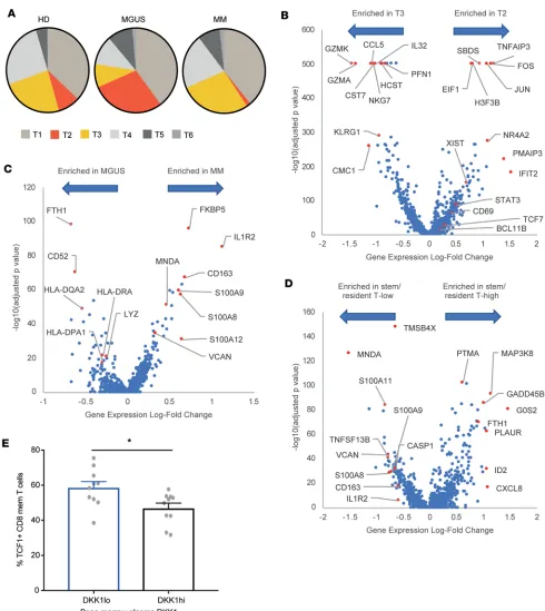
The most notable differences between T cells in MGUS and MM related to 2 distinct T cell clusters (clus-ters T2 and T3) (Figure 4A). T2 cluster (overrepresented in MGUS and reduced in MM) was characterized by increased expression of stem-like genes (TCF1/TCF7) consistent with data from mass cytometry, as well as genes associated with tissue residence (NR4A2, CD69) (15, 16) and reduced expression of lytic genes (granzyme A, granzyme K). In contrast, T3 cluster (increased in MM) was characterized by the expression of lytic genes (e.g., granzyme A), senescence-associated genes (e.g., KLRG1), and proinflammatory cytokines (e.g., IL-32) (Figure 4B). Gene set enrichment analysis (GSEA) of these genes identified pathways related to effector functions and programmed cell death 1–mediated (PD-1–mediated) exhaustion programs as key pathways distinguishing MM-enriched clusters (Supplemental Figure 6). T2 cluster, enriched in MGUS, was also enriched for pathways associated with stem cell memory cells (Supplemental Figure 6) as mass
try data also suggested. Although T3 cluster is present in both healthy donors and patents with MM, the T3 cluster in MM revealed enrichment of PRDM1 (as a marker of exhaustion), Fos, and CCL4 (as a marker of activation) and downregulation of granulysin and lysozyme (as a marker of dysfunction) (Supplemental Fig-ure 7). Taken together with the mass cytometry data (FigFig-ure 1), these studies show that T cells in MGUS are enriched for stem-like memory T cells, overlapping in part with a quiescent tissue-resident phenotype, while T cells in MM show greater expression of lytic genes and senescence markers.
Myeloid cells comprised 3 distinct clusters that correlated with monocytes (M1 cluster), as well as pre-viously described CD1c+ or CD1c– dendritic cell clusters (M2 and M3 clusters) (17). Analysis of
differen-tially expressed genes within myeloid clusters revealed reduced transcripts of HLA-DR in MM, consistent with a less activated phenotype and consistent with the mass cytometry data showing lower CD86 as a marker of reduced activation in MM myeloid cells (Figure 4C). Genes increased in MM myeloid cells also included several genes with possible implications for immunosuppressive properties (such as IL1R2 and calcium-binding proteins S100A8, S100A9, and S100A12), as well as other genes (e.g., versican) previously implicated in this role (18). GSEA of differentially expressed genes identified dendritic cell (DC) activation as a pathway altered in these cells, supporting a role for altered antigen presentation (Supplemental Figure 8). Cells from healthy donors and patients with MM in the M1 cluster were located somewhat differently in the t-SNE plot (Figure 3C). Top differentially expressed genes of cells in this cluster between healthy donors and patients with MM also supported altered myeloid polarization with a more immune-suppres-sive phenotype of myeloid cells in MM (Supplemental Figure 9).
To explore the link between T cell and myeloid changes, we examined the genomic profiles of myeloid cells from patients with a higher proportion of cells in T2 cluster. Myeloid cells from these patients revealed a distinct genomic profile consistent with an enrichment for genes associated with TLR-mediated DC activation on GSEA (Figure 4D and Supplemental Figure 10). Because TCF1 expression is known to be regulated by WNT signaling, we hypothesized that the depletion of TCF1+ cells may be related to increase in WNT inhibitor
Dickkopf-1 previously described in MM (19). Consistent with this possibility, patients with elevated levels of
Dickkopf-1 (DKK1) in the bone marrow plasma had reduced levels of TCF1+ memory T cells (Figure 4E).
Discussion
These data illustrate several changes in the immune landscape in MGUS and MM. The data show progres-sive increase in terminal effector T cells with progresprogres-sive malignancy. However, the finding that terminal effector T cells were increased even in MGUS suggests that alterations in T cell differentiation originate early in carcinogenesis during premalignancy.
An important and potentially novel aspect of this paper is the finding that memory T cells in the bone
marrow in MGUS are enriched for TCF1hi memory T cells that bear a resemblance to stem-like T cells (20).
TCF1hi cells have to date been best studied in the context of models of chronic viral infections, wherein
recent studies have illustrated their importance in preventing attrition of exhausted T cells and thereby maintaining long-term protective immunity (11, 12, 21). TCF1-expressing cells have also been recently identified in human cancer tissue and were linked to outcomes following checkpoint blockade in a small cohort (13, 22). To our knowledge, these are the first studies to identify specific enrichment of these cells in the tumor microenvironment in a human premalignancy. It is now well established that many of the mutations/neoantigens found in cancer that serve as critical targets of tumor immunity originate during the premalignant phase, particularly in MM (1, 2). Therefore, the observed enrichment and persistence of these
cells in MGUS may be key to maintaining tumor immunity during the MGUS phase; loss of TCF1+
mem-ory T cells in MM may lead to inability to maintain protective immunity over time and loss of immune surveillance. Although the cells identified in our studies bear a general resemblance to phenotypes observed in murine models of chronic viral infection, we and others (22) have not detected the expression of CXCR5 on these cells in the tumor tissue. Together, these data suggest that a hierarchy of exhausted clones wherein a small subset of stem-like cells prevent clonal attrition is already evident at the premalignant stage.
An important change in the MM tumor microenvironment relates to phenotype and gene expression in myeloid cells. Prior studies have shown that myeloid cells play an important role in regulating the growth of tumor cells (27, 28). The altered phenotype of tumor-infiltrating myeloid cells, including reduced expres-sion of CD86, CD155, and c-KIT and increased PD-L1, supports an immune-suppressive phenotype. These data, therefore, support prior studies showing enrichment of myeloid cells in MM, including their protumor effects (18). Reduced expression of Fas in MM myeloid cells is of interest because Fas/FasL interactions play a major role in regulating myeloid cell kinetics, as illustrated by increased myeloid progen-itors in mice with mutated Fas/FasL (29), and may contribute to their enrichment in MM.
In addition to changes in T cells, we observed changes in innate cells, with an increase in NK cells. NK cells in MM, however, revealed some functional alterations, such as decline in NKG2D. Therefore, alterations in innate lymphocytes appear to be an early feature in the evolution of gammopathies, which is consistent with prior studies (5, 30). Recent studies suggest that innate cells may be targets for immuno-modulatory drugs currently being explored for prevention of myeloma (5).
The enrichment of stem-like and resident T cells in the MGUS marrow that we observed may be affect-ed by local signals in the tumor baffect-ed. Prior studies have shown that local interactions with antigen-present-ing cells in situ may be critical for tissue retention of murine tissue-resident memory T cells (31, 32). Con-sistent with this, we observed that enrichment of stem-like/resident T cells correlated with distinct genomic features of myeloid cells indicative of TLR-mediated activation. Further studies are needed to dissect the signals needed for retention and persistence of these cells in the bone marrow. Some of the signals that reg-ulate these cells may also be derived from tumors themselves. For example, because TCF1 expression may depend on WNT signaling, WNT inhibitor DKK1, known to be increased in MM (19), may also contribute to the depletion of TCF1-expressing cells in MM. This hypothesis needs further study but is supported by the inverse correlation between DKK1 levels in the marrow and TCF1-expressing cells.
In summary, these studies use complementary high-content single-cell methods to gain novel insights into early changes in the immune microenvironment in MGUS/MM, with potential implications for immune therapy and prevention of MM. They illustrate early and complex alterations in the immune landscape in MGUS, including both innate and adaptive immune cells (Supplemental Figure 11). These data have several implications for immune-based approaches in MM. The presence of stem-like and resident T cells in tumor tissues has recently been linked to responsiveness to checkpoint blockade (22, 33). Depletion of stem-like and resident T cells and predominant accumulation of Ttes in MM may therefore contribute to the lack of tumor regression following PD-1 blockade in MM (34) and may also limit the durability of responses following T cell redirection therapies. The finding that this increase in Ttes begins presymptomatically suggests the need to investigate even earlier stages (e.g., pre-MGUS) (1) to better understand the origins of altered T cell differentiation programs. Further studies in larger cohorts are needed to test whether the loss of stem-like/resident T cells is predictive of the risk of pro-gression to clinical MM or durability of immune therapies and whether preemptive interventions before such depletion might be most effective at achieving durable remissions. Alternatively, strategies that lead to an increase in the stem-like and marrow-resident pool may be effective in delaying both attrition of T cell response in MGUS and its evolution to clinical MM and may enhance the potential of durable remissions by engaging the immune system.
Methods
Patients and samples. Bone marrow samples were obtained from 26 patients with MGUS/MM following receipt
of patients’ informed consent approved by the institutional review board. Mononuclear cells were isolated by density gradient centrifugation. Bone marrow specimens from 12 healthy donors were purchased from All Cells Inc. Cells were cryopreserved in 90% FBS and 10% DMSO for long-term storage in liquid nitrogen.
Mass cytometry. Thawed bone marrow suspensions were stained with a 39- and 37-marker panel using
scRNA-Seq and data analysis. Bone marrow mononuclear cells were thawed and flow sorted to obtain live
cells (healthy donor n = 8, and MGUS n = 14) and CD138– live cells (MM n = 11). Barcoded libraries were
prepared using the manufacturer’s (10× Genomics) protocol and sequenced with Illumina HiSeq. Reads were aligned, filtered, deduplicated, and converted into a digital count matrix using Cell Ranger 1.2 (10× Genom-ics). Additional quality control and data analysis were performed using Seurat (35). Cells with fewer than 200 unique sequenced genes or more than 10% mitochondrial genes were removed, and cells with more than 7000 unique sequenced genes or more than 70,000 unique molecular identifiers were removed to exclude potential doublets. Genes with the greatest dispersion in expression across cells were used in principal component anal-ysis. Identified principal components were used for k-nearest neighbor unsupervised clustering with adjust-ment by the Jaccard similarity, and data were visualized using t-SNE. Differentially expressed genes defining clusters were manually inspected to determine cluster identity. Pathway analysis of differentially expressed genes in T or myeloid cells was performed using GSEA software from the Broad Institute.
Statistics. Statistical analysis of mass cytometry data was performed using 2D graphing and statistics
software GraphPad Prism. Nonparametric Mann-Whitney (for comparing 2 groups) and Kruskal-Wallis (for comparing 3 groups) tests with a significance threshold of P < 0.05 were used to compare different cohorts. Wilcoxon’s rank-sum test with a significance threshold of P < 0.05 after Bonferroni’s correction was used to identify differentially expressed genes between clusters and disease states in the scRNA-Seq data. We used χ2 with a significance threshold of P < 0.005 to identify clusters with differential composition
by disease state. Data in bar graphs were plotted as mean ± SEM.
Study approval. All studies involving human subjects were approved by institutional review boards at
Yale and Emory University.
Author contributions
JKB designed and performed experiments, analyzed data, and wrote the manuscript. SMM analyzed data and wrote the manuscript. DBD, MS, and KP performed experiments and analyzed data. AKN, NN, TLP, NB, JLK, CCH, LHB, and SL performed clinical research and data analysis. MLK supervised data anal-ysis. MVD and KMD designed and supervised research, analyzed data, and wrote the manuscript. All authors reviewed and edited the final manuscript.
Acknowledgments
MVD is supported in part by funds from the NIH (R35CA197603), Leukemia and Lymphoma Society Trans-lational Research Program, International Waldenstrom Macroglobulinemia Foundation, and a Multiple Myeloma Research Foundation-Perelman Family Foundation Early Disease Translational Research grant.
Address correspondence to: Madhav V. Dhodapkar or Kavita M. Dhodapkar, 1760 Haygood Drive, Atlan-ta, Georgia 30322, USA. Phone: 404.778.1900; Email: madhav.v.dhodapkar@emory.edu (MVD). Phone: 404.778.1900; Email: Kavita.dhodapkar@emory.edu (KMD).
1. Dhodapkar MV. MGUS to myeloma: a mysterious gammopathy of underexplored significance. Blood. 2016;128(23):2599–2606. 2. Zhao S, et al. Serial exome analysis of disease progression in premalignant gammopathies. Leukemia. 2014;28(7):1548–1552. 3. Dhodapkar MV, Krasovsky J, Osman K, Geller MD. Vigorous premalignancy-specific effector T cell response in the bone
mar-row of patients with monoclonal gammopathy. J Exp Med. 2003;198(11):1753–1757.
4. Spisek R, et al. Frequent and specific immunity to the embryonal stem cell-associated antigen SOX2 in patients with monoclo-nal gammopathy. J Exp Med. 2007;204(4):831–840.
5. Kini Bailur J, et al. Changes in bone marrow innate lymphoid cell subsets in monoclonal gammopathy: target for IMiD therapy. Blood Adv. 2017;1(25):2343–2347.
6. Dhodapkar MV, Krasovsky J, Olson K. T cells from the tumor microenvironment of patients with progressive myeloma can generate strong, tumor-specific cytolytic responses to autologous, tumor-loaded dendritic cells. Proc Natl Acad Sci U S A. 2002;99(20):13009–13013.
7. Noonan K, et al. Activated marrow-infiltrating lymphocytes effectively target plasma cells and their clonogenic precursors. Can-cer Res. 2005;65(5):2026–2034.
8. Noonan KA, et al. Adoptive transfer of activated marrow-infiltrating lymphocytes induces measurable antitumor immunity in the bone marrow in multiple myeloma. Sci Transl Med. 2015;7(288):288ra78.
9. Dhodapkar MV, et al. Prospective analysis of antigen-specific immunity, stem-cell antigens, and immune checkpoints in mono-clonal gammopathy. Blood. 2015;126(22):2475–2478.
11. Im SJ, et al. Defining CD8+ T cells that provide the proliferative burst after PD-1 therapy. Nature. 2016;537(7620):417–421. 12. Snell LM, et al. CD8+ T cell priming in established chronic viral infection preferentially directs differentiation of memory-like
cells for sustained immunity. Immunity. 2018;49(4):678–694.e5.
13. Brummelman J, et al. High-dimensional single cell analysis identifies stem-like cytotoxic CD8. J Exp Med. 2018;215(10):2520–2535. 14. Kratchmarov R, Magun AM, Reiner SL. TCF1 expression marks self-renewing human CD8. Blood Adv. 2018;2(14):1685–1690. 15. Boddupalli CS, et al. ABC transporters and NR4A1 identify a quiescent subset of tissue-resident memory T cells. J Clin Invest.
2016;126(10):3905–3916.
16. Boddupalli CS, et al. Interlesional diversity of T cell receptors in melanoma with immune checkpoints enriched in tissue-resi-dent memory T cells. JCI Insight. 2016;1(21):e88955.
17. Villani AC, et al. Single-cell RNA-seq reveals new types of human blood dendritic cells, monocytes, and progenitors. Science. 2017;356(6335):eaah4573.
18. Asimakopoulos F, Hope C, Johnson MG, Pagenkopf A, Gromek K, Nagel B. Extracellular matrix and the myeloid-in-myeloma compartment: balancing tolerogenic and immunogenic inflammation in the myeloma niche. J Leukoc Biol. 2017;102(2):265–275. 19. Tian E, et al. The role of the Wnt-signaling antagonist DKK1 in the development of osteolytic lesions in multiple myeloma. N
Engl J Med. 2003;349(26):2483–2494.
20. Hashimoto M, et al. CD8 T cell exhaustion in chronic infection and cancer: opportunities for interventions. Annu Rev Med. 2018;69:301–318.
21. Wu T, et al. The TCF1-Bcl6 axis counteracts type I interferon to repress exhaustion and maintain T cell stemness. Sci Immunol. 2016;1(6):eaai8593.
22. Sade-Feldman M, et al. Defining T cell states associated with response to checkpoint immunotherapy in melanoma. Cell. 2018;175(4):998–1013.e20.
23. Amsen D, van Gisbergen KPJM, Hombrink P, van Lier RAW. Tissue-resident memory T cells at the center of immunity to solid tumors. Nat Immunol. 2018;19(6):538–546.
24. Ganesan AP, et al. Tissue-resident memory features are linked to the magnitude of cytotoxic T cell responses in human lung cancer. Nat Immunol. 2017;18(8):940–950.
25. Milner JJ, et al. Runx3 programs CD8+ T cell residency in non-lymphoid tissues and tumours. Nature. 2017;552(7684):253–257. 26. Milner JJ, Goldrath AW. Transcriptional programming of tissue-resident memory CD8. Curr Opin Immunol. 2018;51:162–169. 27. Zheng Y, et al. Macrophages are an abundant component of myeloma microenvironment and protect myeloma cells from
che-motherapy drug-induced apoptosis. Blood. 2009;114(17):3625–3628.
28. Kukreja A, et al. Enhancement of clonogenicity of human multiple myeloma by dendritic cells. J Exp Med. 2006;203(8):1859–1865. 29. Alenzi FQ, et al. A role for the Fas/Fas ligand apoptotic pathway in regulating myeloid progenitor cell kinetics. Exp Hematol.
2002;30(12):1428–1435.
30. Guillerey C, Nakamura K, Vuckovic S, Hill GR, Smyth MJ. Immune responses in multiple myeloma: role of the natural immune surveillance and potential of immunotherapies. Cell Mol Life Sci. 2016;73(8):1569–1589.
31. Park SL, et al. Local proliferation maintains a stable pool of tissue-resident memory T cells after antiviral recall responses. Nat Immunol. 2018;19(2):183–191.
32. Shin H, Kumamoto Y, Gopinath S, Iwasaki A. CD301b+ dendritic cells stimulate tissue-resident memory CD8+ T cells to pro-tect against genital HSV-2. Nat Commun. 2016;7:13346.
33. Dhodapkar KM. Role of tissue-resident memory in intra-tumor heterogeneity and response to immune checkpoint blockade. Front Immunol. 2018;9:1655.
34. Lesokhin AM, et al. Nivolumab in patients with relapsed or refractory hematologic malignancy: preliminary results of a phase Ib study. J Clin Oncol. 2016;34(23):2698–2704.