ARTICLE OPEN ACCESS
ACO2
homozygous missense mutation associated
with complicated hereditary spastic paraplegia
Christian G. Bouwkamp, PhD,* Zaid Afawi, MD, PhD,* Aviva Fattal-Valevski, MD,* Inge E. Krabbendam, MSc, Stefano Rivetti, PhD, Rafik Masalha, MD, Marialuisa Quadri, PhD, Guido J. Breedveld, BSc, Hanna Mandel, MD, PhD, Muhammad Abu Tailakh, PhD, H. Berna Beverloo, PhD, Giovanni Stevanin, PhD, Alexis Brice, MD, Wilfred F.J. van IJcken, PhD, Meike W. Vernooij, MD, PhD, Amalia M. Dolga, PhD, Femke M.S. de Vrij, PhD, Vincenzo Bonifati, MD, PhD,* and Steven A. Kushner, MD, PhD*
Neurol Genet2018;4:e223. doi:10.1212/NXG.0000000000000223
Correspondence
Dr. S.A. Kushner s.kushner@erasmusmc.nl
Abstract
ObjectiveTo identify the clinical characteristics and genetic etiology of a family affected with hereditary spastic paraplegia (HSP).
Methods
Clinical, genetic, and functional analyses involving genome-wide linkage coupled to whole-exome sequencing in a consanguineous family with complicated HSP.
Results
A homozygous missense mutation was identified in theACO2gene (c.1240T>G p.Phe414Val) that segregated with HSP complicated by intellectual disability and microcephaly. Lympho-blastoid cell lines of homozygous carrier patients revealed significantly decreased activity of the mitochondrial aconitase enzyme and defective mitochondrial respiration. ACO2 encodes mitochondrial aconitase, an essential enzyme in the Krebs cycle. Recessive mutations in this gene have been previously associated with cerebellar ataxia.
Conclusions
Ourfindings nominateACO2as a disease-causing gene for autosomal recessive complicated HSP and provide further support for the central role of mitochondrial defects in the patho-genesis of HSP.
*These authors contributed equally to this work.
From the Department of Psychiatry (C.G.B., S.R., F.M.S.d.V., S.A.K.) and Department of Clinical Genetics (C.G.B., M.Q., G.J.B., H.B.B., V.B.), Erasmus MC, Rotterdam, The Netherlands; Sackler School of Medicine (Z.A., A.F.-V.), Tel-Aviv University, Ramat-Aviv; Pediatric Neurology Unit (A.F.-V.), Dana Children’s Hospital, Tel-Aviv Medical Center, Israel; Department of Molecular Pharmacology (I.E.K., A.M.D.), Groningen Research Institute of Pharmacy, University of Groningen, The Netherlands; Clalit Health Services (R.M.), Sharon-Shomron, Hadera District; Faculty of Health Science (R.M.), Ben-Gurion University of the Negev, Beer Sheva; Metabolic Disease Unit (H.M.), Meyer Children’s Hospital, Rambam Health Care Campus and Technion Faculty of Medicine, Haifa; Nursing Research Unit (M.A.T.), Soroka University Medical Center and Faculty of Health Science, Ben Gurion University of the Negev, Be’er Sheva, Israel; Ecole Pratique des Hautes Etudes (G.S.), PSL Research University, Neurogenetics Laboratory; Institut du Cerveau et de la Moelle Epini`ere (G.S., A.B.), Sorbonne University, Pierre and Marie Curie University UMR_S1127, INSERM u1127, CNRS UMR5225, Paris, France; Center for Biomics (W.F.J.v.I.), Erasmus MC; Department of Epidemiology (M.W.V.) and Department of Radiology (M.W.V.), Erasmus MC, Rotterdam, The Netherlands.
Funding information and disclosures are provided at the end of the article. Full disclosure form information provided by the authors is available with the full text of this article at Neurology.org/NG.
The Article Processing Charge was funded by the authors.
Hereditary spastic paraplegias (HSPs) are a clinically and genetically heterogeneous group of disorders characterized by neuronal degeneration of the corticospinal tracts, typically resulting in progressive weakness in the lower extremities and muscle spasms.1,2Gait difficulties are the most common pre-senting symptom with a mean age at onset of 8 years.3HSP can present as uncomplicated forms, limited to pyramidal tract (plus urinary) dysfunctions, or in complicated forms involving additional neurologic or neuropsychiatric signs and symptoms.4 Complicated HSPs are more often associated with either an autosomal recessive or X-linked pattern of inheritance.5
To date, more than 80 chromosomal loci have been linked to HSP.5–7Nevertheless, in a substantial percentage of patients (38% in autosomal dominant HSP and 53% in autosomal recessive HSP), no causative mutations are identified.6Here, we report the identification, through a genome-wide un-biased approach, of a homozygous mutation in the ACO2 gene (c.1240T>G p.Phe414Val) associated with HSP com-plicated by intellectual disability and microcephaly. ACO2 encodes the mitochondrial isoform of the aconitase enzyme, an iron-sulfur protein that catalyses the stereospecific isom-erization of citrate to isocitrate. Lymphoblastoid cell lines (LCLs) derived from patients carrying the ACO2 p.Phe414Val mutation exhibited a marked decrease in aco-nitase enzyme activity and impaired mitochondrial respira-tion. Recessive mutations in this gene have been previously associated only with cerebellar ataxia in a very small number of patients. Therefore, our data markedly expand the asso-ciated phenotype and nominate ACO2 as responsible for autosomal recessive complicated HSP.
Methods
Family ascertainment
We ascertained a consanguineous Israeli family of Arab-Bedouin descent from Galilee, Northern Israel (figure 1A) with 2 siblings affected with complicated spastic paraplegia. There were no affected relatives in the previous generations, in keeping with an autosomal recessive pattern of disease inheritance.
Standard protocol approvals, registrations, and patient consents
This research was performed in accordance with the Decla-ration of Helsinki and was approved by the Institutional Review Board of the Sourasky University Medical Center, Tel-Aviv University, and the Israeli Ministry of Health. Written informed consent was obtained from the adult
participants, with assent from the minors and written in-formed consent provided by their parents.
Genetic analyses
Genomic DNA was isolated from venous whole blood using standard protocols. Genome-wide search for copy number abnormalities was performed using Illumina HumanOmniExpress-24 BeadChip 700k SNP arrays and NEXUS discovery edition, version 7 (BioDiscovery, El Segundo, CA). Single nucleotide polymorphism (SNP) array data were used to perform a genome-wide linkage scan using Merlin under the assumption of autosomal recessive disease
Figure 1Family pedigree and MRI
(A) Family pedigree. Shaded symbols indicate family members with com-plicated hereditary spastic paraplegia (HSP). Subjects of whom DNA was available are numbered. Males are represented with squares and females with circles. (B) Sagittal weighted MRI (bottom left) and coronal T1-weighted MRI (bottom right) demonstrating mild cerebellar atrophy (arrows) in the proband (Ped ID IV-1).
Glossary
inheritance and full penetrance (penetrance of known HSP genes is estimated at 0.98).
Whole-exome sequencing in the 2 affected siblings was performed at;100× average coverage at the Center for Bio-mics of the Erasmus MC. The exome protocols included in-solution capturing (Agilent SureSelect V4 Human 50 Mb kit; Agilent Technologies) and paired-end sequencing (Illumina Hi-Seq 2000). Reads were aligned to the human reference genome version 19 using Burrows-Wheeler Aligner. SNPs and small insertions or deletions were called using the Genome Analysis Toolkit. Variantfiltering was performed using Cartagenia soft-ware (Cartagenia Bench lab, Agilent Technologies).
Variants were filtered based on the following criteria: (1) located within the genomic loci supported by linkage analysis under an autosomal recessive model of disease inheritance (Logarithm of the Odds score >0); (2) predicted to affect protein coding (missense, nonsense, frameshift, and splice site); (3) called in both affected individuals; (4) absent from dbSNP129; (5) having a minor allele frequency (MAF) < 0.001 in public databases (Exome Variant Server 6500 [EVS6500], 1000 Genomes, and Exome Aggregation Consortium [ExAC]). The resulting candidate variant was confirmed using direct (Sanger) sequencing (PCR primers—forward: TGCTC ACTGTCTCCTCCTGACC and reverse: GGACAATGCC ACCCAGATCC) in all participating family members from whom DNA was available.
Lymphoblastoid cell lines
B lymphoblasts were isolated from venous whole blood of 3 family members (Pedigree ID IV-1, IV-2, and IV-3,figure 1A) and 2 unrelated healthy individuals and immortalized with Epstein-Barr virus using a standardized procedure.9 The resulting LCLs were maintained in Roswell Park Memorial Institute 1640 medium containing 15% fetal calf serum, 1% penicillin/streptomycin, 1% GlutaMAX, and 1% Minimum Essential Medium sodium pyruvate (Thermo Fisher Scien-tific). Functional analyses were performed to quantify aconi-tase enzyme activity, ACO2 protein levels, and cellular respirometry (e-appendix, links.lww.com/NXG/A35).
Results
Clinical studiesThe proband (IV-1) is a 28-year-old man, diagnosed with complicated spastic paraplegia, severe intellectual disability, and microcephaly. He was born following a normal pregnancy with delivery at 40 weeks of gestation. His birth weight was 2,445 g (1st percentile) and head circumference 32 cm (3rd percentile). Throughout infancy, he had failure to thrive and was underweight; however, no vomiting or deterioration re-lated to febrile illness was reported. He had seizures beginning at the age of 3 months, for which he initially received phe-nobarbital and later valproic acid until the age of 5 years, after which his seizures spontaneously remitted. He underwent surgery for a right inguinal hernia. He had recurrent otitis
media as a toddler, and at the age of 4 years, he underwent adenoidectomy and myringotomy. In childhood, he experi-enced walking difficulties due to progressive spasticity of his lower limbs and did not achieve independent walking. At the age of 6 years, he underwent orthopedic surgeries for bilateral iliopsoas, adductor, hamstring, and Achilles tendon release. At no time in his development did he achieve spoken language other than a few words, but he did acquire the ability to com-municate with his family members using vocalized sounds. His cognitive level was evaluated as severely disabled (estimated IQ 40–50). Currently, he is able to walk with assistance albeit with a spastic gait (video 1, links.lww.com/NXG/A36). He is able to eat unassisted, but needs help getting dressed. Hearing and vision are not impaired, including normal fundoscopic exami-nation and auditory event-related potentials. EEG at age 11 was normal. Echocardiography was also normal. At the age of 19 years, MRI of the brain demonstrated mild atrophy of the cer-ebellum (figure 1B), without marked supratentorial abnormal-ities. Bilateral lower extremity EMG and nerve conduction velocity studies were normal.
At the most recent neurologic examination (March 2016), his head circumference measured 52 cm (3rd percentile). His pupils were equal and reactive to light, but eye tracking was abnormal. No facial weakness or tongue fasciculation was observed. He did not have scoliosis and had good control of his back. He had normal muscle strength in his upper limbs and normal deep tendon reflexes. However, supination of the upper limbs was limited, right more severely impaired than left. He had lower limb weakness and spasticity and a foot drop (proximal muscle strength 2/5 and distal strength 1/5). Deep tendon reflexes in the lower limbs were brisk with clonus and a bilateral extensor plantar response. He had limited hip adduction with limited range of knee extension and ankle dorsiflexion bilaterally. Pain, touch, and temperature sensation were normal. Vibration test at the ankle was normal. No cerebellar signs were evident, and his manual ability was normal. He had no increased urinary frequency or urgency.
Mild glycogen accumulation was observed in the liver biopsy, a finding that was considered benign and nonspecific. No abnormalities, including glycogen accumulation, were ob-served in the rectal biopsy tissue.
At the age of 8 years, she underwent orthopedic surgeries for bilateral hamstrings and Achilles tendon lengthening. At the age of 11 years, she was admitted to the hospital with acute mental change and treated with methylphenidate. At present, she walks with assistance and is able to eat unassisted. She has no history of seizures. She has moderate intellectual disability (estimated IQ 50–60), with basic reading, writing, and math-ematics. CT of the head (2005), repeated brain MRI (2005, 2009, 2011), and magnetic resonance spectroscopy (2011) demonstrated a nonspecific isolated subcortical white matter signal abnormality in the left frontal lobe without additional gross abnormal findings. Hearing and visual function were normal, including fundoscopic examination and auditory event-related potentials. Echocardiography was normal. CSF analysis was also normal. Blood tests including blood gases, electrolytes, lactate, ammonia, very-long-chain fatty acids, isotransferrin electrophoresis, amino acids, biotinidase, and thyroid functions were normal. Lysosomal enzyme testing excluded meta-chromatic leukodystrophy, Krabbe disease, Tay-Sachs disease, and GM1 gangliosidosis. Bilateral lower extremity EMG and nerve conduction velocity studies were normal.
At the most recent neurologic examination (March 2016), she presented with no dysmorphologies other than bilateral syn-dactyly of the second and third phalanges of the feet. Both feet were in an equinovarus position. Her head circumference mea-sured 50 cm (3rd percentile). Her pupils were equal and reactive to light, with horizontal end point nystagmus. There was no facial weakness. She exhibited stuttering speech. Hyperreflexia was evident in the upper limbs. Pronation and supination were limited in the upper limbs. She had lower limb weakness and spastic scissor gait and walked on her toes (video 2, links.lww. com/NXG/A37). She also had lordosis without crouch gait. Spasticity was present in both legs with limited adduction in both hips and ankle dorsiflexion bilaterally. Deep tendon hyper-reflexia, as well as contralateral reflexes, was present in the lower limbs with sustained clonus and extensor plantar responses. Vibration and joint position sense were reduced in the distal lower extremities. Lower limb proximal muscle strength was 3/5, and distal strength was 2/5. No cerebellar signs were evident. She had no increased urinary frequency or urgency.
The other 2 siblings of the proband, a 17-year-old brother and 15-year-old sister, were neurologically intact without any evi-dence of HSP. Neither the parents nor any known extended family members exhibited signs or symptoms of HSP.
Genetic studies
No homozygous or compound heterozygous copy number variants were identified, which were shared in both affected siblings. Parametric linkage analysis under an autosomal re-cessive model revealed;23 Mb of candidate genomic regions
distributed across chromosomes 2, 4, 5, 17, and 22 (table e-1, links.lww.com/NXG/A33).
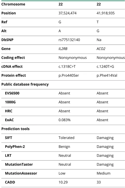
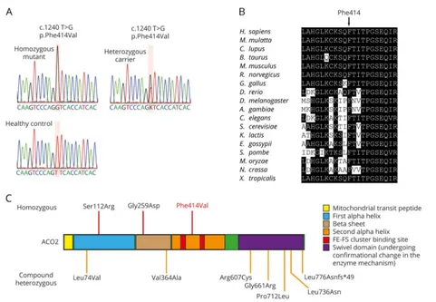
Whole-exome sequencing revealed only 2 homozygous or compound heterozygous rare variants that were located within the linkage regions shared in both affected siblings and pre-dicted to affect protein coding. The first is a homozygous variant in the geneACO2(c.1240T>G p.Phe414Val;figure 1A, table 1, andfigure 2A). This missense variant was absent from all public databases (EVS6500, ExAC, and 1000 Genomes) and predicted to be deleterious by the SIFT, PolyPhen-2, likelihood ratio test (LRT), Mutation Taster, and Combined Annotation Dependent Depletion (CADD) (table 1) with strong evolu-tionary conservation (figure 2B).ACO2is highly expressed in the human brain throughout neurodevelopment and adult-hood (BrainSpan10). Variant genotyping of all available family members was confirmed by Sanger sequencing (figures 1A and 2A).
The other segregating variant was in the IL2RB gene (c.1318C>T p. Pro440Ser), with an MAF of 8 × 10⁻⁵
Table 1Identified exonic variants (GRCh37/hg19)
Chromosome 22 22
Position 37,524,474 41,918,935
Ref G T
Alt A G
DbSNP rs775132140 Na
Gene IL2RB ACO2
Coding effect Nonsynonymous Nonsynonymous
cDNA effect c.1318C>T c.1240T>G
Protein effect p.Pro440Ser p.Phe414Val
Public database frequency
EVS6500 Absent Absent
1000G Absent Absent
HRC Absent Absent
ExAC 0.083% Absent
Prediction tools
SIFT Tolerated Damaging
PolyPhen-2 Benign Damaging
LRT Neutral Damaging
MutationTaster Neutral Damaging
MutationAssessor Low Medium
CADD 10.29 33
(ExAC). This variant is predicted as benign by the SIFT, PolyPhen-2 LRT, MutationTaster, MutationAssessor, and CADD (table 1).IL2RBexhibits very low expression across all developmental ages in the human brain (BrainSpan10).
No additional cases of homozygous or compound hetero-zygous ACO2 mutations were identified by exome se-quencing among subjects with recessive ataxia (n = 319), neurodegenerative disease (n = 2,000), or spastic paraplegia (n = 144).
Functional studies
LCLs were established from the homozygous carrier patients (Pedigree IDs IV-1 and IV-3,ACO2F414V/F414V), a heterozy-gous sibling (Pedigree ID IV-2,ACO2F414V/c), and 2 unrelated controls (ACO2c/c). Aconitase enzyme activity was signifi -cantly reduced in LCLs derived from the homozygous carriers, despite normal protein levels of ACO2 (figure 3, A and B). Given that multiple proteins, including ACO2, contribute to the aconitase enzymatic activity measured in whole-cell lysates, we sought to perform subcellular fractionation to examine mitochondrial aconitase activity, which is determined exclu-sively by ACO2. Consistent with selective impairment of
ACO2 enzymatic activity, cellular fractionation revealed a highly specific defect in mitochondrial aconitase activity, whereas the cytoplasmic fraction that does not contain ACO2 revealed intact aconitase function (figure 3, C and D).
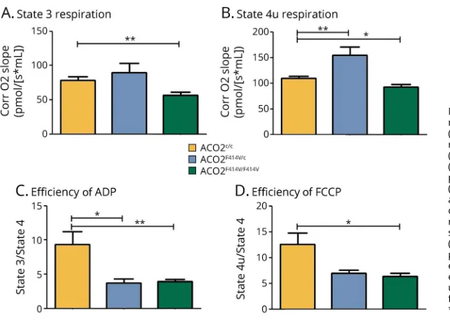
Analysis of cellular respiration in isolated mitochondria revealed that LCLs derived from homozygous carriers have a significant reduction in complex I–linked adenosine diphosphate (ADP)-coupled (state 3) respiration and an at-tenuated maximal respiration following uncoupling with car-bonyl cyanide p-trifluoromethoxyphenylhydrazone (FCCP) (state 4u) (figure 4, A and B). Moreover, the respiratory control ratio (RCR), defined as the ADP-activated flux reflecting coupled oxidative phosphorylation capacity, was attenuated in the mitochondria of LCLs derived from ho-mozygous and heterozygous carriers (figure 4, C and D).
Discussion
We performed genome-wide linkage analysis and exome sequencing in a consanguineous family with 2 siblings aff ec-ted by HSP, complicaec-ted by intellectual disability and
Figure 2Sanger sequencing, conservation, and summary of knownACO2mutations
microcephaly. This led to the identification of 2 candidate homozygous missense variants. On the basis of the variant frequency in public databases, prior association with neurodegenerative disease, in silico pathogenicity prediction, and expression in human brain, we considered theACO2variant (c.1240T>G, p.Phe414Val) as the likely causal mutation. The ACO2p.Phe414Val mutation was absent from 319 patients with recessive ataxia, 2,000 patients with neurodegenerative disease, and 144 patients with spastic paraplegia, indicating the rareness of this form of cHSP.ACO2encodes the protein aconitase 2, a critical enzyme in the tricarboxylic acid cycle (TCA) cycle. Through its enzymatic function, aconitase 2 catalyses the isomerization of citrate to isocitrate. The TCA cycle is the pri-mary source of cellular metabolic energy and therefore strongly conserved evolutionarily. TCA cycle enzymopathies have pre-viously been reported as the cause of a variety of cerebral encephalopathies, also often with muscular hypotonia and de-velopmental delay as prominent presenting features.11
The corticospinal tract contains the longest neurons of the human body, posing strong metabolic demands including the requirement for long-distance transport of proteins and organelles to distal axon terminals. Genes responsible for the
functional integrity of this complex machinery, including mitochondrial proteins, have been implicated in diverse forms of HSP.1Thefinding that a homozygous mutation inACO2is associated with HSP provides further support for the central role of mitochondrial defects in the pathogenesis of spastic paraplegia. Notably, several genetic mutations associated with HSP have been functionally demonstrated to impair mito-chondrial function:SPG7 (PGN),SPG13 (HSPD1), SPG28 (DDHD1), SPG31 (REEP1), SPG55 (C12ORF65), mito-chondrial adenosine triphosphate (ATP) synthase 6 (mt-ATP6), IBA57, and OPA3. SPG7 encodes an m-AAA metalloprotease localized to the inner mitochondrial mem-brane where it functions to guard the integrity of proteins within the respiratory pathway. A mutation in SPG7 was found to cosegregate in a multiplex family with HSP.12 Patients withSPG7 have reduced mitochondrial respiration rates and are more sensitive to oxidative stress.13Patients with mutations inHSPD1 have a pure HSP phenotype14and im-paired mitochondrial integrity.15Patients with SPG28 can be affected with pure or complicated HSP. Mutations inREEP1can cause reduced mitochondrial respiration and ATP production.16,17Patients withSPG31can have a pure or com-plicated form of HSP and have a dysfunction in mitochondrial energy production.18–20A homozygous nonsense mutation in C12ORF65 (SPG55)has been described in a family from Japan with 2 patients affected by HSP complicated by optic atrophy and polyneuropathy. This mutation caused a reduction in mi-tochondrial protein synthesis and respiratory function.21 Fa-milial maternally inherited late-onset HSP was described in association with a homoplasmic mutation inmt-ATP6whereby the clinical severity of symptoms was strongly correlated with biochemical functioning of mt-ATP6.22Recessive mutations in IBA57associated with HSP complicated by optic atrophy and peripheral neuropathy have been found to cause a reduction in mitochondrial 4FE-4S protein expression.23Moreover, induced pluripotent stem cell–derived neurons from patients with HSP resulting fromSPG3Amutations were reported to have reduced mitochondrial motility.24 Lastly, exome sequencing revealed a segregating homozygous missense mutation inOPA3 segre-gating with optic atrophy, chorea, cerebellar ataxia, dystonia, and lower limb pyramidal symptoms.25Heterozygous mutations in this gene are known to affect mitochondrial functioning.26
Although we cannot exclude that the c.1318C>T p.Pro440Ser variant inIL2RBplays a role in the phenotype, the very low gene expression in the human brain, functional prediction as benign, and weak evolutionary conservation argue strongly against the pathogenicity of this variant. Gene-level burden analysis previously demonstrated an association of rare coding variation inIL2RBwith rheumatoid arthritis.27However, to our knowledge, no known associations betweenIL2RBand diseases of the central or peripheral nervous system have been reported. Furthermore, functional evidence from LCLs demonstrates that theACO2p.Phe414Val mutation is responsible for a severe defect in mitochondrial aconitase activity and respiratory function. Notably, basal, maximum, and complex I–linked ADP-coupled (state 3) respiration were all affected only in
Figure 3Mutation carrier–derived lymphoblastoid cell lines (LCLs) show decreased aconitase 2 activity and mitochondrial respiration deficiency compared with control LCLs
(A) Aconitase enzyme activity in LCL lysates of healthy controls (ACOc/c) and
homozygous patient carriers (ACO2F414V/F414V) (p= 1.6 × 10⁻⁸). (B) Western
blot of corresponding LCL lysates showing equal ACO2 protein levels in healthy controls and homozygous carrier patients. (C) Mitochondrial frac-tion of LCL lysates show a significant decrease in aconitase enzyme activity in homozygous patient carrier samples (p= 7.4 × 10⁻⁷). (D) ACO2 enzyme
homozygous carriers. By contrast, the RCR was also decreased in heterozygous carrier LCLs, indicating a subtle intermediate phenotype conferred by heterozygous carriership.
Homozygous and compound heterozygous mutations inACO2 have been previously associated with varying combinations of cerebellar ataxia, retinopathy, and developmental delay (figure 2C and table e-2, links.lww.com/NXG/A34). A p.Ser112Arg missense mutation wasfirst identified in 2 families with infantile cerebellar-retinal degeneration.28Affected individuals exhibited developmental delay including severe psychomotor retardation, with an age at onset between 2 and 6 months. Brain MRI revealed cerebellar degeneration and white matter abnormali-ties (dysmyelination). Optic atrophy and retinal degeneration were readily identifiable in the setting of progressively severe visual impairment. Mitochondrial aconitase enzymatic activity was significantly reduced in lymphoblasts.
Through exome sequencing of sporadic cases of complicated optic neuropathy, 4 patients with homozygous or compound heterozygous mutations in ACO2 were identified. Two patients from 1 family had a compound heterozygous muta-tion (p.Leu74Val and p.Gly661Arg) and presented with de-creased visual acuity in childhood and progression of ophthalmologic symptoms into the fourth decade of life. Brain MRI was not performed. A third patient (homozygous p.Gly259Asp mutation) was born with a low APGAR score, intermittent episodes of central apnea, and moderate cere-bellar atrophy. The fourth patient (compound heterozygous mutation, p.Leu736Asn and p.Leu776Asnfs*49) exhibited ophthalmologic impairments with developmental delay and moderate cerebellar atrophy. Mitochondrial aconitase enzy-matic activity was significantly reduced in patientfibroblasts.29
Furthermore, a 3-year-old sporadic patient with developmen-tal delay, cerebellar dysfunction, and mild auditory neuropa-thy in the absence of retinal degeneration was reported with compound heterozygous mutations inACO2(c.1819C>T p.Arg607Cys and c.2135C>T p.Pro712Leu). Mitochondrial aconitase activity was also significantly reduced in patient
fibroblasts.30
Lastly, an 18-year-old sporadic patient was described with childhood-onset ataxia, profound intellectual disability, in-tractable epilepsy starting at the age of 2 years, cerebellar atrophy peripheral neuropathy, and childhood-onset optic atrophy and pigmentary retinopathy. Metabolic and com-parative genomic hybridization array screening did not identify causative genetic mutations. Using exome se-quencing, a compound heterozygous mutation in ACO2 (c.2328-2331delGGAA p.Lys776Asnfs*49 and c.1091T>C p.Val364Ala) was identified.31
In contrast to previous reports of cases withACO2mutations, the patients in the family described in the current article de-veloped severe HSP without cerebellar signs or retinal ab-normalities, despite a similar decrease in mitochondrial aconitase activity and respiration. Previous reports ofACO2 mutations highlight an important degree of phenotypic het-erogeneity (table e-2, links.lww.com/NXG/A34), with dis-tinct genotypes being associated with optic neuropathy and/ or cerebellar ataxia. The presently reported mutation (c.1240T>G p.Phe414Val) was associated with distinct phe-notypic characteristics (HSP, microcephaly) in the absence of optic neuropathy or cerebellar ataxia. Notably, the proband exhibited mild atrophy of the cerebellar vermis and hemi-spheres, confirming previous reports of discordance between
Figure 4Mitochondrial respiration is affected in lymphoblastoid cell lines (LCLs) ofACO2mutation carriers
Mitochondrial respiration was measured in isolated mitochondria from homozygous patient carriers (ACO2F414V/F414V), heterozygous ACO2mutation
car-rier (ACO2F414V/c), and healthy control cell lines
(ACO2c/c) by high-resolution respirometry. (A) State 3
cerebellar atrophy and cerebellar ataxia among carriers of recessiveACO2 mutations. Moreover, the absence of optic neuropathy and cerebellar ataxia in recessive carriers of the p.Phe414Val mutation may result from its unique localization to the second alpha helix domain, which is distinct from any of the other previously reportedACO2mutations (figure 2C).
Ourfindings nominateACO2as a disease-causing gene for autosomal recessive complicated HSP. Genetic screening of ACO2 should be considered for patients with complicated HSP in an effort to obtain a molecular diagnosis. Moreover, these results provide additional support for the central role of mitochondrial defects in the pathogenesis of HSP.
Author contributions
Dr. Bouwkamp—study concept and design; acquisition, analysis, and interpretation of data; and manuscript writing. Dr. Afawi—study concept and design; acquisition and in-terpretation of data; and critical revision of the manuscript. Dr. Fattal-Valevski—acquisition and interpretation of data and critical revision of the manuscript. Dr. Krabbendam, Dr. Rivetti, and Dr. Masalha—acquisition, analysis, and in-terpretation of data. Dr. Quadri and Dr. Breedveld—analysis and interpretation of data. Dr. Mandel, Dr. Abu Tailakh, and Dr. Beverloo—acquisition of data. Dr. Stevanin, Dr. Brice, Dr. Van IJcken, and Dr. Vernooij—acquisition, analysis, and interpretation of data. Dr. Dolga and Dr. De Vrij—analysis and interpretation of data and critical revision of the manu-script. Dr. Bonifati—study concept and design; analysis and interpretation of data; and manuscript writing. Dr. Kushner—study concept and design; acquisition, analysis, and interpretation of data; and manuscript writing.
Study funding
This study was funded in part by the Netherlands Organiza-tion for Scientific Research (NWO) and Netherlands Orga-nisation for Health Research and Development (ZonMw) to S.A.K. and V.B., the Royal Netherlands Academy of Arts and Sciences (KNAW) Sara van Dam Foundation to C.G.B. and Z.A., the NeuroBasic-PharmaPhenomics consortium to S.A.K., and Stichting ParkinsonFonds (The Netherlands) to V.B.
Disclosure
Dr. Bouwkamp and Dr. Afawi report study funding provided by the Royal Netherlands Academy of Arts and Sciences, Sara van Dam Foundation. Dr. Fattal-Valevski, Dr. Krabbendam, Dr. Rivetti, and Dr. Masalha report no disclosures. Dr. Quadri has received funding for travel from the International Par-kinson & Movement Disorder Society and has served as a consultant of Stichting Centre for Human Drug Research. Dr. Breedveld, Dr. Mandel, Dr. Abu Tailakh, and Dr. Beverloo report no disclosures. Dr. Stevanin has received funding for travel and/or speaker honoraria from the Movement Disorder Society; holds patents with regard to Diagnosis of HSPs by identification of a mutation in theKIAA1840gene or protein and diagnosis of HSPs by identification of a mutation in the ZFYVE26 gene or protein; and has received research
support from European Community’s Seventh Framework Program FP7/2007–2013 under agreement 2012-2017-305121 “Integrated European-omics research project for diagnosis and therapy in rare neuromuscular and neurode-generative diseases (NEUROMICS),” French Agency for Research, Erare Programme 2013–2018, Erare programme 2016–2019, “Investissements d’avenir” ANR-10-IAIHU-06, Association Strumpell-Lorrain France 2016–2018, and As-sociation Connaitre les Syndromes C´er´ebelleux 2015–2017. Dr. Brice has served on the scientific advisory boards of the Research Foundation—Flanders (FWO), European Research Council (ERC), and Bundesministeriums f¨ur Bildung und Forschung (BMBF) Berlin, Germany; has served on the editorial boards of Neurology and Clinical Neuroscience, Parkinsonism and Related Disorders,Brain,Neurodegenerative Diseases,The Cerebellum, andNeurogenetics; and has received research support from the French Research Agency, France Parkinson Association, Roger de Spoelberch Foundation (RDS), Fondation de France (FDF), and Fondation pour la Recherche M´edicale (FRM). Dr. Van IJcken reports no dis-closures. Dr. Vernooij has received research support from the Erasmus MC University Medical Center, Rotterdam, the Netherlands, and Netherlands Organization of Scientific Re-search (NWO)/ZonMW. Dr. Dolga and Dr. De Vrij report no disclosures. Dr. Bonifati has served on a scientific advisory board or Data Safety Monitoring Board of Stichting Parkinson Fonds, International Association of Parkinsonism and Related Disorders (IAPRD), and Erasmus MC Medical Research Advisory Committee (Mrace); has received travel funding and/or speaker honoraria from Sun Pharmaceutical Laboratories Limited; has served on the editorial board of Parkinsonism & Related Disorders, Current Neurology and Neuroscience Reports,Neurogenetics,Journal of Parkinson’s, and Movement Disorders Clinical Practice; has served as a consul-tant of the International Parkinson and Movement Disorder Society and LSP Life Sciences Fund N.V., The Netherlands; and has received research support from the Erasmus Medical Center Rotterdam, The Netherlands, ZonMw, The Nether-lands (under the aegis of the EU Joint Programme— Neurodegenerative Disease Research [JPND]), Stichting Parkinson Fonds (The Netherlands), and the Centre for Human Drug Research (CHDR), Leiden, The Netherlands. Dr. Kushner has received study funding and/or speaker honoraria from the Netherlands Organization for Scientific Research (NWO), Netherlands Ministry of Economic Affairs: Technology Foundation STW, European Research Council (ERC FP7 Marie Curie International Fellowship), and Netherlands Ministry of Finance. Full disclosure form in-formation provided by the authors is available with the full text of this article at Neurology.org/NG.
Received August 13, 2017. Accepted infinal form December 12, 2017. References
1. Salinas S, Proukakis C, Crosby A, Warner TT. Hereditary spastic paraplegia: clinical features and pathogenetic mechanisms. Lancet Neurol 2008;7:1127–1138. 2. Deluca GC, Ebers GC, Esiri MM. The extent of axonal loss in the long tracts in
3. de Bot ST, van de Warrenburg BP, Kremer HP, Willemsen MA. Child Neurology: hereditary spastic paraplegia in children. Neurology 2011;26:75–79.
4. Harding AE. Classification of the hereditary ataxias and paraplegias. Lancet 1983;321: 1151–1155.
5. Lo Giudice T, Lombardi F, Santorelli FM, Kawarai T, Orlacchio A. Hereditary spastic paraplegia: clinical-genetic characteristics and evolving molecular mechanisms. Exp Neurol 2014;261:518–539.
6. Tesson C, Koht J, Stevanin G. Delving into the complexity of hereditary spastic paraplegias: how unexpected phenotypes and inheritance modes are revolutionizing their nosology. Hum Genet 2015;134:511–538.
7. Fink JK. Hereditary spastic paraplegia: clinical principles and genetic advances. Semin Neurol 2014;34:293–305.
8. Fink JK. Hereditary Spastic Paraplegia Overview. Seattle: University of Washington; 2014. Available at: ncbi.nlm.nih.gov/books/NBK1509/. Accessed March 14, 2016. 9. Neitzel H. A routine method for the establishment of permanent growing
lympho-blastoid cell lines. Hum Genet 1986;73:320–326.
10. Hawrylycz MJ, Lein ES, Guillozet-Bongaarts AL, et al. An anatomically comprehen-sive atlas of the adult human brain transcriptome. Nature 2012;489:391–399. 11. Munnich A. Casting an eye on the Krebs cycle. Nat Genet 2008;40:1148–1149. 12. Casari G, De Fusco M, Ciarmatori S, et al. Spastic paraplegia and OXPHOS
impairment caused by mutations in paraplegin, a nuclear-encoded mitochondrial metalloprotease. Cell 1998;93:973–983.
13. Atorino L, Silvestri L, Koppen M, et al. Loss of m-AAA protease in mitochondria causes complex I deficiency and increased sensitivity to oxidative stress in hereditary spastic paraplegia. J Cell Biol 2003;163:777–787.
14. Fontaine B, Davoine C-S, D¨urr A, et al. A new locus for autosomal dominant pure spastic paraplegia, on chromosome 2q24-q34. Am J Hum Genet 2000;66:702–707. 15. Bross P, Naundrup S, Hansen J, et al. The hsp60-(p.V98I) mutation associated with hereditary spastic paraplegia SPG13 compromises chaperonin function bothin vitro
andin vivo. J Biol Chem 2008;283:15694–15700.
16. Bouslam N, Benomar A, Azzedine H, et al. Mapping of a new form of pure autosomal recessive spastic paraplegia (SPG28). Ann Neurol 2005;57:567–571.
17. Tesson C, Nawara M, Salih MAM, et al. Alteration of fatty-acid-metabolizing enzymes affects mitochondrial form and function in hereditary spastic paraplegia. Am J Hum Genet 2012;91:1051–1064.
18. Beetz C, Sch¨ule R, Deconinck T, et al. REEP1 mutation spectrum and genotype/ phenotype correlation in hereditary spastic paraplegia type 31. Brain 2008;131:1078–1086.
19. Goizet C, Depienne C, Benard G, et al. REEP1 mutations in SPG31: frequency, mutational spectrum, and potential association with mitochondrial morpho-functional dysfunction. Hum Mutat 2011;32:1118–1127.
20. Battini R, Fogli A, Borghetti D, et al. Clinical and geneticfindings in a series of Italian children with pure hereditary spastic paraplegia. Eur J Neurol 2011;18: 150–157.
21. Shimazaki H, Takiyama Y, Ishiura H, et al. A homozygous mutation ofC12orf65
causes spastic paraplegia with optic atrophy and neuropathy (SPG55). J Med Genet 2012;49:777–784.
22. Verny C, Guegen N, Desquiret V, et al. Hereditary spastic paraplegia-like disorder due to a mitochondrial ATP6 gene point mutation. Mitochondrion 2011;11: 70–75.
23. Lossos A, St¨umpfig C, Stevanin G, et al. Fe/S protein assembly gene IBA57 mutation causes hereditary spastic paraplegia. Neurology 2015;84:659–667.
24. Zhu PP, Denton KR, Pierson TM, Li XJ, Blackstone C. Pharmacologic rescue of axon growth defects in a human iPSC model of hereditary spastic paraplegia SPG3A. Hum Mol Genet 2014;23:5638–5648.
25. Arif B, Kumar KR, Seibler P, et al. A novel OPA3 mutation revealed by exome sequencing. JAMA Neurol 2013;70:783.
26. Grau T, Burbulla LF, Engl G, et al. A novel heterozygous OPA3 mutation located in the mitochondrial target sequence results in altered steady-state levels and fragmented mitochondrial network. J Med Genet 2013;50:848–858.
27. Diogo D, Kurreeman F, Stahl EA, et al. Rare, low-frequency, and common variants in the protein-coding sequence of biological candidate genes from GWASs contribute to risk of rheumatoid arthritis. Am J Hum Genet 2013;92:15–27.
28. Spiegel R, Pines O, Ta-Shma A, et al. Infantile cerebellar-retinal degeneration asso-ciated with a mutation in mitochondrial aconitase, ACO2. Am J Hum Genet 2012;90: 518–523.
29. Metodiev MD, Gerber S, Hubert L, et al. Mutations in the tricarboxylic acid cycle enzyme, aconitase 2, cause either isolated or syndromic optic neuropathy with en-cephalopathy and cerebellar atrophy. J Med Genet 2014;51:834–838.
30. Sadat R, Barca E, Masand R, et al. Functional cellular analyses reveal energy metab-olism defect and mitochondrial DNA depletion in a case of mitochondrial aconitase deficiency. Mol Genet Metab 2016;118:28–34.
DOI 10.1212/NXG.0000000000000223
2018;4;
Neurol Genet
Christian G. Bouwkamp, Zaid Afawi, Aviva Fattal-Valevski, et al.
paraplegia
homozygous missense mutation associated with complicated hereditary spastic
ACO2
This information is current as of March 21, 2018
Services
Updated Information &
http://ng.neurology.org/content/4/2/e223.full.html including high resolution figures, can be found at:
References
http://ng.neurology.org/content/4/2/e223.full.html##ref-list-1 This article cites 30 articles, 5 of which you can access for free at:
Subspecialty Collections
http://ng.neurology.org//cgi/collection/spastic_paraplegia Spastic paraplegia
http://ng.neurology.org//cgi/collection/mitochondrial_disorders Mitochondrial disorders
http://ng.neurology.org//cgi/collection/genetic_linkage Genetic linkage
following collection(s):
This article, along with others on similar topics, appears in the
Permissions & Licensing
http://ng.neurology.org/misc/about.xhtml#permissions its entirety can be found online at:
Information about reproducing this article in parts (figures,tables) or in
Reprints
http://ng.neurology.org/misc/addir.xhtml#reprintsus Information about ordering reprints can be found online:
reserved. Online ISSN: 2376-7839.
Published by Wolters Kluwer Health, Inc. on behalf of the American Academy of Neurology. All rights an open-access, online-only, continuous publication journal. Copyright Copyright © 2018 The Author(s).
is an official journal of the American Academy of Neurology. Published since April 2015, it is