ISSN: 0976-3031
Research Article
PREVALENCE OF ORAL SOFT TISSUE LESIONS AND DEVELOPMENTAL VARIATIONS AMONG
SCHOOL CHILDREN IN PATNA, INDIA
Shashwat Kumar., Veeranna Ramesh., Suma B.S., Sadananda LD., Garima Mangal.,
Nirmala Kumari and R N P Singh
Department of Public Health Dentistry, Buddha Institute of Dental Sciences and Hospital, Patna
DOI: http://dx.doi.org/10.24327/ijrsr.2017.0809.0842
ARTICLE INFO ABSTRACT
Introduction: Children affected by oral diseases are of great importance and there effects on oral health related quality of life is more of a concern to any Public Health Dentist. The occurrence and distribution of oral soft tissue lesions and developmental variations are very common and wide spread. Oral care and the diseases that occur in the oral cavity are of continuous interest to the dental profession as it is essential for the application of timely intervention for primary prevention, early diagnosis and prompt treatment of these pathologies. Aim is to assess the prevalence of oral soft tissue lesions and developmental variations among school children of Patna. Materials and Methods: A school based approach was used. The study population consisted of 600 school children aged 6-12 years belonging to 5 schools in Patna constituted the study population. Etiological factors were not taken into consideration. Clinical examination was carried out by a single examiner based on WHO criteria’s. Chi-square test was used for statistical analysis. Results: 30.7% of the subjects had one or the other kind of lesions or developmental variations. Angular Chelitis was the most common oral soft tissue lesions with 27.7% among the study population affected, followed by dento-alveolar abscess 27.2%; Aphthous ulcer 10.9%; Traumatic ulcer 10.2%; Herpes Labialis 7.1%; Fissured tongue 7.1% and the remaining formed 9.8% which included Acute necrotizing ulcerative gingivitis, Papilloma, Geographic tongue, focal epithelial hyperplasia and commissural pits. Conclusion: It not only gives additional information on the incidence of oral diseases or conditions but also enhances the responsibility of the dentist in the total care for children.
INTRODUCTION
Dental caries, periodontal diseases, craniofacial defects and oral cancer have been considered the disorders of primary concern regarding oral health. In recent years the health professionals and the public have been made more aware of the importance of oral mucosal pathologies and developmental variations. This has been due to the visibility given to investigations. The growth in knowledge has made serious changes in approach towards its identification. The new diseases with interesting etiological causes, various presentations of cases have led to the improved scope of oral health research. In a country like India, there are few studies available in the literature and there is absolutely no studies done in this part of the country (North Eastern states of India). Nutritional deficiencies, trauma, developmental defects or variations, pathological microorganisms, low birth weight and premature birth all add to the very occurrence of oral soft tissue lesions. Avoidance or control of these factors can control the
very problem and can contribute to better oral health related quality of life. A problem addressed at the early stage can ease the process of discomfort among children.1-3
Epidemiological studies have shown a considerable variation in the prevalence rates of oral mucosal lesions in different parts of the world. Most of the studies carried out have focused on adults. None of the researches have focused on the prevalence of oral soft tissue lesions in children. As little information on the epidemiology of these lesions is available from these regions, it was considered useful to analyze data on this subject. Incidentally this is the first research of its kind in this part of the region. Therefore the aim of this investigation was to estimate the prevalence of oral soft tissue lesions and developmental variations among school children in Patna population which would aid in oral differential diagnosis and provide a base line data for comparison with other populations. Additionally the data collected would provide a baseline for planning and evaluating of community oral health care for school children in Patna 4-5.
International Journal of
Recent Scientific
Research
International Journal of Recent Scientific Research
Vol. 8, Issue, 9, pp. 20175-20179, September, 2017
Copyright © Shashwat Kumar et al, 2017, this is an open-access article distributed under the terms of the Creative Commons Attribution License, which permits unrestricted use, distribution and reproduction in any medium, provided the original work is properly cited.
DOI: 10.24327/IJRSR
CODEN: IJRSFP (USA)
Article History: Received 15th June, 2017 Received in revised form 25th July, 2017
Accepted 23rd August, 2017
Published online 28th September, 2017
Key Words:
Objectives of the Study
To assess the prevalence of oral soft tissue lesions and developmental variations among school children aged 6-12 years in Patna, India.
METHODOLOGY
This present research work is a cross sectional study to assess the prevalence of oral soft tissue lesion and developmental variations among school children belonging to different schools in Patna, Bihar, India. Prior to scheduling the survey, official permission was obtained from Deputy Director, Education Department of Bihar and heads of concerned schools in Patna. The schedule was spread over a period of six months starting June 2016 to November 2016. Ethical clearance was obtained from Ethics committee of Buddha Institute of Dental Sciences and Hospital, Patna. A specially prepared and pre-tested format, exclusively designed for recording all the required and relevant general information and clinical findings was used. All clinical and administrative procedures related to oral soft tissue lesions and developmental variations were carried out before the examinations. Where necessary, colour photographs and transparencies were used to demonstrate the main clinical characteristics of lesions and developmental variations to be considered in the differential diagnosis. Diagnosis was arrived using the clinical criteria’s for the oral soft tissue lesions and developmental variations as per World Health Organization guidelines and standard clinical definitions given by text books, (Table 1) 6-9.
Etiological factors were not taken into consideration. The examiner was calibrated for examination and diagnosis of oral soft tissue lesions and developmental variations. A small sample was selected and the examiner coded the lesions or variations and these findings were subjected to kappa statistics in order to limit the examiner variability. The Kappa co-efficient value of intra-examiner reliability for oral soft tissue lesions and developmental variations was 0.87. This value reflected high degree of conformity in observations. A pilot survey was undertaken to test the feasibility of the study including the assessment of clarity, validity, and applicability of the questionnaire followed by the procedure to be employed for examination and recording of oral soft tissue lesions and developmental variations.
A school based approached was used. A total of 600 available school children in the age group of 6-12 years belonging to five different schools constituted the study population. The schools were selected by using simple random sampling after zone wise division of Patna map. Children of all socio-economic class were included in the study. Informed consent was obtained from every student’s parents participating in the study. Children with severe systemic illness and those who did not consent for the study were not included. Examinations were conducted outdoors within the premises of the school under available natural daylight. Artificial illumination was also used when required using torchlight. The scores were recorded in the proforma by a trained recorder.
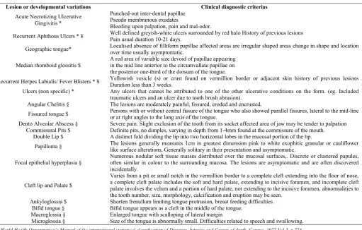
Table 1 Clinical Diagnosis for Oral Soft Tissue Lesions and Developmental Variations
Lesion or developmental variations Clinical diagnostic criterias
Acute Necrotizing Ulcerative Gingivitis *
Punched-out inter-dental papillae Pseudo membranous exudates
Bleeding upon palpation, pain and mal-odor.
Recurrent Aphthous Ulcers * ¥ Well defined greyish-white ulcers surrounded by red halo History of previous lesions
Pain usual duration 10-21 days.
Geographic tongue* Localised absence of filliform papillae affected areas are irregular shaped areas change in shape and location
over time usually asymptomatic.
Median rhomboid glossitis $
A red area of variable size devoid of papillae appearing in the mid line anterior to the circumvallate papillae on the posterior one-third of the dorsum of the tongue.
Recurrent Herpes Labialis/ Fever Blisters * ¥ Yellowish vesicle (s) or crust found on vermillion border or adjacent skin history of previous lesions
Duration less than 3 weeks.
Ulcers (non specific) * Any ulcers that cannot be attributed to one of the other ulcerative conditions on the form. (eg. Included
traumatic ulcers and an ulcer due to tooth brush abrasion).
Angular Chelitis § The lesions are moderately painful, fissured, eroded and encrusted.
Fissured tongue $ Persons with or without central fissure of the tongue who also showed parallel fissures, lateral to the mid-line
or at right angles to the long axis of the tongue.
Dento Alveolar Abscess § Severe pain. Slight exclusion of the tooth from its socket affected area of jaw may be tender to palpation
Commissural Pits $ Definite pits, no dimples, varying in depth from 1-4mm found at the commissure of the mouth.
Double Lip $ A distinct fold dividing the lip into two horizontal lobes in the mucosal portion of the lip.
Papilloma § The lesions generally measures 1cm in greatest dimension pink to white exophitic granular or cauliflower
like surface alterations, Generally solitary in their presentation and asymptomatic.
Focal epithelial hyperplasia §
Numerous nodular soft tissue masses distributed over the mucosal surfaces,. Discrete or clustered papules, often similar in colour to the surrounding mucosa. The lesions are asymptomatic and are often discovered incidentally.
Cleft lip and Palate $
Varies from a pit or small notch in the vermillion border to a complete cleft extending into the floor of nose, a complete cleft palate includes the soft and hard palate, extending to incisive foramen, and incomplete cleft palate involves the velum and a portion of hard palate, not extending to the incisive foramen, abnormalities to the tooth number, size, morphology, calcification and eruption may be seen.
Ankyloglossia $ Shorten frenullum limiting tongue protrusion, breast feeding difficulties.
Bifid tongue § Bifid tongue appears as a cleft in the middle of the tongue.
Macroglossia § Enlarged tongue with scalloping of lateral margin
Microglossia § Size of the tongue is abnormally small. Difficulties related to speech and swallowing.
* World Health Organization’s Manual of the international statistical classification of Diseases, Injuries and Causes of death. Geneva, 1977 Vol 2, p 774.
$ Sawyer DR, Taiwo EO, Mosadomi A; Oral anamolies In Nigeria Children. Community Dent Oral Epidemiol.1984;12:269-73.
§ Joseph A Regezi James J Sciubba, Richard CK Jordan Oral Pathology. Clinical Pathologic Coorelation.5th Edn, Saunders Elsevier Inc;2008.
On an average 30-40 subjects were interviewed and examined in any given day during the survey period. Duration for data collection for each subject ranged from 5-10 minutes. All necessary aseptic precautions were taken before and during the examination procedure. The data obtained was compiled, tabulated and analysed by applying Chi-square test for assessing statistical significance between groups and significance for statistical tests were predetermined at probability values of 0.05 or less.
RESULTS
A total of 600 school children in the age group of 6-12 years participated in the study, of which 376 (62.7) were males and 224(37.3%) were females.
Of the total 600 subjects, 30.7 % of the subjects had one or the other kind of oral soft tissue lesions or developmental variations of which 59.8% were present with males and 40.2% were with females. The results was statistically significant with p<0.05. Angular Chelitis was the most common oral soft tissue lesions with 27.7% among the study population affected, followed by dento-alveolar abscess 27.2%; Aphthous ulcer 10.9%; Traumatic ulcer 10.2%; Herpes Labialis 7.1%; Fissured tongue 7.1% and the remaining formed 9.8% which included Acute necrotizing ulcerative gingivitis, Papilloma, Geographic tongue, focal epithelial hyperplasia and commissural pits. Other conditions like cleft lip and palate, Median rhomboid glossitis, Ankyloglossia, Bifid tongue, Macroglossia and Microglossia were also evaluated but there was not a single case recorded in this study.
DISCUSSION
The purpose of this study was to assess the prevalence of oral soft tissue lesions and developmental variations among school
children from five schools in Patna which would aid in oral differential diagnosis and also provide a basis for comparison with other population. Additionally the data collected will provide a baseline for planning and evaluation of community oral health preventive programs for school children in Patna.
Most of the studies carried out were primarily aimed at adults and there is little data related to school children aged between 6-12 years. It is first study in northeast region of India. The schools selected for this study included school children from all socio-economic status and the main staple food in this part of the region is roti and rice.
The overall prevalence of oral soft tissue lesions and developmental variations was 30.7%. This increase in prevalence among children can be attributed to lesions being asymptomatic and unaware of its existence which may have lead to failure to seek any preventive or curative care. The result of this study is similar to the studies of Veeranna et al (2012) 10 and Knapp MJ et al. 1 Also increased visibility given to the newer diagnostic methods have added to increased prevalence of oral soft tissue lesions and developmental variations.
Of all the oral soft tissue lesions and developmental variations, the prevalence of angular Chelitis was high (27.7 %). Considering that this condition is usually seen in denture wearers with decreased vertical height and as none of the subjects in this study used dentures, the most likely cause for this lesion is various nutritional deficiencies. Although we were not able to test for these, the socio economic status of these children suggests that they are more likely to be deficient of iron, riboflavin, folic acid and Vitamin B12. 11-12 Wray and Dagg state a prevalence figure of 15 %for angular Chelitis in patients with iron deficiency which is less as that seen in the present study. No pattern emerged regarding school attended or their area of residence. 13
In the Witkop and Barros Chilean study, 5.1 % of this population was found to manifest fissured tongue with the anomaly being slightly more frequently seen in females. In this present study a prevalence of 7.1 % was found and the males were affected more when compared to females. The prevalence of this anomaly entity in the Indian population studied is more in keeping with the prevalence values reported in other population groups studied. (Chosack et al, Halperin et al, Sawyer et al and Redman) 7, 14-18
Dentoalveolar abscess is sequelae in the cycle of untreated dental caries and periodontal problems. It was observed that in the present study the prevalence of dentoalveolar abscess was 27.2 %. It can be reasoned that increased number of untreated dental caries and poor oral hygiene affecting the supporting periodontium can contribute to the risk of developing dento-alveolar abscess. 14 When compared with the results of Veeranna et al (2012), the results of the present study showed higher prevalence of dentoalveolar abscess. 10
The prevalence of traumatic ulcers was 10.2 %. It was more commonly observed among males. It can be reasoned that sporting activities and other forms of physical activities (fights or falls) can significantly influence these outcomes. Some studies have reported that poor physical coordination (neuro Table 2 Shows the distribution of oral soft tissue lesions
and developmental variations according to Gender
Gender
Oral soft tissue lesions and developmental
variations Present
Oral soft tissue lesions and developmental
variations Absent
Total
Males 110(59.8%) 266 (63.9%) 376 (62.7)
Females 74 (40.2%) 150 (36.1%) 224(37.3%)
Total 184 (30.7%) 416 (69.3%) 600 (100)
p<0.05 (Sig), X2 = 4.14, df = 1
Table 3 Shows the distribution of oral soft tissue lesions and developmental variations
Sl no
Oral soft tissue lesions and developmental
variations.
Males Females Total
1 Angular Chelitis 33(30%) 18(24.3%) 51(27.7%)
2 Fissured tongue 8(7.3%) 5(6.8%) 13(7.1%)
3 Dento-alveolar abscess 28(25.5%) 22(29.7%) 50(27.2%)
4 Traumatic ulcer 11(10%) 8(10.8%) 19(10.2%)
5 Geographic tongue 3(2.7%) 2(2.7%) 5 (2.7%)
6 Commissural pits 0 (0%) 1(1.4%) 1 (0.5%)
7 Aphthous ulcer 13(11.8%) 7(9.5%) 20(10.9%)
8 Acute necrotizing ulcerative
gingivitis 4(3.6%) 2(2.7%) 6 (3.3%)
9 Papilloma 2(1.8%) 2(2.7%) 4 (2.2%)
10 Herpes labialis/Fever blisters 7(6.4%) 6(8.1%) 13(7.1%)
11 Focal epithelial hyperplasia 1(0.9%) 1(1.4%) 2 (1.1%)
muscular) can also contribute to increase in trauma affecting oral structures. 11
Geographic tongue was found to affect both the sexes and the prevalence was 2.7 % of those examined. This figure was similar to studies of Chosack et al14, Witkop and Barros 15, Sedano 19 and Veeranna et al (2012) 10. One exception to the study done by Rahmimoff and Mahsam gives an exceptionally high frequency (15 %) of this condition in Jaffe soon after the Arab-Israel war in 1948. Since etiological factors were not assessed in this study, predisposing factors like stress, nutritional deficiency may have contributed to this increased prevalence. 20
Commissural pits were seen in 0.5% of school children. The result of this study was less when compared to the study of Veeranna et al (2012), 10 which was 3.4 %. We could find very few epidemiological studies with the same age range as the present one. These conditions are genetically influenced and can be attributed to autosomal dominant trait which is commonly seen among blacks. 15, 19-21.
Recurrent Aphthous ulcers and Herpes Labialis have been the subject of a number of epidemiological investigations in India and abroad. Because the conditions have similar clinical courses, they have often been investigated together. The prevalence of recurrent aphthous ulcers and herpes labialis are 10.9 % and 7.1 % respectively. Because of the natural history and recurrent nature of these lesions, cross sectional surveys under estimate the true prevalence of the underlying conditions, as active lesions may not be present at the time of examinations. It is difficult to compare the results of many studies because of the differences in population characteristics and case definition. Study groups have often been selected for convenience rather than representativeness (medical or dental student for example). Only a few studies have comprised subjects of a broad age range that included children. 2, 22-23. A variety of risk factors have been investigated to rule out the possible cause. Emotional stress, socio economic status, denture wearers and smoking have been observed to be associated with recurrent aphthous ulcers. As many of these factors are commonly associated with adults, its influence on children are less which substantiate why the low prevalence among children. 2 In comparison, the results of Axell studies show less prevalence rates when compared to present study 24. For general discussion of other aspects of the epidemiology of recurrent aphthous ulcers, the paper by Ship is recommended. In most of the studies observed, the prevalence of herpes labialis is similar as compared to the present study. Associations with a positive history of aphthous ulcers were studied by SHIP et al who reported that subjects with a history of either condition were at increased risk of having the other. 25
Acute Necrotising Ulcerative Gingivitis (ANUG) was observed in 3.3% of the subjects. These results are similar to studies of Veeranna et al (2012). Enwonwu 1994 has reasoned that poor nutritional status is one of the contributing factors for the occurrence. Very few studies were available for comparison which showed similar results. 26 It was observed that this condition has seasonal variations and surveys conducted during warm months yielded higher prevalence rate (Sheiham 1966 and Kardarchi et al 1974). 27-28 We could find no comparable epidemiological study which mentions the remaining oral soft
tissue lesions and developmental variations referred to in our study.
CONCLUSION
The prevalence of oral soft tissue lesions and developmental variations are interestingly high (20.7 %) but in contrast we could only find very few comparable epidemiological studies which mentions the oral lesions referred to in our study among school children. This emphasizes that a broader set of standardized clinical criteria’s are needed to facilitate inter-researcher comparison in epidemiological studies in India or elsewhere and such data would not only give additional information concerning the incidence of oral diseases but also enhances the responsibility of the dentist in planning and implementing total oral care for children.
References
1. Knapp MJ. Oral diseases in 181,338 consecutive oral examinations. J. Am Dent Assoc 1971; 83:1288-93. 2. Kleinman DV, Swango PA, Niessen LC:
Epidemiological studies of oral mucosal conditions – methodological issues. Community Dent Oral Epidemiol. 1991; 19:129-40.
3. Kleinman DV, Swang PA , Pindborg JJ. Epidemiology of Oral Mucosal Lesions in United States school children 1986-87. Community Dent Oral Epidemiol. 1994; 22:243-53.
4. Axell T, Zain RB, Siwamogstham P, Tantiniran Thampipit J. Prevalence of oral soft tissue lesions in out-patients at two Malaysian and Thai Dental schools. Community Dent Oral Epidemiol, 1990; 18:95-9. 5. Bouquot JE. Common oral lesion founding during a
mass screening. J. Am Dent Assoc.1986; 112:50-7. 6. World Health Organization’s Manual of the
International Statistical Classification of Diseases, Injuries and Causes of Death, Geneva, 1977 Vol 2,p.774.
7. Sawyer DR, Taiwo EO. Mosadomi A; Oral anomalies In Nigeria Children. Community Dent Oral Epidemiol.1984; 12:269-73.
8. Joseph A Regezi James J Sciubba, Richard CK. Jordan Oral Pathology. Clinical Pathologic Coorelation.5th edition, Saunders Elsevier Inc; 2008.
9. World Health Organization Guide to Epidemiology and diagnosis of oral-mucosal diseases and conditions. Community Dent Oral Epidemiol.1980; 8:126.
10. Veeranna Ramesh, Pallavi H N and Santosh b Sakri. Oral soft tissue lesions and developmental variations among school children in Malebennur, South India. Journal of IAPHD, Vol 2012 Issue 19, p no 63-70. 11. Arendor TM. Van Der Ross R. Oral soft tissue lesions in
black pre-school South African population. Community Dent Oral Epidemiol.1996; 24:296.
12. Soames JV, Southam JC. Oral Pathology 2nd edition. Oxford University Press, 1993; 188-9.
14. Shafer WG Hine MK, Leyy BM. A text book of oral pathology India 4th Edition, WB Saunders Company 1993.
15. Witkop CI, Barros L. oral and genetic studies of Chileans, 1990.1. Oral anomalies. AM.J. Phy Anthropol 1963; 21:15-24.
16. Chosack A, Zadik D. E. The prevalence of scrotal tongue and geographic tongue in 70.359 Israeli school children. Community Dent Oral Epidemiol.1974; 2:253-7.
17. Halperin V, Kolas S. Jefferis KR. Huddleston SO. Robinson HBG. The occurrence of Fordyce Granules, benign migratory glossitis, median rhomboid glossitis and fissured tongue in 2,478 dental patients. Oral Surg.1953; 6:1072-7.
18. Redman RS. The prevalence of geographic tongue, fissured tongue, median rhomboid glossitis and hairy tongue among 3,611. Minnesota school children. Oral Surg.1970; 30:390-5.
19. Sedano HO. Congenital oral anomalies in Argentinean children. Community Dent Oral Epidemiol 1975; 3:61-3. 20. Rahaminoff P. Muhsain HV. Some observation on 1246
cases of geographic tongue AM J. Dis Child.1957; 93:519-25.
21. Everett FG Westcott WB. Commissural Lip Pits. Oral Surg Oral Med Oral Pathol.1961; 14:202-209.
22. Sireus W. Church R, Kellher J Recurrent Aphthous ulceration of the mouth. A study of natural history, etiology and treatment. QJ Med 1957; 26:235-49. 23. Fahmy MS. Recurrent aphthous ulceration in a mixed
Arab community. Community Dent Oral Epidemiol.1976; 4:160-4.
24. Axell T. A prevalence study of oral mucosal lesion in an adult. Swedish population. Odontol Revy Suppl.1976; 36:1-103.
25. Ship H, Brightman VJ. Laster W. The patient with recurrent aphthous ulcer and the patients with recurrent herpes labialis: a study of two population samples, J. Am Dent Assoc.1967; 75:645-54.
26. Enwonwn CO. Cellular and molecular effects of malnutrition and their prevalence to periodontal disease J Clin Peridontol.1994;21:643-657.
27. Sheiham A. An epidemiological survey of acute ulcerative gingivitis in Nigerians Arch Oral Biol.1996; 11:937-42.
28. Kardarchi BJR, Charke NG. Aetiology of acute necrotizing ulcerative gingivitis, a hypothetical explanation. J Periodontal.1974; 45:830-832.
*******
How to cite this article: