A STUDY OF ZETA POTENTIAL OF PLASMA
SPRAYED HYDROXYAPATITE COATING IN FOUR
SIMULATED PHYSIOLOGICAL SOLUTIONS
A. Afshar, M. Ghorbani, N. Ehsani and M. R. Saeri
Department of Materials Science and Engineering, Sharif University of Technology Azadi Avenue, P. O. Box 11365- 9466, Tehran, Iran
[email protected] - [email protected] - [email protected]
C. C. Sorrell
School of Materials Science and Engineering, University of New South Wales Sydney, NSW 2052, Australia, [email protected]
(Received: December 3, 2002 – Accepted in Revised Form: November 5, 2003)
Abstract The zeta potential magnitude and the duration of its changes have been thought to be directly related to the surface reactivity, the governing of osteoconductivity and availability of valuable information in determining the anticipated in-vivo performance of implants. In this study the zeta potential of plasma sprayed hydroxyapatite (HA) was evaluated in various simulated physiological solutions. For this purpose the prepared samples were immersed in SBF, Ringers, Tyrodes and Hanks solutions for 3 weeks and kept at 37°C and the time dependent variations of zeta potential results were compared with calcined (2 hr at 850°C) and as-precipitated HA powders. Furthermore, following immersion test, the coating surfaces were examined with SEM and XRD as well as FTIR methods. The results showed that in majority of solutions, the zeta potential increased in the following order: calcined, as precipitated and plasma sprayed. Moreover, the ion concentrations and types in the solutions have significant effects on the zeta potential values. Following the immersion test, the morphology of the samples were determined a function of the thermal history of HA, duration of immersion and the type of the test solution. The results indicated that the surface of the HA coated samples were unstable with significant changes in the observed charge.
Key Words Hydroxyapatite, Zeta potential, Plasma spray coat, Corrosion
ﻩﺪﻴﻜﭼ
ﻩﺪﻴﻜﭼ
ﻩﺪﻴﻜﭼ
ﻩﺪﻴﻜﭼ
ﻦﻴﻴﻌﺗﺕﺍﺮﺛﺍ،ﺎﺗﺯ ﻞﻴﺴﻧﺎﺘﭘﺕﺍﺮﻴﻴﻐﺗ ﻩﻮﺤﻧﻭﺭﺍﺪﻘﻣﻪﻛﺪﻨﻫﺪﻴﻣﻥﺎﺸﻧﻩﺪﺷﻡﺎﺠﻧﺍ ﺕﺎﻘﻴﻘﺤﺗ ﺮﺑﻱﺍﻩﺪﻨﻨﻛﺕﺎﻌﻄﻗ ﺪﻧﻮﻴﭘ ﻞﻴﻜﺸﺗ ﺖﻴﻠﺑﺎﻗ
ﺪﻧﺭﺍﺩ ﻥﺪﺑ ﻞﺧﺍﺩ ﺭﺩ ﻩﺪﺷ ﺖﺷﺎﻛ
.
ﺎﺗﺯ ﻞﻴﺴﻧﺎﺘﭘ ﺮﺿﺎﺣ ﻖﻴﻘﺤﺗ ﺭﺩ ﺱﺎﺳﺍ ﻦﻳﺍ ﺮﺑ
ﻲﺴﻛﻭﺭﺪﻴﻫﻚﻴﻣﺍﺮﺳﻮﻴﺑ ﻪﺑﻩﺪﺷﻩﺩﺍﺩﺶﺷﻮﭘ ﺖﻴﺗﺎﭘﺁ
ﻤﺳﻼﭘﺵﻭﺭ ﻪﻴﺒﺷﻲﻜﻳﮊﻮﻟﻮﻳﺰﻴﻓﻱﺎﻬﻟﻮﻠﺤﻣﺭﺩﻱﺮﭙﺳﺍﺎ
ﻱﺯﺎﺳ
ﺖﺳﺍ ﻩﺪﺷﻲﺳﺭﺮﺑﻩﺪﺷ
.
ﻪﻧﻮﻤﻧ ﻪﻄﺑﺍﺭﻦﻳﺍﺭﺩ ﻩﺩﺎﻣﺁﻱﺎﻫ
ﺕﺪﻤﺑﻩﺪﺷ ٣
ﻱﺎﻣﺩ ﺭﺩﻪﺘﻔﻫ °C
٣٧ ﻱﺎﻬﻟﻮﻠﺤﻣﻞﺧﺍﺩﺭﺩ
ﻪﻴﺒﺷﻲﻜﻳﮊﻮﻟﻮﻳﺰﻴﻓ ﻩﺪﺷﻱﺯﺎﺳ
SBF ﻪﻃﻮﻏ ﻚﻧﺎﻫﻭﺪﻳﻭﺮﻴﺗ،ﺯﺮﮕﻨﻳﺭ، ﻲﺳﺭﺮﺑﺎﻬﻧﺁﺎﺗﺯﻞﻴﺴﻧﺎﺘﭘ ﺕﺍﺮﻴﻴﻐﺗﻭﻩﺪﺷﺭﻭ
ﺪﻳﺩﺮﮔ
.
ﺕﺍﺮﺛﺍﻲﺳﺭﺮﺑﺖﻬﺟ ﻲﺴﻛﻭﺭﺪﻴﻫﻱﻭﺭﺮﺑﻪﺑﺎﺸﻣﺕﺎﺸﻳﺎﻣﺯﺁﻚﻴﻣﺍﺮﺳﻮﻴﺑﻦﻳﺍﻱﻭﺭﺮﺑﻱﺮﭙﺳﺍﺎﻤﺳﻼﭘ
ﺖﻴﺗﺎﭘﺁ
ﻩﺪﺷﻩﺩﺍﺩﺏﻮﺳﺭ
)
ﻡﺎﺧ
(
ﺕﺪﻤﺑﻭ ٢ ﻱﺎﻣﺩﺭﺩﺖﻋﺎﺳ °C
٨٥٠ ﻩﺪﺷﻩﺩﺍﺩﺕﺭﺍﺮﺣ
)
ﻪﻨﻴﺴﻠﻛ
(
ﺪﻳﺩﺮﮔﻡﺎﺠﻧﺍﺰﻴﻧ
.
ﻩﻭﻼﻋ
ﻱﺎﻬﺷﻭﺭﺯﺍﻩﺩﺎﻔﺘﺳﺍﺎﺑﻦﻳﺍﺮﺑ SEM ، XRD ﻭ FTIR ، ﻲﺴﻛﻭﺭﺪﻴﻫﺶﺷﻮﭘﺢﻄﺳﻱﻭﺭﺮﺑﻲﮔﺩﺭﻮﺧﺕﺍﺮﺛﺍ ﺗﺎﭘﺁ
ﺖﻴ
ﺪﺷﻲﺳﺭﺮﺑ
.
ﻪﻧﻮﻤﻧﺢﻄﺳﻪﻛﺪﻨﻫﺪﻴﻣﻥﺎﺸﻧﻩﺪﻣﺁﺖﺳﺪﺑﺞﻳﺎﺘﻧ ﻲﺴﻛﻭﺭﺪﻴﻫﺎﺑﻩﺪﺷﻩﺩﺍﺩﺶﺷﻮﭘﻱﺎﻫ
ﻞﺧﺍﺩﺭﺩﺖﻴﺗﺎﭘﺁ
ﺩﻮﺸﻴﻣﻩﺪﻳﺩﺎﻬﻧﺁﺎﺗﺯﻞﻴﺴﻧﺎﺘﭘﺮﻳﺩﺎﻘﻣﺭﺩﻱﺩﺎﻳﺯﺕﺍﺮﻴﻴﻐﺗﻭﻩﺩﻮﺑﺭﺍﺪﻳﺎﭘﺮﻴﻏ،ﻪﺘﻓﺭﺭﺎﻜﺑﻱﺎﻬﻟﻮﻠﺤﻣ
.
ﺮﺘﺸﻴﺑﺭﺩﻩﻭﻼﻌﺑ
ﻪﻧﻮﻤﻧ ﻱﺍﺮﺑﺎﺗﺯ ﻞﻴﺴﻧﺎﺘﭘﺭﺍﺪﻘﻣ،ﺎﻬﻟﻮﻠﺤﻣ ﺎﻤﺳﻼﭘﻱﺎﻫ
ﻪﻧﻮﻤﻧﻪﻴﻘﺑﺯﺍﺮﺗﺩﺎﻳﺯﻩﺪﺷﻱﺮﭙﺳﺍ ﺐﻴﺗﺮﺘﺑﻥﺁﺯﺍﺲﭘﻭ ﻩﺩﻮﺑﺎﻫ
ﻪﻧﻮﻤﻧ ﺪﻧﺭﺍﺩ ﺭﺍﺮﻗ ﻩﺪﺷﻪﻨﻴﺴﻠﻛ ﻭ ﻩﺪﺷﻩﺩﺍﺩ ﺏﻮﺳﺭ ﻱﺎﻫ
.
ﺭﺩ ﺩﻮﺟﻮﻣﻱﺎﻬﻧﻮﻳ ﺖﻈﻠﻏﻭ ﻉﻮﻧﺕﺍﺮﺛﺍ ﻪﻄﺑﺍﺭﻦﻳﺍ ﺭﺩ
ﺖﺳﺍ ﻪﺘﻓﺮﮔ ﺭﺍﺮﻗ ﻲﺳﺭﺮﺑ ﻭ ﺚﺤﺑ ﺩﺭﻮﻣ ﺰﻴﻧ ﻪﺘﻓﺭ ﺭﺎﻜﺑ ﻱﺎﻬﻟﻮﻠﺤﻣ ﻞﺧﺍﺩ
.
ﻪﻃﻮﻏ ﻝﺎﺒﻧﺪﺑ ﻦﻴﻨﭽﻤﻫ ﻞﺧﺍﺩ ﺭﺩ ﻱﺭﻭ
ﻟﻮﻓﺭﻮﻣ،ﻝﻮﻠﺤﻣ ﻪﻧﻮﻤﻧ ﺡﻮﻄﺳ ﻱﮊﻮ
ﻲﺴﻛﻭﺭﺪﻴﻫ ﻱﺎﻫ ﻪﻘﺑﺎﺳﺱﺎﺳﺍ ﺮﺑﺖﻴﺗﺎﭘﺁ
ﻪﻃﻮﻏ ﺕﺪﻣ،ﻲﺗﺭﺍﺮﺣ ﻉﻮﻧﻭ ﻥﺪﺷﺭﻭ
ﻲﻣﺮﻴﻴﻐﺗﻪﺘﻓﺭﺭﺎﻜﺑﻝﻮﻠﺤﻣ ﺪﻳﺎﻤﻧ
.
1. INTROCUDTION
Phenomena such as electrode kinetics, electro
porous media) cannot be properly treated without knowledge of the distribution of charges and dipoles in the interfacial region [1]. Initially, when a single negative colloid is in a solution, attraction from the negative colloid causes some of the positive ions to form a firmly attached layer around the surface of the colloid; this layer of counter ions is known as the Stern layer. A diffuse secondary layer is formed by the other positive ions that are also trying to approach the colloid. The attached counter-ions in the Stern layer and the charged atmosphere in the diffuse layer are what we refer to as the double layer. The boundary where the Stern layer and the diffuse layer meet is called the slip plane. The slip plane [1] or the surface of shear [2] is an imaginary surface, which is considered to lie close to the solid surface, and within which the fluids is stationary [1]. The zeta potential ( ) of HA bioceramic, which is the value of the electrical potential of the double layer at the slip plane between HA and surrounding fluids, is related to bone formation and bone tissue attachment. [3,4]. The zeta potential measurement as a function of pH establishes one factor responsible for the observed physicochemical bonding between bone and HA noted by many in the orthopedic community [5]. Suzuki and his co-workers clarified the effects of zeta potential of ceramics on cell adhesiveness [6]. In the presence of an adsorbed layer the sign of the zeta potential depends on the polarity of the adsorbed ions, the surface charge of the solid, and the ion concentration in the fluid. Therefore, the sign of zeta potential can differ from the surface charge of the solid [3]. Some of the results from the important studies on the effects of ions on zeta potential amounts of calcium phosphates are summarized below:
1- K+ and NO3- ions do not affect the isoelectric
point. [1, 3] 2- H+, OH-, PO
43-, Ca2+ [2, 3] and F- [2, 7] and the
ions formed by their reactions such as CaOH+ and
CaH2PO4+ are the main potential determining ions
for the system [1].
3- The negatively charged ions such as OH-, HPO
2-and H2PO4- determine the net negative charge in
the beginning of the immersion as well as Ca2+,
CaOH+ and CaH
2PO4+ for the net positive charge
on the surface at steady state [3].
4- Fluoride addition causes an increase in negative zeta potential of apatite but the effect is less than that produced by the addition of phosphate [2]. 5- The charge at the surface of HA depends on the extent of hydrolysis of each ion. Hydrolysis is greatest for calcium ions at high pH and for phosphate ions at low pH, making the surface more negative in the former case and more positive in the latter [8].
Few electro kinetic studies, including zeta determinates have been performed on calcium phosphate materials in the simulated physiological solutions [6,9]. Investigations on changes of microstructure and the chemical behavior during immersion in simulated physiological solutions give valuable information about the anticipated in-vivo performance [3]. In this study, zeta potential variations with time for plasma sprayed HA coat were monitored. There are various simulated physiological solutions, which are commonly used as test media. In order to study the effect of the solution type on the results, some of the most common solutions (SBF, Ringers, Tyrodes and Hanks) were used, simultaneously [10,11,14,15,16]. It is also the purpose of this work to characterize the solution-mediated changes in the plasma sprayed HA as well as the starting HA powders. The results can reveal further structural information for dissolution mechanism TABLE 1. The Molar Ca/P (Measured Analytically) Ratios of Hydroxyapatite Powders, which were used for Experiments.
[For more details see References 12 and 13].
Name Treatment Molar Ca/P ratio
As-precipitate - 1.685
Calcined Heated for 2hr at 850°C 1.666
TABLE 2. Ion Concentrations (mM) of the Physiological Solutions used for Experiments [According to References 11, 15 and 16].
Solutions Na
+K
+Ca
2+Mg
2+Cl
-HCO3
-HPO4
2-SO4
2-Ringers
158.70 5.64 2.16
- 168.65 2.40
-
-
Tyrodes
141.85
2.68 1.80 0.49
147.84
12.00 0.26 -
Hanks
142.00 5.81 1.26 0.90 146.00 4.17 0.78 0.41
SBF
142.00 5.00 2.50 1.50 148.80 4.20 1.00 0.50
Human
Plasma
142.00
5.00 2.50 1.50
103.00
27.00 1.00 0.50
TABLE 3. Composition (g/L) of the Physiological Solutions used for the Experiments [10,11,14].
Solutions SBF
Ringers
Tyrodes
Hanks
NaCl
7.996 9.000 8.000 8.000
KCl
0.224 0.420 0.200 0.400
CaCl
20.278 0.240 0.200 0.140
NaHCO
30.350 0.200 1.000 0.350
MgCl
2,6H
2O
0.305 - 0.100
0.100
Na
2HPO
4,2H
2O
- - -
0.048
NaH
2PO
4- -
0.050
0.100
MgSO
4,7H
2O
- - -
0.027
K
2HPO
4,3H
2O
0.228 -
-
-
Na
2SO
40.071 -
-
-
NH
2C(CH
2OH)
36.057 -
-
-
Glucose
- - 1.00
1.00
pH
7.40 7.40 7.40 7.40
of the coated samples by combination of the characterization techniques with the zeta potential dissolution test itself.
2. MATERIALS AND METALS
The materials used, their designation and the Ca/P
plates (30×30×1.6 mm) were plasma sprayed to have an HA coat with 100 micrometers thickness. For zeta potential measurements, the plasma coatings were scraped and milled with a vibration rotary silicon carbide mill for 20 minutes. The milling was continued to obtain a grain size less than 10 micrometers for all the samples. The same Ringers, Tyrodes and Hanks solutions were prepared by dissolving reagent grade of chemical components in distilled water (Table2). The ion procedure was used in order to prepare for as- precipitated and calcined samples. The SBF, concentrations of the solutions were selected to be close to those in human blood plasma (Table 3).
2.1 Surface Characterization of Coated
Samples The
HA coated samples was immersed 37°C for 3 weeks. The changes in characteristics of HA-coated plates were in SBF, Ringers, Tyrodes and Hanks solutions at investigated. After 2, 10 and 21 days, the specimens were taken out from the solutions washed in distilled water and subsequently in acetone. The washed specimens were dried in room temperature. After drying, the specimens were examined for coating characteristics including microstructure, phase purity and crystallinity. The surface morphology of HA coated samples was examined by scanning electron microscope (SEM, Hitachi-4500, USA), at an accelerating voltage of 2 KV. The morphology and size distribution of synthesized HA powder were investigated using SEM. A monochromatic copper Ka (Wave length = 1.5418 Å) was selected for XRD studies. The operational tube voltage and current were 30 kV and 30 mA respectively (Siemens diffractometer D5000, Germany). The chemical nature and molecular bond structure of the synthesized HA were determined using FTIR. (AT I MATT SON, Genesis series FT IR spectrometer, USA). Routinely, 50 interferograms collected at 4 cm-1 resolutions were co-added and the resultant the resultant interferograms Fourier
transformed.
2.2 Zeta Potential Measurements
The immersion tests were performed on powder samples in SBF, Ringers, Tyrodes and Hanks solutions (Table 2) at pH 7.4 with a 1-mg/ml weight to volume ratio without stirring. Theelectrophoretic mobility measurements were performed at 37°C using a zeta Potential Analyzer (Zeta Plus, Brookhaven Instrument corporation, USA) with 15 mV solid-state lasers operated at a laser wavelength of 635 nm. It uses electrophoretic light scattering and the Laser Doppler Velocimetry (LDV) method to determine particle velocity and, from this calculates the zeta potential of the particles using the Smoluchowski equation [1]. The factors that can influence the value of the zeta potential have been described before [3]. In this study, the tendency for different equilibrium condition due exclusively to particle concentration, shape and size were minimized. The effect of particle size and shape was also kept in check, for only the intense peak in the histogram was used to determine the electrophoretic mobility, and hence the zeta potential. For each test, at least three samples were used. For each time point examined, the zeta potential data (n = 5) presented here were calculated from average electrophoretic mobility values based on five separate histograms and standard error of the mean were calculated.
3. RESULTS AND DISCUSSION
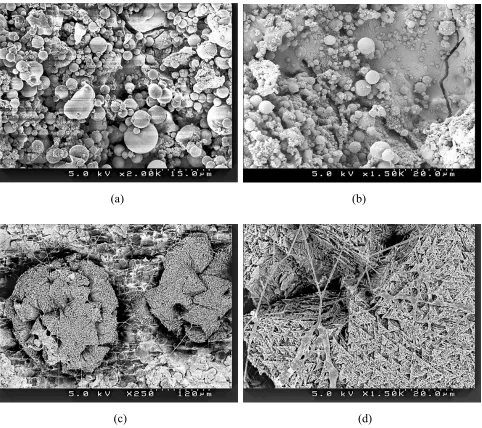
micrometers were isolated from the surface matrix (Figure1c). The samples which, were soaked in other solutions, were not significantly different from these mentioned sample. But after 21 days, it seemed that the SBF-soaked samples appeared sparser to be composed of globular material with smaller coating patches compared to the samples,
which had been soaked in the other solutions. The net like structure of these surfaces actually is the individual HA crystals (Figure 1c), which can be seen in higher magnification monograph (Figure 1d). The amount of the above-mentioned petal like crystal was less for the sample soaked in Ringers solution compared to other ones.
(a) (b)
(c) (d)
Figure 1. The SEM micrographs of the surface of the plasma sprayed HA sample before (a) and after 2 days (b) and 21 days (c,d) soaking in SBF at 37°C .SEM micrographs obtained at various magnitudes. Notice more microcracks in
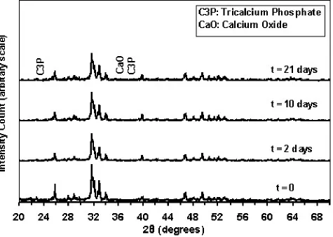
Figure 2 shows typical X-ray diffractions of an HA coated sample before and after 2, 10 and 21 days of immersion in SBF solution respectivly. The crystalline phases of as-plasma coated are primarily a mixture of HA with very small amount of alpha-TCP and calcium oxide [13] which are labeled in this figure. The relative amount of this impurity decreased with the increase of the time of immersion for all solutions.
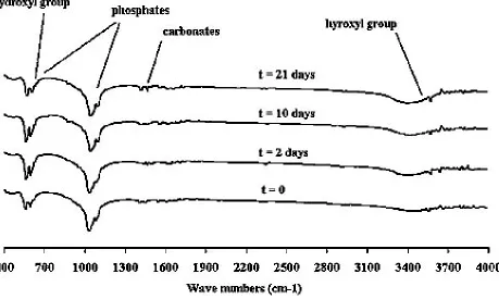
Figure 3 illustrates the typical infrared analysis of this sample before and after 2, 10 and 21 days immersion in SBF solution. The characteristic features like a very weak OH- band at 3570 cm–1
and the absence of hydroxyl band at 633 cm–1 in
as-plasma sprayed sample are indications of
dehydration of HA [13,18]. After 21 days immersion the intensity of the above mentioned peeks [13] increased. This result shows that the dehydroxylation degree [11,19] of the coat decreases with an increase in the time of immersion. The 961 cm-1 bands, characteristic of
symmetric stretching of PO4 group also shows
some changes that can be related to crystallinity changes [20,21] or dissolving of secondary phases [22]. Some little peaks also appear at 1450-1550 cm-1 wave numbers that are characteristics of
carbonated apatite [22], as deposited on the surface of samples. These findings are compatible with those Li and his co-worker [23]. The result taken from the samples which were soaked in other Figure 2. Typical XRD patterns for the plasma sprayed HA coatings before (t = 0) and
solutions are also the same. Joint XRD and FTIR analysis indicated that after 10 days of immersion, a more crystalline structure is formed due to loss of the more soluble secondary and amorphous phases [11]. Also, after 21 days immersion, a bone-like apatite layer precipitates on the coat which is found to be poorly crystallized and contained carbonate. These results are similar to those of the samples which had been soaked in other solution.
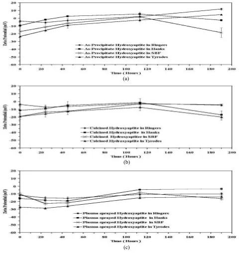
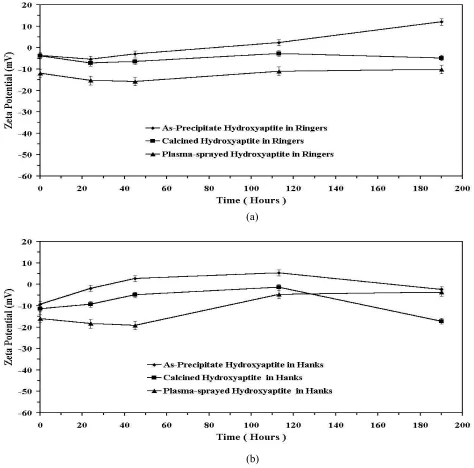
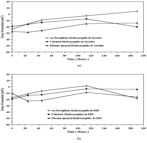
Time-dependent changes in as-precipitate, calcined and plasma sprayed HA’s zeta potentials are shown in Figures 4a to 4c, respectively. Figures 5 and 6 show the variation of zeta potential of several HA powders as a function of immersion time in Ringers, Tyrodes, Hanks and SBF solutions. Each data point represents the mean of five zeta potential values calculated based on average EPM of five histograms (n = 5).
The error bar represents the standard deviation of the mean values. The data showed that the
surface was unstable with significant changes in the charge being observed. As shown in Figures 5 and 6, for most solutions, after 21 days immersion, the zeta potential values increase in the following order: calcined, as-precipitated and as-plasma. A comparison between the zeta potential values and the Ca/P ratio values of the samples (Table 1) reveals that with decrease of Ca/P ratio, the zeta potential values decrease toward more negative amounts. This result is compatible with that of Suzuki and his co-workers who have worked on the effects of the Ca/P ratios on zeta potential of calcium phosphates [6]. Also, as the results of XRD showed, the amount of secondary phases have increased in the same order [13]. This reveals that dissolution of secondary phases have significant effects on the zeta potential value of HA.
The zeta potential variations with time are similar for most of the samples since the differences Figure 3. Typical FTIR spectra for the plasma sprayed HA coatings before (t = 0) and
between them slightly decreased after a long immersion time. The kinetic zeta potential
variations corresponded to, and might directly influence, a Ca-P layer formation. Furthermore, it Figure 4. Variation of the zeta Potential as a function of time of immersion of as precipitate (a), Calcined
(b) and Plasma sprayed (c) Hydroxyapatite in Ringers, Tyrodes, Hanks and SBF solutions at 37°C .
(a)
(b)
may be presumed that the precipitated layers which have been deposited on the surface of sample particles, is a determining factor for the Stern layer`s potential.
For the Ringers solution, the zeta potential
amount is more positive in comparision with the other solutions, especially at the beginning of immersion. According to Table 3, Ringers solution does not have any phosphate ions by itself. Thus at the beginning of immersion, in the case of Ringers Figure 5. Variation of the zeta Potential as a function of time of immersion of as precipitate, Calcined and Plasma sprayed
Hydroxyapatite in (a) Ringers and (b) Tyrodes solution at 37°C.
(a)
solution, the effective ions integrated on the surface of the Stern layer and then dissolution of HA followed this reaction [11]:
Ca10 (PO4)(OH) 2 → Ca(OH) 2 + H3PO4
+ H2O
OH- and PO
43- ions integrated on the surface so
calcium ions could be absorbed to this layer and zeta potential lightly decreased. Consequently these calcium ions could produce Ca(OH)
-complexes here and make zeta potential more negative.
The effects of solution compound on the zeta potential of HA are shown in Figure 4a to 4c. Tyrodes and Ringers solutions show respectively Figure 6. Variation of the zeta Potential as a function of time of immersion of as precipitate, Calcined and Plasma sprayed
Hydroxyapatite in (a) Hanks and (b) SBF solution at 37°C .
the lowest and the highest zeta potential values, especially at the beginning of immersion. As shown in Table 3, Tyrodes solution has the most HCO3- ions concentration compared to other
solutions. On the other hand Ringers solution has more Ca2+ ions and less HCO
3- ions and as a result
the zeta potential would be more positive.
4. CONCLUSION
The results of XRD and FTIR were shown, the
amount of amorphous and secondary phases
(TCP, Tet-CP and CaO) in the coat decreased
after soaking in the solutions. On the other
hand, the increased degree of hydroxylation of
the coat after immersion shows that the coat
surface absorbs hydroxyl ions. As a result, a
relatively large amount of dissolution products
as well as hydroxyl ions were found available
near the surface of the coat at the beginning of
soaking. The negative zeta potential values at
the beginning of soaking show the negative
charge at the Stern layer. This is caused by the
negative ions that were adsorbed on surface of
the samples such as hydroxyl ions. These
adsorbed hydroxyl ions can also perform
hydrogen bonding with phosphate groups [23].
This bonding can form a local saturation [15]
of HA at the surface that leads to precipitation
of amorphous HA. Furthermore, as the FTIR
results show, in the presence of carbon dioxide
in air, the deposed HA is carbonated (type
A-B) [11]. SEM studies showed that, this
precipitate layer became smoother as time
progressed. With formation of this layer, the
concentration of the adsorbed negative ions
(Ca(OH)
-HCO
3-OH
-and PO
43-) decreased
and so the zeta potential values started to
increase. The decrease enhancement of zeta
potential amount, as time progressed, is due to
diffusion of the ions from a longer distance.
On the other hand, this observation is
reasonable considering that the solution had
considerable amounts of sodium bicarbonate
as a buffer. Thus, the possibility exists that
some type of dissolution-precipitation reaction
would have occurred. Another possibility for
the increase in the carbonate vibrational band
in the FTIR spectra for the immersed HA, is
CO
32-ions substitution into the apatite lattice.
Such a substitution will decrease the solubility
of the HA coating and has been proposed for
plasma sprayed HA by Whitehead et al. [24].
This may be the reason for the smoother
changes of zeta potential and it seems that this
reaches a steady state condition. The amount
and speed of each of change of zeta potential
is dependant upon the properties of HA as well
as the ion concentration in the test solutions.
5. REFRENCES
1. Hunter, R. J., "Zeta Potential in Colloid: Science, Principles and Applications", Academic Press, (1981). 2. Somasundaran, P., "Zeta Potential of Apatite in Aqueous
Solution and its Change During Equilibration", Journal of Colloid Interface Science, Vol. 27, (1968), 659-667.
3. Duchyne, P., Kim, C. S. and Pollack, S. R., "The Effect of Phase Difference on the Time Dependent Variation of the Zeta Potential of Hydroxyapatite", J. Biomed. Mater. Res., Vol. 26, (1992), 147-168.
4. Watzek, G., “Endossous implants: Scientific and clinical aspects”, Quintessene Publicating Co. Inc, (1996). 5. Oppermann, D. A., Crimp, M. J. and Bement, D.
M., “In-vitro Stability Predictions for the Bone/ Hydroxyapatite Composite System”, J. Biomed. Mater. Res., Vol. 42, (1998), 412-416.
6. Suzuki, T., Yamamoto, T., Toriyama, M., Nishizawa, K., Mucalo, M. R., Kawamoto, Y., Nagata F. and Kameyama, T., "Surface Instability of Calcium Phosphate Ceramics in Tissue Culture Medium and the Effect on Adhesion and Growth of Anchorage-Dependent Animal Cells", J. Biomed. Mater. Res., Vol.
34, (1997), 507-517.
7. Gasser, P., Haikel, Y., Voegel, J. C. and Gramain, P. H., “Surface Reactions of Hydroxyapatite in the Presence of Fluoride Ions-2, Effects of Calcium and Phosphate in Saturated Solutions”, Colloids Surf., A., Vol. 88, (1994),
157-168.
8. Kibby, C. L. and Hall, W. K., “Surface Properties of Calcium Phosphates”, Hair, M. L. (Ed.), The Chemistry of Surfaces, Vol. 2, Marcel Decker Inc., New York, (1972), 663-730.
Environment for the Testing of Metallic Biomaterials”, in Francis, P. E. and Lee, T. S. (Eds.), “The Use of Synthetic Environments for Corrosion Testing”, STP970, ASTM, Philadelphia, (1988), 79-97.
11. Hench, L. L. and Wilson, J. (Eds.), “An Introduction to Bioceramics”, World Scientific Pub., (1993).
12. Afshar, A., Ghorbani, M., Ehsani, N., Saeri, M. R. and Sorrell, C. C., “Importance Effects of some Parameters in the Wet Precipitation of HA”, Mater. Des., Vol. 24,
(2003), 197-202.
13. Ghorbani, M., Afshar, A., Ehsani, N., Saeri, M. R. and Sorrell, C. C., “Interface Characterization of Plasma Sprayed HA Coat on Ti-6Al-4V”, International Journal of Engineering Transactions B: Applications, Vol. 15, No. 2, (July 2002), 173-182.
14. Kukubo, T. in “CRC Handbook of Bioactive Ceramics”, Yamamura, T, Hench, L. L. and Wilson, J. (Eds.), Vol. 1, CRC Press, Boca Raton Florida, (1990), 41-48. 15. Lee, T. M., Chang, E., Wang, B. C. and Yang, C. Y.,
“Charecteristics of Plasma-Sprayed Bioactive Glass Coatings on Ti-6Al-4V Alloy: an Invitro Study”, Surf. Coat. Technol., Vol. 79, (1996), 170-177.
16. Wen, H. B., de-Wijn, J. R., Cui, F. Z. and de-Groot, K., “Preparation of Bioactive Ti-6Al-4V Surfaces by a Simple Method”, Biomaterials, Vol. 19, (1998),
215-224.
17. Duchyne, P., Cucker, J., Rabin, S. and Nazar, E., “Plasma Sprayed Calcium Phosphate Ceramic Lining on Porous Metal Coating for Bone in Growth”, in Yamamura, T., Hench, L. L. and Wilson, J. (Eds.), CRC Handbook of Bioactive Ceramics, Vol. 2, CRC Press, Boca Raton, FL., USA, (1990), 123-131.
18. Radin, S. R. and Ducheyne, P., “Plasma Spraying Induced Changes of Calcium Phosphate Ceramic Characeristics and the Effects on In-vitro Stability”, J. Mater. Sci.-Mater. Med., Vol. 3, (1992), 33-42.
19. Haman, J. D., Lucas, L. C. and Crawmer, D., “Characterization of High Velocity Oxy-fuel Combustion Sprayed Hydroxyapatite”, Biomaterials,
Vol. 16, (1995), 229-237.
20. Flohr, K. W., “Techniques for Characterization and Quality Control of Hydroxyapatite Raw Materials and Coating”, in Horowtiz E., et. al (Ed.), Characterization and Performance of Calcium Phosphate Coatings for Implants, STP 1196, ASTM, Philadelphia, (1994), 16-24.
21. Heimann, R. B., Kurzweg, H., Ivey, D. G. and Wayman, M. L., “Microstructural and In-vitro Chemical Investigations into Plasma-Sprayed Bioceramic Coatings”, J. Biomed. Mat. Res., (Appl. Biomater.), Vol.
43, (1998), 441-450.
22. Cleries, L., Fernandez-Pradas, J. M., Sardin, G. and Moreza, J. L., “Application of Dissolution Experiments to Characterize the Structure of Pulsed Laser-Deposited Calcium Phosphate Coatings”, Biomaterials, Vol. 20,
(1999), 1401-1405.
23. Li, P. and Duchyne, P., “Quasi-Biological Apatite Film Induced by Titanium in a Simulated Body Fluid”,
Journal of Biomedical Mat. Res., Vol. 41, (1998),
341-348.
![TABLE 2. Ion Concentrations (mM) of the Physiological Solutions used for Experiments [According to References 11, 15 and 16]](https://thumb-us.123doks.com/thumbv2/123dok_us/244048.2019110/3.595.56.543.296.554/table-ion-concentrations-physiological-solutions-experiments-according-references.webp)