Open Access
Hypothesis
Primary sclerosing cholangitis – The arteriosclerosis of the bile
duct?
Peter Fickert, Tarek Moustafa and Michael Trauner*
Address: Laboratory of Experimental and Molecular Hepatology, Division of Gastroenterology and Hepatology, Department of Internal Medicine, Medical University of Graz, Austria
Email: Peter Fickert - peter.fickert@klinikum-graz.at; Tarek Moustafa - tarek.moustafa@klinikum-graz.at; Michael Trauner* - michael.trauner@meduni-graz.at
* Corresponding author
Abstract
Primary sclerosing cholangitis (PSC) is a chronic inflammatory disease of unknown aetiology affecting the large bile ducts and characterized by periductal fibrosis and stricture formation, which ultimately result in biliary cirrhosis and liver failure. Arteriosclerosis involves the accumulation of altered lipids and lipoproteins in large arteries; this drives inflammation and fibrosis and ultimately leads to narrowing of the arteries and hypoperfusion of dependent organs and tissues.
Knowledge of the causative factors is crucial to the understanding of disease mechanisms and the development of specific treatment. Based on pathogenetic similarities between PSC and arteriosclerosis, we hypothesize that PSC represents "arteriosclerosis of the bile duct" initiated by toxic biliary lipids. This hypothesis is based on common molecular, cellular, and morphological features providing the conceptual framework for a deeper understanding of their pathogenesis. This hypothesis should stimulate translational research to facilitate the search for novel treatment strategies for both diseases.
Background
Arteriosclerosis, a disease of the large arteries, represents the single most important contributor to total disease bur-den in developed countries [1,2]. From a pathomorpho-logical point of view, arteriosclerosis is characterized by smooth muscle cell hyperplasia or hypertrophy and matrix protein accumulation in the vessel intima and media, with lipid deposition leading to thickening and induration of the arterial wall and subsequently to arterial stenosis. Over the past decade, attention has become focused on the critical link between modified lipids and lipid products and the initiation and perpetuation of inflammation in arteriosclerosis [3,4].
PSC, a disease of the large bile ducts with a poor median survival of 12 years, is characterised by chronic bile duct
inflammation leading to biliary fibrosis and finally cirrho-sis. It is often complicated by cholangiocarcinoma (10– 15%) [5]. Due to the overall costs to society related to its morbidity, mortality, and need for liver transplantation, PSC in young adults and cholangiopathies in general are increasingly recognized as very important liver diseases [6]. The lack of effective medical treatments for PSC may be deeply rooted in the lack of understanding of the dis-ease mechanisms of PSC.
Based on similar pathogenetic features and mediators found in both entities (summarized in Table 1 and dis-played in Figure 1 and 2, we postulate that PSC shares common disease mechanisms with arteriosclerosis vice versa which may become therapeutic targets in the near future in both diseases.
Published: 25 January 2007
Lipids in Health and Disease 2007, 6:3 doi:10.1186/1476-511X-6-3
Received: 17 November 2006 Accepted: 25 January 2007
This article is available from: http://www.lipidworld.com/content/6/1/3
© 2007 Fickert et al; licensee BioMed Central Ltd.
Common Pathways in the Pathogenesis of Arteriosclerosis and Primary Sclerosing Cholangitis
Although Virchow first described arteriosclerosis as an inflammatory disorder in 1885 it has taken a long time to become recognised in modern medicine as an active inflammatory disease rather than simply a problem of lipid storage in the vessel wall. In the following we present analogies and potential common pathogenetic mecha-nisms in the pathobiology of arteriosclerosis and PSC based on recent findings derived from animal models for both diseases [7-10]. Even when it is taken into account that the human body may have a limited repertoire of reactions to noxious and/or infectious stimuli, the
simi-larities between PSC and arteriosclerosis in regard to their pathobiology are stunning and deserve further explora-tion.
Exposure to abnormal luminal lipid content: blood versus bile – toxic serum lipids versus cholephiles
All known critical molecular and cellular events underly-ing the pathogenesis of arteriosclerosis are apparently linked to alterations in lipid homeostasis [3]. As such, the induction of endothelial vascular cell adhesion molecules (Table 1), currently believed to represent the major initia-tors in arteriosclerosis, is related to the accumulation of oxidized LDL (oxLDL) and phospholipids on the
Table 1: Factors common to arteriosclerosis and sclerosing cholangitis
Arteriosclerosis Sclerosing Cholangitis
Co-factors
-Gender: Age below 60, men develop coronary heart disease twice as frequently as women
-Infectious agents: e.g. Chlamydia pneumoniae
-Systemic inflammation: elevated CRP levels, associated rheumatoid arthritis
Co-factors
-Gender: Males are affected twice as often as females
-Infectious agents: e.g. increased prevalence of chlamydial antibodies, pos. immunostaining for LPS in bile duct epithelial cells
-Systemic inflammation: elevated CRP levels, strongly associated with inflammatory bowel disease
Abnormal luminal content (blood composition)
-Lipoproteins -Oxidized LDL -Oxidized phospholipids -Cholesterol
Abnormal bile composition
-Bile acids
-Cholesterol (supersaturated bile, oxidized?) -Phospholipids(reduced, oxidized?, metabolites?)
Activated endothelial cells
-Adhesion molecules and receptors: VCAM-1, ICAM-1, PCAM-1, E-selectin, P-E-selectin, CD39, CD40, CD44, P2Y, CXCRs, ADAMs, NTFs/ NTRKs
-Cyto- and chemokines: CXCL, MCP-1, TNF-α, IL-1β, IL-6, IL-8, fractalkine, osteopontin
-Growth factors: CTGF, PDGF-β, VEGF, EGF, ET-1, TGF-β
-Tight junction alterations: ZO-1, claudin-1, induction of NOS (iNOS)
Activated bile duct epithelial cells
-Adhesion molecules and receptors: VCAM-1, ICAM-1, E-selectin, CD39L1, CD40, CD44, P2Y, CXCRs, ADAMs, NTFs/NTRKs
-Cyto- and chemokines: CXCL, MCP-1, TNF-α, IL-1β, IL-6, IL-8, fractalkine, osteopontin
-Growth factors: CTGF, PDGF-β, VEGF, EGF, ET-1, TGF-β
-Tight junction alterations: ZO-1, claudin-1, induction of NOS (iNOS)
Inflammatory cells
-Monocytes/macrophage/foam cells: ROS, IL-6, IL-1, TNF-α, MMPs
-T cells: INF-γ, TNF-α, IL-1, IL10
Inflammatory cells
-Neutrophil granulocytes: ROS, IL-6, IL-1, TNF-α, MMPs
-T cells: INF-γ, TNF-α, IL-1, IL10
Smooth muscle cells
-MMPs: 1, 2, 3, 8, 9, 13,14
-ADAMTS: 1, 13
-TIMPs: 1, 2, 3
-Growth factors and chemokines: HGF, ET-1, TGF-β, ACE2I, RANTES
-Matrix deposition: collagen, elastic fibres, glycoproteins, proteoglycanes
Periductal myofibroblasts
-MMPs: 1, 8, 13
-ADAMTS: 13
-TIMPs: 1, 2, 3, 4
-Growth factors and chemokines: HGF, ET-1, TGF-β, MCP-1, MIP-2, ACE2, RANTES
-Matrix deposition: collagen, elastic fibres, glycoproteins, proteoglycanes
ACE2, angiotensin converting enzyme II; ADAM, a disintegrin and metalloproteinase domain; ADAMTS, a disintegrin-like metallopeptidase with thrombospondin type 1 motif; CD40-antigen, TNF receptor superfamily; CD44, CD44 antigen; CLDN1; claudin 1; CTGF, connective tissue growth factor; CXCLs chemokines, C-X-C motif ligand; CXCRs, chemokine C-X-C motif receptors; CD39/NTPDase-1, ectonucleoside triphosphate diphosphohydrolase 1;CD39L1/NTPDase-2, ectonucleoside triphosphate diphosphohydrolase 2; EGF, epidermal growth factor;ET-1, endothelin 1; HGF; hepatocyte growth factor; ICAM-1 intercellular adhesion molecule 1; INF-γ, interferon gamma; IL1-β, interleukin 1 beta; IL-6; interleukin 6; IL-8; interleukin 8; Mcp-1; monocyte chemoattractant protein-1; MIP-2, chemokine (C-X-C motif) ligand 2;
Model of the Development of Arteriosclerosis Figure 1
Model of the Development of Arteriosclerosis. (1) Low-density lipoprotein (LDL) moves into the subendothelial space and becomes oxidized. (2) Inflammatory cells are recruited into the vessel wall via induced endothelial adhesion molecules (VCAM, ICAM) and (3) take up oxidized LDL and become foam cells. (4) Activation of macrophages leads to the release of inflammatory cytokines, chemokines, inflammatory molecules and reactive oxygen species leading to (5) the perpetuation of inflammation and tissue damage. (6) Antigens presented by macrophages and dendritic cells trigger the activation of cytotoxic
T-cells, which produce Th-1 cytokines and heat-shock proteins as well as TGF-β, which activates smooth-muscle cells (7).
1 LDL reaches the subendothelial space
2 Recruitment of inflammatory cells via VCAM-1/ ICAM-1
3 Foam cell formation
4 ROS, Cytokines & Chemokines
5 Perpetuation of inflammation and tissue damage
6 Interaction between macrophages & T-cells
?
Progenitor cell HPCs:
SMCs
Arteriosclerosis
1
2
LDL
3
4 5
6
7
oxLDL
CD36
15 LO iNOS
4 5
T-Lymphocytes
TGF-ß
7 Smooth muscle cell activation
Fibrous cap: Extracellular matrix
•Proteoglycans •Collagen
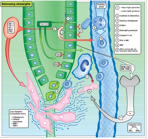
Model of the Development of Primary Sclerosing Cholangitis Figure 2
Model of the Development of Primary Sclerosing Cholangitis. (1) Toxic bile (e.g. toxic bile acids, oxidized/modified
cholesterol and phospholipids) causes (2) the induction of a reactive phenotype of cholangiocytes characterized by de novo
expression or overexpression of adhesion molecules, inflammatory and profibrogenetic cytokines, and receptors (e.g. TNF-α,
TGF-β, toll-like receptors, VCAM, CD44). (3) Proinflammatory cytokines induce leakiness of the bile duct epithelium leading
to regurgitation of bile into the portal space, leading to (4) transmigration of (5) neutrophils and (6) lymphocytes and their
acti-vation. Bile duct epithelial cell-derived growth factors and cytokines (e.g. TGF-β, PDGF) and reactive molecules released from
neutrophils (7) (e.g. ROS, 4-HNE) stimulate extracellular matrix production, accumulation, and proliferation of (8) periductal myofibroblasts, leading to periductal fibrosis and in consequence to vascular deprivation of the bile duct system itself, causing (9) death of bile duct epithelial cells. Bile duct epithelial cell-derived growth factors (e.g. EGF, HGF, PDGF, BDNF) may also activate and recruit bone marrow derived progenitor cells into the portal field probably engaged in ductal reaction and prolif-eration of periductal MFBs.
1 – Intact tight junctions
1
2
2 Cytokines & Chemokines
3
3 Reactive cholangiocyte
4
4 VCAM-1
5 Neutrophil granulocyte
6
6 Cytotoxic T-cell
7
8
? BDNF, EGF HGF, PDGF Peribiliary
plexus
Progenitor cell HPCs:
BECs MFBs TNF-α
TGF-β INF-γ MCP1 PDGF
TLRs
CD44
Scar formation: Extracellular matrix
•Proteoglycans
•Collagen •Lipoproteins
MMPs TIMPs
9 Sclerosing cholangitis
-- Leaky tight junctions
5
7
8
9
ROS, 4-HNE
MFB
endothelial surface (Figure 1) [3]. As such, oxidized lipo-protein particles (e.g. oxidized phospholipids and short-chain aldehydes arising from lipoprotein oxidation) can induce endothelial VCAM-1 expression via TNF-α- or IL1-β-induced nuclear factor-κB activation [4].
Since the liver is the major organ responsible for the pro-duction and degradation of apoB-100-containing lipo-proteins, it is most likely that these molecules may also be engaged in the pathogeneses of liver diseases. Moreover, the fact that bile secretion is the most effective means of cholesterol elimination from the human body leads to the suggestion that altered biliary lipid secretion and/or abnormal lipid oxidation leading to oxidative stress may also be critically involved in the initiation and/or perpet-uation of PSC. In parallel to arteriosclerosis, enhanced cholangiocellular VCAM-1 expression was recently dem-onstrated in multidrug resistance gene (Mdr2/Abcb4) knockout mice (Mdr2-/-), a well characterized mouse model that closely mirrors the macroscopic and micro-scopic pathology of PSC [8,11]. Potentially toxic bile acids synthesized from cholesterol may represent first-line can-didates for the induction of cholangitis in Mdr2-/- mice [12]. Bile acids are normally packed into mixed micelles together with phospholipids and cholesterol to protect cholangiocytes from potentially toxic bile acids, which may otherwise cause necrosis or apoptosis of cholangi-ocytes [6]. Absence of phospholipids as a consequence of Mdr2 defects not only results in unopposed bile acid tox-icity but also in cholesterol supersaturated bile, which could facilitate oxidation. The critical role of biliary lipo-toxicity in the sense of deranged and consequently toxic bile composition in the development of cholangitis was also underlined by the enhanced cholestatic phenotype in bile acid-fed Mdr2-/- mice [13]. Despite the apparent dis-crepancy between decreased biliary cholesterol concentra-tion in Mdr2-/- mice and increased serum cholesterol levels in arteriosclerosis, the common denominator may be abnormal cholesterol oxidation in both conditions. Alter-natively, abnormal metabolic products of phosphatidyl-cholines (e.g. lysoPC) could also be excreted into bile in Mdr2-/- mice or reach the cholangiocytes also from the per-biliary plexus or lymph vessels instead from the luminal side. Moreover, bile acids may be able to induce a reactive phenotype of cholangiocytes characterized by the produc-tion of several proinflammatory and profibrogenic cytokines and chemokines as well as their corresponding receptors (summarized in Table 1 and shown in Figure 2) [6]. Conversely, some bile acids, their receptors, and syn-thetic agonists were recently shown to influence the degree of arteriosclerosis in several animal models [14-18]. This fits well with the rapidly progressing insights into the transcriptional regulation by nuclear receptors (e.g. FXR, LXR) of bile acid synthesis/transport on the one hand and lipid metabolism/transport on the other
[17,19,20]. Finally, toxic bile acids can activate endothe-lial cells [21]. Taken together, exposure to abnormal lumi-nal lipid content and composition, ultimately resulting in lipid oxidation, may be critically involved in both condi-tions.
Activated endothelial cells versus reactive cholangiocytes – mechanisms of chemoattraction in arteriosclerosis and PSC
Endothelial and bile duct epithelial cells may not only be the victims/targets but also the culprits/effectors that actively participate in inflammatory processes in arterio-sclerosis and PSC, respectively. The endothelium repre-sents the first-line contact, where the accumulation of oxLDL induces the expression of adhesion molecules (e.g., VCAM-1) and proinflammatory cytokines/chemok-ines (e.g., MCP-1, see also Table 1) critically involved in the initiation and perpetuation of arteriosclerosis, making them potential therapeutic targets [4]. Most interestingly, the cytokine/chemokine/receptor armamentarium of reactive endothelial cells and reactive cholangiocytes appears to be largely comparable, as summarized in Table 1[6].
Altered endothelial versus altered epithelium barrier function – mechanisms of leukocyte penetration and activation
Endothelial binding and penetration of monocytes and T lymphocytes in nascent atheromas by adhesion molecules is pivotal in the development of arteriosclerosis [4]. More-over, induction of endothelial-derived adhesion mole-cules reduces barrier function via alterations of the delicately regulated tight junction system, allowing oxi-dized lipids and marginalized/attached inflammatory cells to migrate into subendothelial regions of the vessel [22]. The MCP-1/CCR2 interaction together with the action of M-CSF represents a critical step for migrated monocytes to differentiate into activated macrophages/ foam cells, which themselves are able to produce numer-ous cytokines, reactive oxygen species (ROS), and growth factors (Table 1). Penetrating T lymphocytes further per-petuate this inflammatory response in a complex orches-trated interplay of cytokines and tissue factors (Table 1, Figure 1).
with enhanced expression and secretion of adhesion mol-ecules may then induce the migration of neutrophils and T lymphocytes from the peribiliary plexus out into the subepithelial space and into the bile duct epithelium, leading to cholangitis (Figure 2). Despite the fact that cytotoxic T cells were shown to induce cholangiocyte apoptosis in humans [26], their role in PSC is still a matter of debate since these cells are virtually absent in precir-rhotic PSC stages. The hypothesis becomes questionable that cholangiocytes are the primary target of the immune system in PSC. The role of portal and/or penetrated neu-trophils (probably leading to ROS formation and destruc-tion of the basement membrane) in PSC also deserves more detailed studies because these cells may be critically involved in the activation of periductal myofibroblasts by aldehydic end-products of lipid peroxidation.
Endothelial cell – smooth muscle cell interactions in arteriosclerosis versus bile duct epithelial cell – periductal myofibroblast interactions in PSC
Differentiated monocytes/macrophages and T cells that find their way out through the leaky endothelial barrier exert their pro-atherogenic effects by the secretion of cytokines and growth factors driving smooth muscle cell (SMC) migration and proliferation as well as extracellular matrix (ECM) deposition (Table 1). The development of fibrous plaques results from proliferating SMCs, choles-terol ester and ECM components. This complex interplay is mediated by extensive crosstalk between T-cells and macrophages (CD40/CD40L interactions). Increased homocystein and angiotensin II levels in the vessel wall were also demonstrated to be important mediators of SMC proliferation as well as their activation. In addition, molecules such as osteopontin, CD44 and TGF-β were demonstrated to be critically involved in SMC activation. Reduction of arteriosclerosis in corresponding athero-genic diet-fed knock-out mice [27-29] further underlines the importance of these factors for the development of arteriosclerosis.
A number of these central mediators of arteriosclerosis were also demonstrated to originate from activated cholangiocytes in cholangiopathies [30-32] (Table 1 and Figure 2). Cholangiopathies are frequently accompanied by ductular proliferation/reaction, which is a potential stimulus for biliary fibrosis. The question whether ductu-lar proliferates induce/stimulate proliferation and activa-tion of periductal myofibroblasts and how this might happen (e.g., via stimulation with cytokines such as TGF-β or PDGF, brain-derived peptides such as NGF and BDNF, epithelial-mesenchymal transformation) is critical and still to be resolved.
Remodelling of the extracellular matrix as self-perpetuating events in arteriosclerosis and PSC
Little is known about the potential importance of the extracellular matrix composition in the pathogenesis of both entities and how modifications (e.g. binding of oxLDL/phospholipids to proteoglycans, cleavage and sig-nalling through proteinases, collagenases, gelatinases, resulting advanced glycosylated endproducts or trans-glutamination of collagen fibres) could influence the per-petuation of both diseases. In arteriosclerosis, diminished stability of the matrix scaffold is driven by MMPs, colla-genases, and the recently identified ADAMTSs (a disin-tegrin-like metalloprotease with thrombospondin type-1 motifs) [33]. These enzymes could either play a role in the resolution of vessel stenoses or also destabilize plaques [34,35]. In parallel, these enzymes could also be engaged in the chronic persistent wound-healing process in PSC. It is attractive to speculate that chronic activation of MMPs could further increase duct permeability. In addition, per-sistent activation of ADAMTSs could further increase the inflammatory response through their thrombospondin-1 motive (TSP-1) on TGF-β [36,37] or in the sense of a self-perpetuating process by generating chemotactic/immuno-genic ECM products.
Limitations and caveats using Mdr2-/- mice as a model system for PSC
The exact role of MDR3 variants (the human orthologue of rodent Mdr2) in the pathogenesis of PSC is still open to question. Two studies (with a total number of 80 PSC patients) found no differences in the number of genetic
MDR3 variants between healthy individuals and PSC
pericholangitis and periductal fibrosis) of sclerosing cholangitis in humans [8,42]. An additional advantage of this model system is the high reproducibility and low var-iance in the cholestatic phenotype of these animals mak-ing them also an ideal model for therapeutic interventional studies [43]. Irrespective of the underlying cause of sclerosing cholangitis, the Mdr2-/- model is an appropriate animal model for sclerosing cholangitis since it reliably reproduces the final common pathways of bile duct injury and (peri)biliary fibrosis. However, Mdr2 -/-mice do not show some of the (pheno)typical features observed in human PSC such as the association with cholangiocarcinoma and inflammatory bowel disease. finally, the Mdr2-/- model may also have general validity as model for cholangiopathies resulting from bile toxicity with a deranged biliary phospholipid/bile salt secretion.
Differences between PSC and artieriosclerosis
Despite the stunning similarities between PSC and arteri-osclerosis in regard to their pathobiology as outlined above, there are also obvious differences between both entities such as the age of onset, different risk factors, and different co-factors (outlined in Table 1) which deserve attention. The exact definition and discrimination of sim-ilarities and differences between PSC and artiosclerosis should further help to understand the pathogenesis of both diseases.
How to test the hypotheses
(i) The toxic bile hypothesis could be tested by studying bile composition with particular emphasis on biliary lip-ids (e.g. oxidized/modified cholesterol and phospholip-ids) and other candidate molecules (e.g. CD66a, E-selectin, cyto-, and chemokines), either in bile sampled by endoscopic techniques or assessed via non-invasive modalities such as advanced MR technologies (MR spec-troscopy).
(ii) In addition, detailed immunohistochemical studies in PSC livers (either explanted organs or liver biopsy mate-rial) using antibodies against adhesion molecules recently identified on cholangiocytes and other well defined medi-ator molecules in arteriosclerosis such as the receptor for advanced glycosilated end products (RAGE) and oxidized lipids/phospholipids should answer the question of whether these molecules are indeed engaged in the patho-genesis in PSC.
(iii) Ulcerated bile ducts are frequently observed in explanted PSC livers and remind us, at least in some mor-phological aspects, of plaque rupture in arteriosclerosis. The frequency and importance of duct ulcerations in PSC might be underestimated due to sampling errors inherent to the technique of liver biopsy. Serial sections of explanted PSC livers should help to answer the question
of whether concentric bile duct stenoses are the result of a perpetuated wound-healing process of ulcerated/ruptured bile ducts.
(iv) Basic research in PSC should determine the potential role of new inflammatory mediators (e.g. adipokines) known to be critically engaged in the pathogenesis of the metabolic syndrome and consequently arteriosclerosis.
Conclusion
We postulate a pathogenetic link between PSC and arteri-osclerosis. Our model provides a structured and unifying pathogenetic model, based on biological, clinical, and experimental evidence and a network of molecular simi-larities between both entities. Scientific exchange between researchers/scientists working either in the field of arteri-osclerosis or cholangiopathies and cholestatic liver dis-eases in general should be fruitful and stimulating. Research on PSC may greatly benefit from translating pathogenetic concepts from advanced work in arterioscle-rosis to the study of cholestasis. However, this could also work the other way around since it is becoming increas-ingly apparent that arteriosclerosis may represent a kind of "liver disease of the heart" [44].
Acknowledgements
This work was supported by a GenAU grant (GaTib) from the Austran Min-istry of Education, Science and Culture
References
1. Lusis AJ: Atherosclerosis. Nature 2000, 407(6801):233-241. 2. Murray CJ, Lopez AD: Global mortality, disability, and the
con-tribution of risk factors: Global Burden of Disease Study. Lan-cet 1997, 349(9063):1436-1442.
3. Berliner JA, Watson AD: A role for oxidized phospholipids in atherosclerosis. N Engl J Med 2005, 353(1):9-11.
4. Libby P: Inflammation in atherosclerosis. Nature 2002, 420(6917):868-874.
5. Mendes FD, Lindor KD: Primary sclerosing cholangitis. Clin Liver Dis 2004, 8(1):195-211.
6. Lazaridis KN, Strazzabosco M, Larusso NF: The cholangiopathies: disorders of biliary epithelia. Gastroenterology 2004, 127(5):1565-1577.
7. de Winther MP, Hofker MH: New mouse models for lipoprotein metabolism and atherosclerosis. Curr Opin Lipidol 2002, 13(2):191-197.
8. Fickert P, Fuchsbichler A, Wagner M, Zollner G, Kaser A, Tilg H, Krause R, Lammert F, Langner C, Zatloukal K, Marschall HU, Denk H, Trauner M: Regurgitation of bile acids from leaky bile ducts causes sclerosing cholangitis in Mdr2 (Abcb4) knockout mice. Gastroenterology 2004, 127(1):261-274.
9. Vierling JM: Animal models for primary sclerosing cholangitis. Best Pract Res Clin Gastroenterol 2001, 15(4):591-610.
10. Xu Q: Mouse models of arteriosclerosis: from arterial injuries to vascular grafts. Am J Pathol 2004, 165(1):1-10.
11. O'Mahony CA, Vierling JM: Etiopathogenesis of primary sclero-sing cholangitis. Semin Liver Dis 2006, 26(1):3-21.
Publish with BioMed Central and every scientist can read your work free of charge "BioMed Central will be the most significant development for disseminating the results of biomedical researc h in our lifetime."
Sir Paul Nurse, Cancer Research UK
Your research papers will be:
available free of charge to the entire biomedical community
peer reviewed and published immediately upon acceptance
cited in PubMed and archived on PubMed Central
yours — you keep the copyright
Submit your manuscript here:
http://www.biomedcentral.com/info/publishing_adv.asp
BioMedcentral
14. Beaven SW, Tontonoz P: NUCLEAR RECEPTORS IN LIPID METABOLISM: Targeting the Heart of Dyslipidemia. Annu Rev Med 2006, 57:313-329.
15. Gutierrez A, Ratliff EP, Andres AM, Huang X, McKeehan WL, Davis RA: Bile acids decrease hepatic paraoxonase 1 expression and plasma high-density lipoprotein levels via FXR-mediated signaling of FGFR4. Arterioscler Thromb Vasc Biol 2006, 26(2):301-306.
16. Hanniman EA, Lambert G, McCarthy TC, Sinal CJ: Loss of func-tional farnesoid X receptor increases atherosclerotic lesions in apolipoprotein E-deficient mice. J Lipid Res 2005, 46(12):2595-2604.
17. Kalaany NY, Mangelsdorf DJ: LXRs and FXR: The Yin and Yang of Cholesterol and Fat Metabolism. Annu Rev Physiol 2005. 18. Tontonoz P, Mangelsdorf DJ: Liver X receptor signaling
path-ways in cardiovascular disease. Mol Endocrinol 2003, 17(6):985-993.
19. Shih DM, Kast-Woelbern HR, Wong J, Xia YR, Edwards PA, Lusis AJ: A role for FXR and human FGF-19 in the repression of paraoxonase-1 gene expression by bile acids. J Lipid Res 2006, 47(2):384-392.
20. Duran-Sandoval D, Cariou B, Percevault F, Hennuyer N, Grefhorst A, van Dijk TH, Gonzalez FJ, Fruchart JC, Kuipers F, Staels B: The far-nesoid X receptor modulates hepatic carbohydrate metabo-lism during the fasting-refeeding transition. J Biol Chem 2005, 280(33):29971-29979.
21. Qin P, Tang X, Elloso MM, Harnish DC: Bile acids induce adhesion molecule expression in endothelial cells through activation of reactive oxygen species, NF-kappaB, and p38. Am J Physiol Heart Circ Physiol 2006, 291(2):H741-7.
22. Alevriadou BR: CAMs and Rho small GTPases: gatekeepers for leukocyte transendothelial migration. Focus on "VCAM-1-mediated Rac signaling controls endothelial cell-cell con-tacts and leukocyte transmigration". Am J Physiol Cell Physiol 2003, 285(2):C250-2.
23. Lamireau T, Zoltowska M, Levy E, Yousef I, Rosenbaum J, Tuchweber B, Desmouliere A: Effects of bile acids on biliary epithelial cells: proliferation, cytotoxicity, and cytokine secretion. Life Sci 2003, 72(12):1401-1411.
24. Hanada S, Harada M, Koga H, Kawaguchi T, Taniguchi E, Kumashiro R, Ueno T, Ueno Y, Ishii M, Sakisaka S, Sata M: Tumor necrosis fac-tor-alpha and interferon-gamma directly impair epithelial barrier function in cultured mouse cholangiocytes. Liver Int 2003, 23(1):3-11.
25. Sakisaka S, Kawaguchi T, Taniguchi E, Hanada S, Sasatomi K, Koga H, Harada M, Kimura R, Sata M, Sawada N, Mori M, Todo S, Kurohiji T: Alterations in tight junctions differ between primary biliary cirrhosis and primary sclerosing cholangitis. Hepatology 2001, 33(6):1460-1468.
26. Aoki CA, Bowlus CL, Gershwin ME: The immunobiology of pri-mary sclerosing cholangitis. Autoimmun Rev 2005, 4(3):137-143. 27. Bruemmer D, Collins AR, Noh G, Wang W, Territo M, Arias-Magal-lona S, Fishbein MC, Blaschke F, Kintscher U, Graf K, Law RE, Hsueh WA: Angiotensin II-accelerated atherosclerosis and aneu-rysm formation is attenuated in osteopontin-deficient mice. J Clin Invest 2003, 112(9):1318-1331.
28. Cuff CA, Kothapalli D, Azonobi I, Chun S, Zhang Y, Belkin R, Yeh C, Secreto A, Assoian RK, Rader DJ, Pure E: The adhesion receptor CD44 promotes atherosclerosis by mediating inflammatory cell recruitment and vascular cell activation. J Clin Invest 2001, 108(7):1031-1040.
29. Kalinina N, Agrotis A, Antropova Y, Ilyinskaya O, Smirnov V, Tararak E, Bobik A: Smad expression in human atherosclerotic lesions: evidence for impaired TGF-beta/Smad signaling in smooth muscle cells of fibrofatty lesions. Arterioscler Thromb Vasc Biol 2004, 24(8):1391-1396.
30. Brown LF, Berse B, Van de Water L, Papadopoulos-Sergiou A, Per-ruzzi CA, Manseau EJ, Dvorak HF, Senger DR: Expression and dis-tribution of osteopontin in human tissues: widespread association with luminal epithelial surfaces. Mol Biol Cell 1992, 3(10):1169-1180.
31. Cruickshank SM, Southgate J, Wyatt JI, Selby PJ, Trejdosiewicz LK: Expression of CD44 on bile ducts in primary sclerosing cholangitis and primary biliary cirrhosis. J Clin Pathol 1999, 52(10):730-734.
32. Whitington PF, Malladi P, Melin-Aldana H, Azzam R, Mack CL, Sahai A: Expression of osteopontin correlates with portal biliary proliferation and fibrosis in biliary atresia. Pediatr Res 2005, 57(6):837-844.
33. Wight TN: The ADAMTS proteases, extracellular matrix, and vascular disease: waking the sleeping giant(s)! Arterioscler Thromb Vasc Biol 2005, 25(1):12-14.
34. Jonsson-Rylander AC, Nilsson T, Fritsche-Danielson R, Ham-marstrom A, Behrendt M, Andersson JO, Lindgren K, Andersson AK, Wallbrandt P, Rosengren B, Brodin P, Thelin A, Westin A, Hurt-Camejo E, Lee-Sogaard CH: Role of ADAMTS-1 in atherosclero-sis: remodeling of carotid artery, immunohistochemistry, and proteolysis of versican. Arterioscler Thromb Vasc Biol 2005, 25(1):180-185.
35. Suzuki M, Murata M, Matsubara Y, Uchida T, Ishihara H, Shibano T, Ashida S, Soejima K, Okada Y, Ikeda Y: Detection of von Wille-brand factor-cleaving protease (ADAMTS-13) in human platelets. Biochem Biophys Res Commun 2004, 313(1):212-216. 36. Breitkopf K, Sawitza I, Westhoff JH, Wickert L, Dooley S, Gressner
AM: Thrombospondin 1 acts as a strong promoter of trans-forming growth factor beta effects via two distinct mecha-nisms in hepatic stellate cells. Gut 2005, 54(5):673-681. 37. Uemura M, Tatsumi K, Matsumoto M, Fujimoto M, Matsuyama T,
Ishikawa M, Iwamoto TA, Mori T, Wanaka A, Fukui H, Fujimura Y: Localization of ADAMTS13 to the stellate cells of human liver. Blood 2005, 106(3):922-924.
38. Pauli-Magnus C, Kerb R, Fattinger K, Lang T, Anwald B, Kullak-Ublick GA, Beuers U, Meier PJ: BSEP and MDR3 haplotype structure in healthy Caucasians, primary biliary cirrhosis and primary sclerosing cholangitis. Hepatology 2004, 39(3):779-791. 39. Rosmorduc O, Hermelin B, Boelle PY, Poupon RE, Poupon R,
Chazouilleres O: ABCB4 gene mutations and primary scleros-ing cholangitis. Gastroenterology 2004, 126(4):1220-2; author reply 1222-3.
40. deVree JM: Defects in hepatobiliary transport. Genetics and therapy of progressive familial intrahepatic cholestasis type 3. University of Amsterdam; 1999.
41. Kaw M, Silverman WB, Rabinovitz M, Schade RR: Biliary tract cal-culi in primary sclerosing cholangitis. Am J Gastroenterol 1995, 90(1):72-75.
42. Fickert P, Zollner G, Fuchsbichler A, Stumptner C, Weiglein AH, Lammert F, Marschall HU, Tsybrovskyy O, Zatloukal K, Denk H, Trauner M: Ursodeoxycholic acid aggravates bile infarcts in bile duct-ligated and Mdr2 knockout mice via disruption of cholangioles. Gastroenterology 2002, 123(4):1238-1251.
43. Fickert P, Wagner M, Marschall HU, Fuchsbichler A, Zollner G, Tsybrovskyy O, Zatloukal K, Liu J, Waalkes MP, Cover C, Denk H, Hofmann AF, Jaeschke H, Trauner M: 24-norUrsodeoxycholic acid is superior to ursodeoxycholic acid in the treatment of sclerosing cholangitis in Mdr2 (Abcb4) knockout mice. Gas-troenterology 2006, 130(2):465-481.