R E S E A R C H A R T I C L E
Open Access
Association of systemic and ocular risk factors
with neurosensory retinal detachment in diabetic
macular edema: a case
–
control study
Aditi Gupta
1, Rajiv Raman
1, Vaitheeswaran Kulothungan
2and Tarun Sharma
1*Abstract
Background:Diabetic macular edema (DME) with neurosensory retinal detachment (NSD) remains an important cause of visual loss in patients with diabetes. The aim of the study was to elucidate the association of systemic and ocular risk factors with NSD in DME.
Methods:In a retrospective case–control study, we reviewed clinical records of all the subjects with DME seen between January 2010 and December 2010. Cases and controls were selected based on optical coherence tomography and stereoscopic biomicroscopy review. NSD was defined as subfoveal fluid accumulation under detached retina with or without overlying foveal thickening. The association between the presence of NSD, blood pressure, lipid status and various other biochemical parameters was evaluated.
Results:Group I (cases) included 37 eyes of 33 patients having DME with NSD and Group II (controls) included 30 eyes of 21 patients having DME without NSD. Patients ranged in age (mean ± SD) from 50 to 62 years (56.6 +/-6.78) for cases and from 51 to 65 years (58.4+/-7.84) for controls. The duration of diabetes ranged from 4 to 15 year (mean 9.45+/-6.08) among cases and 4 to 14 years (9.7+/-5.12) among controls. Significant risk factors for NSD were high values of systolic and diastolic blood pressure (p = 0.039 and 0.043 respectively).
Conclusion:High systolic and diastolic blood pressures are independent and significant risk factors for NSD in DME.
Keywords:Diabetic macular edema, Neurosensory retinal detachment, Risk factors, Blood Pressure
Background
Diabetic macular edema (DME) remains a major cause of visual loss in patients with diabetes [1]. Optical coher-ence tomography (OCT) has specifically been used for characterizing the morphological features of DME, and five OCT patterns of DME have been described: diffuse retinal thickening (DRT), cystoid macular edema (CME), neurosensory retinal detachment (NSD) without poster-ior hyaloidal traction, posterposter-ior hyaloidal traction (PHT) without tractional retinal detachment (TRD) and PHT with TRD [2-5]. NSD under the fovea has been reported in 3–31% of patients with DME [2,4-10].
The prognosis of DME is decided by many factors, such as the presence of NSD, inner segment/outer segment
(IS/OS) conjunction and integrity of the external limit-ing membrane (ELM) line [11-13]. IS/OS integrity and intact ELM are important indicators in the evaluation of foveal photoreceptor layer integrity, and correlate strongly with best-corrected visual acuity (BCVA) after medical or surgical treatment of DME [11,12].
The presence of NSD is found to adversely affect the prognosis of DME. NSD can significantly limit effective laser treatment of the macula [14]. The presence of NSD in DME associated with subretinal exudation has been reported to be associated with poor visual prognosis after vitrectomy [2]. Likewise, in macular edema secondary to branch retinal vein occlusion, the presence of subfoveal NSD was shown to retard the absorption of macular edema and recovery of vision after grid laser photocoagu-lation [14]. The high percentage of NSD in CRVO [9] may have played a role in the poor response of macular edema to grid laser photocoagulation in the multicenter trial on * Correspondence:drtaruns@gmail.com
1
Shri Bhagwan Mahavir Department of Vitreoretinal Services, Sankara Nethralaya, 18, College Road, Chennai 600 006, Tamil Nadu, India Full list of author information is available at the end of the article
CRVO by the Central Retinal Vein Occlusion Study Group [15]. Hence, the need to better understand the pathogenesis of NSD has been stressed [15]. Although previous studies have extensively reported the systemic and ocular risk factors for the presence of DME [16-20], the risk factors associated with NSD in DME have been rarely studied in detail [21]. A recent study indicated the presence of high glycosylated hemoglobin (HbA1c) as a risk factor for NSD, suggesting the role of systemic factors in the causation of NSD in DME [21]. The aim of this study was to elucidate the association of various systemic and ocular risk factors with NSD in Indian subjects with DME.
Methods
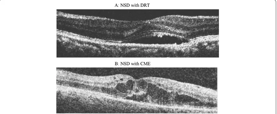
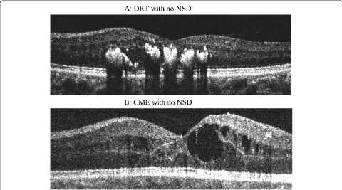
This study was a retrospective chart review of patients with diagnosed DME seen between Jan 2010 to Dec 2010. Cases and controls were selected based on SD-OCT re-view. Group I (cases) included 37 eyes of 33 patients who were diagnosed as DME with NSD on OCT and Group II (controls) included 30 eyes of 21 patients who had DME without NSD. The macular edema was diagnosed by bio-microscopy according to the criteria reported by ETDRS. NSD type DME was defined as subfoveal fluid accumula-tion with distinct outer border of detached retina with or without overlying foveal thickening (Figure 1). All cases had NSD associated with DRT or CME (Figure 1). All controls had DRT or CME without any NSD (Figure 2). Patients with posterior hyaloidal traction (PHT) without TRD and PHT with TRD as documented on OCT, and media opacities such as corneal opacity, dense cataract, vitreous or preretinal hemorrhage, uveitis were excluded from the study. This study was approved by the Institu-tional Review Board, Vision Research Foundation and
adhered to the Declaration of Helsinki. The medical re-cords of all the patients including cases and controls were
reviewed, with documentation of patients’ age, gender,
duration and type of diabetes mellitus, history of any asso-ciated systemic disease like hypertension, hyperlipidemia, nephropathy, ischemic heart disease status post coronary artery bypass surgery, details of systemic medications, his-tory of ocular surgeries including intravitreal injections and laser treatment in the past. The details of patients’ ocular examination findings including BCVA on Snellen chart (later converted to logMAR for statistical analyses), lens status, stage of diabetic retinopathy, OCT findings, and systemic biochemical parameters were also noted. A prototype SD-OCT system (Topcon 3D1000, Tokyo, Japan) was used with an axial resolution of 6u and ac-quisition rate of approximately 18,000 scans per second. All OCT images were acquired through a dilated pupil. During the OCT examination, macula was scanned on six radial sections including the horizontal, vertical and oblique planes through the centre of the fovea. The ret-inal thickness was measured as distance of vitreoretret-inal interface and inner edge of retinal pigment epithelium (RPE) at the maximum point of edema. Central subfield retinal thickness was also noted on OCT.
The blood pressure was recorded, in the sitting position, in the right arm to the nearest 2 mm Hg using the mer-cury sphygmomanometer (Diamond Deluxe BP apparatus, Pune, India). Two readings were taken with a five minutes interval and their mean indication was taken as the blood pressure. In most of the subjects, the blood pressure measurement was taken on the day of complete ophthal-mic examination which included OCT. In the remaining subjects, blood pressure was measured on the day of OCT review which was 1 or 2 days later. The biochemical
parameters noted were fasting and post-prandial blood sugar levels, hemoglobin, glycosylated hemoglobin, blood pressure and renal function tests (serum urea and serum creatinine). All biochemical parameters were done at same laboratory using standard techniques. The grading of diabetic retinopathy was done based on modified klein classification as mild, moderate and severe nonproli-ferative diabetic retinopathy and prolinonproli-ferative diabetic retinopathy [22]. The modification was proposed as a standardized alternative to the more detailed Early Treat-ment Diabetic Retinopathy Study (ETDRS) system. It in-volves grading seven stereoscopic standard fields as a whole, and assigning a level of severity for the eye accord-ing to the greatest degree of retinopathy usaccord-ing a modified Airlie House Classification scheme [22].
Statistical analysis
A computerized database was created for all the records. Statistical analyses were performed using SPSS Windows version 14.0 (SPSS Science, Chicago, IL, USA). All the data were expressed as mean ± S.D or as percentage. The normality of distribution was checked for all factors by Kolmogorov–Smirnov analysis. The data in the study followed normal distribution, hence we used the para-metric tests to determine significance. Chi-square test was used to compare proportions among neurosensory detachment status with the independent categorical
vari-ables and the Student’s t-test was used to compare
proportions among neurosensory detachment status with the independent measured (continuous) variables in Uni-variate analyses. P value less than 0.05 was considered sig-nificant. Multivariate analyses could not be done because of small sample size in both the groups.
Results
A total of 37 eyes of 33 patients who had NSD with DME (Group 1, cases) and 30 eyes of 21 patients who had DME without NSD (Group 2, controls) were included in our study. All patients included were of type 2 diabetes melli-tus. The patients ranged in age from 50 to 62 years among the cases (mean 56.6 +/-6.7) and 51 to 65 among the con-trols (mean 58.4+/7.84). The duration of diabetes ranged from 4 to 15 years (mean 9.45+/-6.08) among cases and 4 to 14 years (9.7+/-5.12) among controls. Group 1 included 28 males (84.8%) and 5 females (15.2%) and Group 2 in-cluded 14 males (67.7%) and 7 females (33.3%).
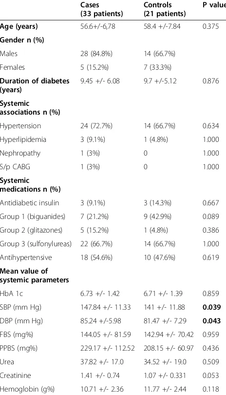
Table 1 depicts the comparison of systemic factors as-sociated with cases and controls. There were no signifi-cant differences in the two groups in terms of the mean age (p = 0.375), duration of diabetes (p = 0.876), fasting blood sugar and post prandial blood sugar levels (p = 0.959 and 0.436 respectively), glycosylated hemoglobin (p = 0.859), hemoglobin (p = 0.118) and presence of hyper-tension (p = 0.634). However, the mean systolic and dia-stolic blood pressures were significantly higher (p = 0.039
and 0.043 respectively) in the NSD group than the control group.
Table 2 compares the ocular factors among eyes with NSD and control eyes. The mean log MAR BCVA was 0.822 +/-0.421 in NSD group and 0.61+/-0.47 in the con-trol group (p 0.056). The mean central macular thickness as determined by OCT was higher in cases than in the controls (485.7 +/-189.81 versus 332.2+/-134.14 microns, p = 0.0004). Cystoid macular edema was more commonly seen than diffuse edema in NSD group, although the dif-ference was not significant (p = 0.085). 35.1% of eyes with NSD had associated proliferative diabetic retinopathy compared to 23.3% in the control group (p = 0.820). There were no significant differences in the two groups in terms
of pseudophakic status (p = 0.370). As expected, ocular surgery including anti- VEGF injections were performed more frequently in Group 1 (p = 0.008).
Discussion
DME remains the leading cause of visual loss among pa-tients with diabetes mellitus. Among the various patterns of DME, NSD under the fovea has been reported in 3– 31% of patients. The pathogenesis of NSD is linked not only to the limitations of the draining vascular system, but also to impairment in the function of the RPE. Kang et al. reported that in diabetic eyes, the incidence of CME and NSD increasd with the existence of retinal vascular hyper-permeability and the pathology of these two phenomena might share a common pathogenesis in this regard [4].
Various systemic factors have been associated with in-creased incidence of DME like severity of diabetic retin-opathy, poor glycemic control and duration of diabetes. Hypertension, proteinuria, dyslipidemia, uncontrolled renal parameters, and PRP for PDR (causing acute choroidal ischemia), have also been associated with increased risk of
Table 1 Comparison of systemic features amongst cases and controls
Cases (33 patients)
Controls (21 patients)
P value
Age (years) 56.6+/-6,78 58.4 +/-7.84 0.375
Gender n (%)
Males 28 (84.8%) 14 (66.7%)
Females 5 (15.2%) 7 (33.3%)
Duration of diabetes (years)
9.45 +/- 6.08 9.7 +/-5.12 0.876
Systemic associations n (%)
Hypertension 24 (72.7%) 14 (66.7%) 0.634
Hyperlipidemia 3 (9.1%) 1 (4.8%) 1.000
Nephropathy 1 (3%) 0 1.000
S/p CABG 1 (3%) 0 1.000
Systemic medications n (%)
Antidiabetic insulin 3 (9.1%) 3 (14.3%) 0.667
Group 1 (biguanides) 7 (21.2%) 9 (42.9%) 0.089
Group 2 (glitazones) 5 (15.2%) 1 (4.8%) 0.386
Group 3 (sulfonylureas) 22 (66.7%) 14 (66.7%) 1.000
Antihypertensive 18 (54.6%) 10 (47.6%) 0.619
Mean value of systemic parameters
HbA 1c 6.73 +/- 1.42 6.71 +/- 1.39 0.859
SBP (mm Hg) 147.84 +/- 11.33 141 +/- 11.88 0.039
DBP (mm Hg) 85.24 +/-5.98 81.47 +/- 7.29 0.043
FBS (mg%) 144.05 +/- 81.59 142.94 +/- 70.42 0.959
PPBS (mg%) 229.17 +/- 112.52 208.15 +/- 60.97 0.436
Urea 37.82 +/- 17.0 34.52 +/- 19.0 0.509
Creatinine 1.41 +/- 0.74 1.07 +/- 0.331 0.053
Hemoglobin (g%) 10.71 +/- 2.36 11.77 +/- 2.44 0.118
S/P CABG: Status post coronary artery bypass graft, HbA1c: Glycosylated hemoglobin, SBP: Systolic blood pressure, DBP: Diastolic blood pressure, FBS: fdasting blood sugar, PPBS: Post prandial blood sugar.
P value <0.05 considered significant (boldface).
Table 2 Comparison of ocular findings amongst cases and controls
Cases (37 eyes)
Controls (30 eyes)
P value
Mean BCVA (logMAR)
0.822 +/- 0.421 0.61+/- 0.470 0.056
Mean CMT (microns)
485.7 +/- 189.81 332.2 +/- 134.14 0.0004
Type of edema n (%)
CME 25 (67.6%) 14 (46.7%) 0.085
Diffuse 12 (32.4%) 16 (53.3%)
Stage of DR n (%)
Mild NPDR 0 6 (20%) 0.820
Moderate NPDR 15 (40.5%) 15 (50%)
Severe NPDR 9 (24.3%) 2 (6.7%)
PDR 13 (35.1%) 7 (23.3%)
Phakic status n (%)
Phakic 33 (89.2%) 29 (96.7%) 0.370
Aphakic 0 0
Pseudophakic 4 (10.8%) 1 (3.3%)
Ocular surgery (includes anti VEGF injections) n (%)
19 (51.4%) 6 (20%) 0.008
S/P PRP n (%) 12 (32.4%) 6 (20%) 0.254
Bcva: Best corrected visual acuity, CMT: Central macular thickness, CME: Cystoid macular edema, DR: Diabetic retinopathy, NPDR: Non proliferative DR, VEGF: Vascular endothelial growth factor, S/P PRP: status post pan
retinal therapy.
DME. Although all these factors are known to correlate with increased incidence of DME, very few studies have correlated the presence of uncontrolled systemic disease and biochemical parameters with increased incidence of NSD in DME. Poor control of systemic factors could be related to increased leakage from the capillaries with loss of vascular integrity as well to an impaired function of RPE. One study demonstrated the presence of high HbA1c levels in the patients with diabetic CME and NSD, compared to those with diabetic CME and no associated NSD [21]. In our study, we did not find a significant asso-ciation between HbA1c and presence of NSD. Instead, only high mean systolic and diastolic blood pressures were found to be independent and significant risk factors for NSD in DME.
Increased blood pressure has been implicated, through the effects of increased blood flow, to cause damage to the retinal capillary endothelial cells in eyes of diabetic patients [23]. Elevated blood pressure also alters the ret-inal arteriolar hemodynamics, causing a reduction in the compliance (i.e., an increase of vascular rigidity) of the arteriolar circulation with increasing risk of DME [20]. Hypertension is a well recognized cause of NSD preferen-tially affecting the macular region, although NSD is more commonly accompanied with malignant hypertension [24,25]. The occurrence of NSD in DME can be secondary to excessive leakage in retina or to a poorly functioning RPE. Raised blood pressure can lead to increased retinal leakage as well as ischemic damage to RPE. Another possi-bility is that diabetes may have caused subclinical chor-oidal vascular damage in diabetic subjects, rendering the circulatory system more susceptible to further ischemic insult by raised blood pressure.
Choroidal vascular damage causes ischemic damage to the RPE and leads to breakdown of the blood-retinal barrier with transudation of fluid into subretinal space. Hayreh observed that the presence of NSD was corre-lated to the degree of choroidal circulation disruption. Fluid overload has also been implicated as a cause of NSD [26].
Anemia is another known risk factor for DME. Low hemoglobin levels can occur in diabetic patients secondary to renal disease or can occur independently. However, the renal disease as measured by serum urea and creatinine was not found to be associated with NSD in this study. Futhermore, anemia was not found as an independent risk factor for formation of NSD. Low hemoglobin has been described as an independent baseline risk factor in the EDTRS for the development of DME and severe visual loss [27]. Other studies have corroborated this finding [18] and have also found improvement in the DME status fol-lowing correction of anemia [28-30]. Correction of anemia (and also supplementation of erythropoietin) was noted to decrease the effects of retinopathy with structural
improvement, possibly through improved oxygenation of the macula [28]. Singh et al. noted spontaneous clos-ure of microaneurysms in diabetic retinopathy with treatment of co-existing anemia [30]. Friedman et al. re-ported that increased hematocrit may improve visual acuity due to resolution of macular edema in diabetic retinopathy [29]. Over the past few years, growing evi-dence supports the hypothesis that hypoxia contributes to progression of tissue injury in diabetic individuals [31]. In our study, although the patients with NSD had lower hemoglobin, none of them had significantly low hemoglobin levels which could be clinically called as anemia. This could be the reason why we were unable to find any significant effect of anemia on NSD.
Conclusion
In conclusion, we found high systolic and diastolic blood pressures to be independent and significant risk factors for NSD in DME. The present study suggests that the treating ophthalmologists should get a complete systemic workup done for the presence of co-morbidities especially high blood pressure in subjects with NSD in DME. Achieving adequate metabolic control of associated condi-tions should be aimed in such subjects. Prospective studies are warranted to see whether decreasing the blood pressure of the patient will help in the resolution of NSD in DME.
Competing interests
The authors declare that they have no competing interests.
Authors’contributions
AG carried out the data collection, data analysis, and drafted the manuscript. RR conceived the study, and participated in its design and coordination and helped to draft the manuscript. VK helped in data collection, data analysis and performed the statistical analysis. TS helped to conceive the study, supervised the entire study and helped to draft the manuscript. All authors read and approved the final manuscript.
Acknowledgements
We acknowledge the support of RD Tata Trust, Mumbai, for this project.
Author details
1Shri Bhagwan Mahavir Department of Vitreoretinal Services, Sankara
Nethralaya, 18, College Road, Chennai 600 006, Tamil Nadu, India.
2Department of Preventive Medicine and Biostatistics, Sankara Nethralaya, 18,
College Road, Chennai 600 006, Tamil Nadu, India.
Received: 7 October 2013 Accepted: 4 April 2014 Published: 9 April 2014
References
1. Fong DS, Ferris FL, Davis MD, Chew EY:Causes of severe visual loss in the early treatment diabetic retinopathy study: ETDRS report no. 24. Early Treatment Diabetic Retinopathy Study Research Group.Am J Ophthalmol 1999,127:137–141.
4. Kang SW, Park CY, Ham DI:The correlation between fluorescein angiographic and optical coherence tomographic features in clinically significant diabetic macular edema.Am J Ophthalmol2004,137:313–322. 5. Kim NR, Kim YJ, Chin HS, Moon YS:Optical coherence tomographic
patterns in diabetic macular oedema: prediction of visual outcome after focal laser photocoagulation.Br J Ophthalmol2009,93:901–905. 6. Network DRCR, Browning DJ, Glassman AR, Aiello LP, Beck RW, Brown DM,
Fong DS, Bressler NM, Danis RP, Kinyoun JL, Nguyen QD, Bhavsar AR, Gottlieb J, Pieramici DJ, Rauser ME, Apte RS, Lim JI, Miskala PH:Relationship between optical coherence tomography-measured central retinal thickness and visual acuity in diabetic macular edema.Ophthalmology 2007,114:525–536.
7. Yeung L, Lima VC, Garcia P, Landa G, Rosen RB:Correlation between spectral domain optical coherence tomography findings and fluorescein angiography patterns in diabetic macular edema.Ophthalmology2009, 116:1158–1167.
8. Ozdek SC, Erdinc MA, Gürelik G, Aydin B, Bahçeci U, Hasanreisoğlu B: Optical coherence tomographic assessment of diabetic macular edema: comparison with fluorescein angiographic and clinical findings.
Ophthalmologica2005,219:86–92.
9. Ozdemir H:Serous macular detachment in diabetic cystoid macular oedema.Acta Ophthalmol Scand2005,83:63–66.
10. Koleva-Georgieva D, Sivkova N:Assessment of serous macular detachment in eyes with diabetic macular edema by use of spectral-domain optical coherence tomography.Graefes Arch Clin Exp Ophthalmol 2009,247:1461–1469.
11. Shin HJ, Lee SH, Chung H, Kim HC:Association between photoreceptor integrity and visual outcome in diabetic macular edema.Graefes Arch Clin Exp Ophthalmol2012,250:61–70.
12. Chhablani JK, Kim JS, Cheng L, Kozak I, Freeman W:External limiting membrane as a predictor of visual improvement in diabetic macular edema after pars plana vitrectomy.Graefes Arch Clin Exp Ophthalmol2012, 250:1415–1420.
13. Otani T, Yamaguchi Y, Kishi S:Correlation between visual acuity and foveal microstructural changes in diabetic macular edema.Retina2010, 30:774–780.
14. Ohashi H, Oh H, Nishiwaki H, Nonaka A, Takagi H:Delayed absorption of macular edema accompanying serous retinal detachment after grid laser treatment in patients with branch retinal vein occlusion.Ophthalmology 2004,111:2050–2056.
15. Kokame GT:Editorial. Newly recognized serous macular detachment in retinal vascular disease.Retina2006,26:493–494.
16. Sachdev N, Sahni A:Association of systemic risk factors with the severity of retinal hard exudates in a north Indian population with type 2 diabetes.J Postgrad Med2010,56:3–6.
17. Asensio-Sánchez VM, Gómez-Ramírez V, Morales-Gómez I, Rodríguez-Vaca I: Clinically significant diabetic macular edema: systemic risk factor.
Arch Soc Esp Oftalmol2008,83:173–176.
18. Ajoy Mohan VK, Nithyanandam S, Idiculla J:Microalbuminuria and low hemoglobin as risk factors for the occurrence and increasing severity of diabetic retinopathy.Indian J Ophthalmol2011,59:207–210.
19. Singh R, Abhiramamurthy V, Gupta V, Gupta A, Bhansali A:Effect of multifactorial intervention on diabetic macular edema.Diabetes Care 2006,29:463–464.
20. Guan K, Hudson C, Wong T, Kisilevsky M, Nrusimhadevara RK, Lam WC, Mandelcorn M, Devenyi RG, Flanagan JG:Retinal hemodynamics in early diabetic macular edema.Diabetes2006,55:813–818.
21. Turgut B, Gul FC, Ilhan N, Demir T, Celiker U:Comparison of serum glycosylated hemoglobin levels in patients with diabetic cystoid macular edema with and without serous macular detachment.Indian J Ophthalmol2010,58:381–384.
22. Kohner EM:Diabetic retinopathy.Br Med Bull1989,45:148–173. 23. Olson JL, Prall FR, Ciardella AP:Bilateral foveal neurosensory detachment
in hypertensive retinopathy demonstrated by optical coherence tomography.Eye2006,20:1370–1371.
24. Wolfensberger TJ, Tufail A:Systemic disorders associated with
detachment of the neurosensory retina and retinal pigment epithelium.
Curr Opin Ophthalmol2000,11:455–461.
25. Klein R, Klein BE, Magli YL, Brothers RJ, Meuer SM, Moss SE, Davis MD:An alternative method of grading diabetic retinopathy.Ophthalmology1986, 93:1183–1187.
26. Ciardella AP:Partial resolution of diabetic macular oedema after systemic treatment with furosemide.Br J Ophthalmol2004,88:1224–1225. 27. Davis MD, Fisher MR, Gangnon RE, Barton F, Aiello LM, Chew EY, Ferris FL
3rd, Knatterud GL:Risk factors for high-risk proliferative diabetic retinopathy and severe visual loss: Early Treatment Diabetic Retinopathy Study Report #18.Invest Ophthalmol Vis Sci1998,39:233–252.
28. Berman DH, Friedman EA:Partial absorption of hard exudates in patients with diabetic end-stage renal disease and severe anemia after treatment with erythropoietin.Retina1994,14:1–5.
29. Friedman EA, Broom CD, Brown MD, Berman DH:Erythropoietin in diabetic macular edema and renal insufficiency.Am J Kidney Dis1995, 26:202–208.
30. Singh R, Gupta V, Gupta A, Bhansali A:Spontaneous closure of micro-aneurysms in diabetic retinopathy with treatment of co-existing anemia.Br J Ophthalmol2005,89:248–249.
31. Williamson JR, Chang K, Frangos MY:Hyperglycemic pseudohypoxia and diabetic complications.Diabetes1993,42:801–813.
doi:10.1186/1471-2415-14-47
Cite this article as:Guptaet al.:Association of systemic and ocular risk factors with neurosensory retinal detachment in diabetic macular edema: a case–control study.BMC Ophthalmology201414:47.
Submit your next manuscript to BioMed Central and take full advantage of:
• Convenient online submission
• Thorough peer review
• No space constraints or color figure charges
• Immediate publication on acceptance
• Inclusion in PubMed, CAS, Scopus and Google Scholar
• Research which is freely available for redistribution