CrossMark
Published by DiscoverSys
ABSTRACT
Background: The new anticancer is urgently needed due to the high resistance and recurrence of breast cancer. The previous study reported that a new chalcone derivative, (e)-1-(4’-aminophenyl)-3-phenylprop-2-en-1-on, has a potential cytotoxicity against T47D breast cancer cell line. In this study we investigated the anticancer activity of (e)-1-(4’-aminophenyl)-3-phenylprop-2-en-1-on against DMBA-induced mammary cancer in Sprague-Dawley rat and its effect on microRNA-21 expression.
Methods: Twenty-four female rats were divided into six groups. The first group, G1 received corn oil. The groups 2 to 6 (G2, G3, T1, T2, and T3) were induced by DMBA (dissolved in corn oil) 20 mg/kgBW for five weeks. After breast nodule had observed, G2 received the vehicle, and G3 received tamoxifen. Whereas, T1, T2, T3 received (e)-1-(4’-aminophenyl)-3-phenylprop-2-en-1-on with different doses, ie. 5,
15, and 45 mg/kgBW/day for 21 days, respectively. Tumor size was measured every week for 21 days, and on day 22, plasma and breast tissues were collected to examine the miR-21 expression by qRT-PCR and histopathological feature, respectively.
Result: The result showed that significantly decreased tumor growth (p<0.05) and better histopathological malignant grading in G3, T1, T2, T3 were observed. Moreover, significantly reduced miR-21 relative expression (p<0.05) in G3, T1, and T2 were also observed.
Conclusion: (e)-1-(4’-aminophenyl)-3-phenylprop-2-en-1-on has potential anticancer activity on DMBA-induced mammary cancer in Sprague-Dawley rat through its activity on miR-21 expression. Hence, (e)-1-(4’-aminophenyl)-3-phenylprop-2-en-1-on might be a new anticancer candidate in the future.
Keywords: (e)-1-(4’-aminophenyl)-3-phenylprop-2-en-1-on, tumor growth, histopathology, microRNA-21 expression, breast cancer
Cite This Article: Wahyuniari,I.A.I., Arijana, I.G.K.N., Sriwidyani, N.P., Wiryanthini, I.A.D., Suwito, H., Widyarini, S., Ghufron, M., Mustofa, Mubarika, S. 2017. The anticancer activity of (e)-1-(4’-aminophenyl)-3-phenylprop-2-en-1-on against DMBA-induced mammary cancer in Sprague Dawley rat through the regulation of microRNA-21 expression. Bali Medical Journal 6(3): 589-594. DOI:10.15562/bmj.v6i3.697
The anticancer activity of
(e)-1-(4’-aminophenyl)-3-phenylprop-2-en-1-on against DMBA-induced
mammary cancer in Sprague Dawley rat through
the regulation of microRNA-21 expression
Ida Ayu Ika Wahyuniari,1 I G K Nyoman Arijana,1 Ni Putu Sriwidyani,2 Ida Ayu Dewi
Wiryanthini,3 Hery Suwito,4 Sitarina Widyarini,5 Muhammad Ghufron,6 Mustofa,7
Sofia Mubarika6
IINTRODUCTION
Breast cancer is the most prevalent cancer, and the leading cause of cancer death among women in the world.1 Approximately 70% of breast cancer express
estrogen receptor alpha-positive (ERα-positive).2,3,4
Hormonal therapy is the most effective therapies for a patient with ERα-positive breast cancer. However, the resistance and recurrence of breast cancer cells to the hormonal therapy is relatively high.5,6
Therefore, the need of new anticancer agents that will be more sensitive is necessary.
Chalcone belongs to flavonoid subgroup7,8 that
have been reported to have a wide range of biolog-ical activities including as anticancer.9 Previous
studies reported that chalcone derivatives had potential cytotoxicity effect against breast cancer cell lines.10,11,12 As part of our program to discover
and develop new anticancer, some chalcone deriv-atives had been successfully synthesized and tested their cytotoxicity against T47D breast cancer cell
line. Among the chalcone derivatives tested, (e)-1-(4’-aminophenyl)-3-phenylprop-2-en-1-on was reported to have a potential cytotoxicity against the cancer cell line with an inhibitory concentra-tion 50% (IC50) value 5,28 μg/mL.13 However, the
in vivo anticancer activity of this derivative has not been investigated yet. This current study was conducted to investigate the effect of (e)-1-(4’-aminophenyl)-3-phenylprop-2-en-1-on against 7,12-Dimethylbenz(a)anthracene (DMBA)-induced mammary cancer in Sprague-Dawley rat.
Some studies reported that flavonoid affects epigenetic mechanism in cancer treatment. Flavonoid could inhibit DNA methyltransferase and histone deacetylase activities14 as well as
regu-late microRNA (miRNA) expression to modu-late gene expression.15 Most of the breast cancer
(90 – 95%) cases are thought to be caused by
epigenetic inactivation.3,16 In mammals, miRNAs
1Department of Histology, Faculty
of Medicine, Udayana University, Bali,
2Department of Anatomical
Pathology, Faculty of Medicine, Udayana University, Bali,
3Department of Biochemistry,
Faculty of Medicine, Udayana University, Bali,
4Department of Chemistry,
Faculty of Science and Technology, Universitas Airlangga, Surabaya,
5Department of Anatomical
Pathology, Faculty of Veterinary Science, Universitas Gadjah Mada, Yogyakarta 6Department of
Histology and Cell Biology, Faculty of Medicine, Universitas Gadjah Mada, Yogyakarta, 7Department of
Pharmacology and Therapy, Faculty of Medicine, Universitas Gadjah Mada, Yogyakarta, INDONESIA.
*Correspondence to: Ida Ayu
Ika Wahyuniari, Department of Histology, Faculty of Medicine, Udayana University, Jalan PB. Sudirman, Denpasar, Bali, 80237, Indonesia
HP. 08123614856, 0361-222510
ikawahyuniari@yahoo.com
Volume No.: 6
Issue: 3
First page No.: 589
P-ISSN.2089-1180
E-ISSN.2302-2914
are predicted to control protein-coding genes
activities more than 60%.17 Generally, miRNAs
have multiple target mRNAs, therefore enhance the
possibility to inhibit tumor growth.18 MicroRNAs
can be detected in circulation in a remarkably stable form. It can serve as a potential non-invasive proce-dure to detect screening biomarker, prognosis, and cancer therapies.19,20
Micro-RNA expression profile in MCF-7 human breast cancer cell line showed that miR-21 was the highest expressed after miRNA microarray analy-sis of a total of 871 human miRNAs deposited in
the miRNA database miRBase.21 MicroRNA-21
targeted mRNAs that were involved in every
hall-mark of cancer.22 The high-level expression of
miR-21 was significantly correlated with clinical stage, lymph node metastasis, and shortens survival of the patients, indicating that miR-21 can be used as a prognostic marker for breast cancer and disease
progression.23 This current study also aimed to
evaluate the effect of the (e)-1-(4’-aminophenyl)-3-phenylprop-2-en-1-on on plasma miRNA-21 expression of the DMBA-induced mammary cancer in Sprague-Dawley rat.
MATERIAL AND METHODS
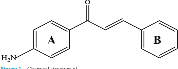
Tested compound and Animals. This was an exper-imental study with the randomized post test-only control group design. Tested compound, (e)-1-(4’-aminophenyl)-3-phenylprop-2-en-1-on, was synthesized at Department of Chemistry, Faculty of Science and Technology, Universitas Airlangga, Surabaya, Indonesia by Suwito et al.24 The chemical
structure of (e)-1-(4’-aminophenyl)-3-phenylprop-2-en-1-on is presented in Figure 1.
Female Sprague-Dawley rats aged 3-4 weeks with body weight 32-90 g were obtained from the Integrated Research and Testing Laboratory
(LPPT), Universitas Gadjah Mada, Yogyakarta.
Each animal was housed in an individual cage and maintained at 12 hours light-dark cycle
and temperature 24°C at animal house of the Department of Pharmacology and Therapy, Faculty
of Medicine, Universitas Gadjah Mada, Yogyakarta
and were fed and had accessed to water ad libitum. The protocol of the study has been approved by Medical and Health Research Ethics Committee,
Faculty of Medicine, Universitas Gadjah Mada,
Yogyakarta. Twenty-four female Sprague-Dawley rats were divided into six groups by random allo-cation. Group G1 (normal) received corn oil, group G2, G3, T1, T2, T3 were induced by 20 mg/ kgBW DMBA dissolved in corn oil (Sigma-Aldrich, St Louis) twice a week, for five weeks (modified by Meiyanto et al.).25 Body weight was measured, and
the breast was palpated to observe tumor growth every week. After mammary tumor had appeared, group G2 received vehicle (DMSO: Tween 80: saline = 1: 1: 8), and G3 received tamoxifen 6,6 mg/ kgBW. Group T1, T2, T3 received intraperitoneal (e)-1-(4’-aminophenyl)-3-phenylprop-2-en-1-on everyday with dosage of 5, 15, and 45 mg/kgBW dissolved in vehicle for 21 days, respectively. Tumor size was measured every week during 21 days. On day 22, plasma samples were collected then the rats were sacrificed by ketamine HCl with a dosage of 0.2 cc/100gBW intramuscularly.
Tumor Growth. Tumor size was measured by caliper on day 0, 7, 14, and 21. In order to prevent observation differences, only one person measured the entire tumor in the study. The formula for tumor
volume calculation V = ½ (Length x (Width)2).26
Tumor growth was assessed from tumor volume
(cm3) by repeated measures ANOVA analysis using
SPSS 17.0 Software.
Histopathological Evaluation. The mammary gland was excised and fixed in 10% PBS-Formalin for 24 hours then replaced with 70% alcohol and processed for histopathological evaluation with hematoxylin-eosin. The mammary tumor was classified as recommended by Goldschmidt et al.27
and was based on the most pronounced histological pattern observed in more than 50% of the tumor mass.28
Tumor Grade. Criteria for the histopathologi-cal grade was determined according to Histologic Malignant Grade (HMG) modified by Clemente et al.,29 based on the tubule formation, nuclear
pleo-morphism, and mitotic rate.
The point of all three features was added together to give a total of the point, called HMG. HMG I, well differentiated (3 – 5 point); HMG II, moder-ately differentiated (6 – 7 point); HMG III, poorly differentiated (8 – 9 point).29 First researcher and
anatomic pathologist assessed tumor grade (HMG) and histopathological pattern with double-blind Figure 1 Chemical structure of
experiment used image raster three software. It used inter-observer agreement with kappa statistic.
RNA Isolation and Quantitative Real-Time PCR Analysis. Total RNA including microRNA was isolated from plasma according to Qiagen miRNeasy plasma kit (Qiagen, Germany, Cat#217184). It required miRNeasy plasma spike-in control (Cat#219610). Reverse transcription was performed using the Qiagen miscript II RT Kit (Cat#218160). Real-time PCR MyGo Mini (IT-IS Life Science, UK) was carried out using Qiagen miScript SYBR Green PCR Kit (Cat#218073). The primers for PCR were a miScript universal primer and rno-miR-21 specific primer, 5’-TAGCTTATCAGACTGATGTTGA-3’ (IDT, Singapore). All samples were normalized by internal control C.elegans miR-39. The relative expression of miR-21 used livak method (2 -[Ct(miR-21)-Ct(miR-39)target]-[Ct(miR-21)-Ct(miR-39)control]) and G1 as control. Data were analyzed by one-way ANOVA using SPSS 17.0 Software.
RESULTS
Mammary tumor growth
The result showed that (e)-1-(4’-aminophenyl)-3-phenylprop-2-en-1-on inhibited tumor growth. Tumor growth was lower in rat that received (e)-1-(4’-aminophenyl)-3-phenylprop-2-en-1-on than rat that did not receive
(e)-1-(4’-aminophenyl)-3-phenylprop-2-en-1-on as shown on Figure 2.
There was a significant main effect of (e)-1-(4’-aminophenyl)-3-phenylprop-2-en-1-on in tumor growth on day 0 and 21 (p<0.05) between DMBA + vehicle group and treatment group that analyzed by repeated measured ANOVA. The significant
Table 1 The relationship between histological grading and type from each group.
Group Histopathological type
Number and percentage of HMG
I II III
G2 (DMBA + vehicle) Tubular 1 (25%) 0 0
Cribriform 0 1 (25%)
0
Comedo Ca 0 0 1 (25%)
Lipid-rich 0 0 1 (25%)
G3 (DMBA + tamoxifen) Tubular 3 (75%) 0 0
Cribriform 0 1 (25%) 0
T1 (DMBA + chalcone 5 mg) Tubulopapillary 1 (25%) 0 0
Cystic-papillary 2 (50%) 1 (25%) 0
T2 (DMBA + Chalcone 15 mg) Tubular 1 (25%) 0 0
Cribriform 1 (25%) 0 0
Tubulopapillary 1 (25%) 0 0
Solid 0 0 1 (25%)
T3 (DMBA + Chalcone 45 mg) Tubular 3 (75%) 1 (25%) 0
Note: G1 (Normal group) showed normal breast without tumor
Figure 2 Mammary tumor growth assessed from tumor volume (cm3) on day 0, 7, 14, and 21. Tumor growth was lower in the treatment group (G3, T1, T2, T3) compared to G2. Group G1: Normal, G2: DMBA + vehicle, G3: DMBA + tamoxifen, T1: DMBA + chalcone 5 mg, T2: DMBA + chalcone 15 mg, T3: DMBA + chalcone 45 mg.
effect of (e)-1-(4’-aminophenyl)-3-phenylprop-2-en-1-on to the tumor growth could be seen from partial eta squared value (0.404). Tumor volume showed significant differences on day 14 between DMBA + vehicle group and DMBA + chalcone dosage 5 mg/kgBW. On day 21, the significant different showed between DMBA + vehicle group and treatment group, i.e. G3, T1, and T2 (p<0.05).
Histopathological evaluation
All samples from rat mammary tumor that induced by DMBA were malignant. The most frequent tumor type was carcinoma-simple. The histology malignant grade (HMG) of rat mammary tissue that received (e)-1-(4’-aminophenyl)-3-phenyl-prop-2-en-1-on were better than group which did not receive
(e)-1-(4’-aminophenyl)-3-phenylprop-2-en-1-on (Figure 3). The treatment groups that
belonged to HMG I were 75%, and the DMBA + vehicle group was 25%, the other was HMG II and HMG III.
Prognosis of mammary cancer from a combi-nation of histopathological grade and type is more accurate than histopathological type alone. In this study, most of the histopathological type was carcinoma-simple (85%). It consists of tubular carcinoma (45%), cribriform (15%), cystic-papil-lary (15%), and tubulopapilcystic-papil-lary (10%). Carcinoma-simple types of mammary gland were HMG I and II, while others (comedo carcinoma, solid carcinoma, and lipid-rich carcinoma) were HMG III. Most of the tubular carcinoma (88.9 %) and tubulopapillary carcinoma (100 %) belonged to HMG I. Cribriform carcinoma type were graded I in DMBA + chalcone dosage 15 mg/KgBW, while graded II in DMBA +
vehicle group and DMBA + tamoxifen (Table 1).
MicroRNA-21 expression
(E)-1-(4’-aminophenyl)-3-phenylprop-2-en-1-on inhibited tumor growth and repaired histopatho-logical feature were parallel to the reduction level of miR-21 relative expression. The result indicated that miR-21 was overexpressed (5.79±2.24 fold) in DMBA + vehicle group compares to normal rat (p<0.05). Figure 4 showed the relative expression of miR-21, i.e. 1.41±1.13 fold (G3); 0.5±0.1 fold (T1); 2.0±1.8 fold (T2); 3.3±2.1 fold (T3). These result indicated a significantly lower in its expression in treatment groups (G3, T1, and T2) compared to that DMBA + vehicle (G2) (p<0.05). The miR-21 expression was not significant in DMBA + chalcone dosage 45 mg/kgBW (T3) compared to other groups.
DISCUSSION
New chalcone derivatives, (e)-1-(4’-aminophenyl)-3-phenylprop-2-en-1-on was synthesized and eval-uated its cytotoxic effect against T47D breast cancer cell line. This compound showed a potent cytotoxic effect with an IC50 value of 5.28 μg/mL.13 Further
study to evaluate the in vivo anticancer activity of the (e)-1-(4’-aminophenyl)-3-phenylprop-2-en-1-on against DMBA-induced mammary cancer in Sprague-Dawley rat was conducted. DMBA is used for an animal model to study estrogen receptor positive-breast cancer.30
The result showed this compound could reduce tumor growth and repair histopathology feature (Figure 2 and Table 1). It was demonstrated that the (e)-1-(4’-aminophenyl)-3-phenylprop-2-en-1-on has a potential in vivo anticancer activity. Anticancer activities of some chalcone derivatives against animal cancer models have been reported in previous studies. Loch-Neckel et al.31 reported
in vivo anticancer activity of a hybrid chalcone- quinoxaline compound in a murine xenograft model of U87-MG by a significant reduction of tumors volume. Hayashi et al.32 reported in vivo
anticancer activity of quercetin chalcone against colon-25 tumors implanted in Balb-c mice by a significant reduction in tumor volume.
In the present study, histopathological feature showed a majority of the cancer cells in the DMBA + vehicle group were at a higher grade than the treatment group. It revealed that the most common mammary cancer type was simple carci-noma (85%). Some studies showed the same result. They were reported the most frequently represented
cancer type was carcinoma-simple (87%) in rat33
and (56.8%) in canine28 mammary cancer.
This compound belongs to flavonoid
subgroups7,8 that have been reported to exert
some biological activities, including anticancer.9
It was reported that flavonoid affected epigenetic
mechanism including microRNA.14,15 Flavonoids
regulate miRNAs expression through epigenetic modification, transcription factor modulation, and
by interfering miRNA maturation process.34 In
this study, we investigated the miR-21 expression. Overexpression of miR-21 was found in DMBA + vehicle group compared to normal rat (G1 group). Yan et al.23 also found up-regulated of miR-21
expression in human breast cancer. In this current study (e)-1-(4’-aminophenyl)-3-phenylprop-2-en-1-on could decrease miR-21 expression. This result is similar to other flavonoid derivatives, 3,
6- dihydroxyflavone that revealed significantly
downregulated miR-21 expression and increased miR-34a expression on 1-methyl-1-nitrosourea- induced breast cancer.15 Epigallocatechin-3-gallate
(EGCG) in green tea was found a significant down-regulation of androgen-regulated miR-21 and up-regulation of miR-330 in tumors of mice treated with EGCG.35
MicroRNA-21 can be used as a prognostic marker for breast cancer and disease progression due to the high-level expression of miR-21 was significantly correlated with clinical stage, lymph node metastasis, shorten survival of the patients.23
A Recent report provided evidence that
knock-down of miR-21 increased the sensitivity of ER+
breast cancer cell to tamoxifen.36 In addition, Wang
et al.37 reported that miR-21 might play important
roles in the formation of chemoresistance of breast cancer cells by targeting PTEN. PTEN is a tumor suppressor gene that antagonizes the phosphatidy-linositol 3-kinase (PI3K) pathway to repress tumor
cell growth and survival.38 The PI3K/Akt pathway
aberration occurs in 70% of breast cancers
irre-spective of subtype.4 Those evidence showed the
important roles of miR-21 in cancer therapy. In this study, the reduction of miR-21 expres-sion had no difference between G2 and T3. Those results showed the effectiveness of chalcone dosage 45 mg/kgBW was lower compared to the chalcone dosage 5 and 15 mg/kgBW. Furthermore, for the next research, it will be better to use a lower dosage that was more efficient to reduce tumor growth and miR-21 expression. It is important to determine the therapeutic windows for this compound to avoid its toxicity.
CONCLUSION
In conclusion,
(e)-1-(4’-aminophenyl)-3-phenyl-prop-2-en-1-on has potential in vivo anticancer
activities on DMBA-induced mammary cancer in rats as indicated by decreasing tumor growth and repairing histopathological grade after treatment
with this compound. These activities might be attributed to its effect in decreasing plasma miR-21 expression. Thus, the (e)-1-(4’-aminophenyl)-3-phenylprop-2-en-1-on can be developed as new anticancer in the future.
ACKNOWLEDGEMENTS
Part of this study was supported by a grant from Research & Development Unit, Faculty of Medicine, Udayana University, Bali-Indonesia.
REFERENCES
1. Moradi R, Roudi MA, Kiani MM, Abdelhossein S, Rigi M, Mohammadi M, et al. Investigating the relation-ship between self-efficacy and quality of life in breast cancer patients receiving chemical therapy. Bali Med J. 2017;6(1):6-11.
2. Bianco S and Gevry N. Endocrine resistance in breast cancer. Transcription. 2012;3(4):165-170.
3. Hofstatter EW, Chung GG, Harris LN. Molecular biology of breast cancer in cancer principles & practice of oncol-ogy. Ed. Devita V.T., Lawrence T.S., Rosenberg S.A. 9thEd. Lippincott Williams & Wilkins. Philadelphia.
2011;1392-1400.
4. Martin HL, Smith L, Tomlinson DC. Multidrug-resistant breast cancer: current perspective. Breast Cancer: targets and therapy. 2014;(10)6:1-13.
5. Osborne CK and Schiff R. Mechanisms of endocrine resis-tance in breast cancer. Annu Rev Med. 2011;62:233-247. 6. Sas L, Lardon F, Vermeulen PB, Hauspy J, Dam PV,
Pauwels P. The interaction between ER and NFκB in resistance to endocrine therapy. Breast Cancer Res. 2012;14(4):212-226.
7. Ferreyra MLC, Rius SP, Casati P. Flavonoids: biosynthesis, biological function, and biotechnological applications. Front Plant Sci. 2012;3(222):1-15.
8. Orlikova B, Tasdemir D, Golais F, Dicato M, Diederich M. Dietary chalcones with chemopreventive and chemothera-peutic potential. Genes Nutr; 2011;6(2):125-147. 9. Jin C, Liang YJ, He H, Fu L. Synthesis and antitumor
activ-ity of novel chalcone derivates. Biomed Pharmacother. 2013;67(3):215-217.
10. Kamal A, Mallareddy A, Suresh P, Shaik TB, Nayak VL, Kishor C, et al. Synthesis of chalcone-amidobenzothiazole conjugates as antimitotic and apoptotic inducing agents. Bioorg Med Chem. 2012;20(11):3480-3492.
11. Kong Y, Wang K, Edler MC, Hamel E, Mooberry SL, Paige MA, et al. A boronic acid chalcone analog of com-bretastatin A-4 as a potent anti-proliferation agent. Bioorg Med Chem. 2010;18(2):971-977.
12. Mai CW, Yaeghoobi M, Rahman NA, Kang YB, Pichika MR. Chalcone with electron-withdrawing and electron-donating substituents: anticancer activity against TRAIL resistent cancer cells, structure-activity relation-ship analysis and regulation of apoptotic proteins. Eur J Med Chem. 2014;77:378-387.
13. Suwito H, Jumina, Mustofa, Matuzahroh N, Puspaningsih NNT. Anticancer and antimicrobial activ-ity of methoxy amino chalcone derivatives. Der Pharma Chemica. 2015;7(3):89-94.
14. Busch C, Burkand M, Leischner C, Lauer UM, Frank J, Venturelli S. Epigenetic activities of flavonoids in the prevention and treatment of cancer. Clin Epigenetics. 2015;7(1):64-81.
16. Davis JD and Lin SY. DNA damage and breast cancer. World J Clin Oncol. 2011;2(9):329-338.
17. Melo SA and Esteller M. Dysregulation of microR-NAs in cancer: playing with fire. FEBS Lett. 2011;585(13):2087-2099.
18. Guo L, Zhao Y, Yang S, Cai M, Wu Q, Chen F. Genome-wide screen for aberrantly expressed miRNA reveals miRNA profile signature in breast cancer. Mol Biol Rep. 2013;40(3):2175-2186.
19. Badr FM. Potential role of miR-21 in breast cancer diagno-sis and therapy. SciMed Central. 2016;3(5):1068-1075. 20. Komatsu S, Ichikawa D, Takeshita H, Morimura R,
Hirajima S, Tsujiura M, et al. Circulating miR-18a: a sensi-tive cancer screening biomarker in human cancer. In vivo. 2014;28(3):293-298.
21. Fix LN, Shah M, Efferth T, Farwell MA, Zhang B. MicroRNA expression profile of MCF-7 human breast cancer cells and the effect of green tea polyphenon-60. Cancer Genomic and Proteomics. 2010;7(5):261-278. 22. Buscaglia LEB and Li Y. Apoptosis and the target genes of
microRNA-21. Chin J of Cancer. 2011;30(6):371-380. 23. Yan L, Huang X, Shao Q, Huang M, Deng L, Wu Q, et al.
MicroRNA miR-21 overexpression in human breast can-cer is associated with advanced clinical stage, lymph-node, metastasis, and patient poor prognosis. RNA. 2008;14(11):2348-2360.
24. Suwito H, Jumina, Mustofa, Pudjiastuti, P, Fanani MZ, Kimata-Ariga Y, Katahira R, Kawakami T, Fujiwara T, Hase T, Sirat HM, Puspaningsih NNT. Design and synthe-sis of chalcone derivatives as inhibitors of the ferredoxin —
ferredoxin-NADP+ reductase interaction of Plasmodium
falciparum: pursuing new antimalarial agents. Molecules. 2014;19(12):21473-21488.
25. Meiyanto E, Tasminatun S, Susilowati S, Murwanti R, Sugiyanto. Penghambatan karsinogenesis kanker payudara tikus terinduksi DMBA pada fase post inisiasi oleh ekstrak etanolik daun Gynura procumbens (Lour), Merr. Indonesian J Pharm. 2007;18(4):169-175.
26. Jensen MM, Jorgensen JT, Binderup T, Kjaer A. Tumor volume in subcutaneous mouse xenografts measured by microCT is more accurate and reproducible than
deter-mined by 18F-FDG-microPET or external caliper. BMC
Med Imaging. 2008;8:16.
27. Goldschmidt M, Pena L, Rasotto R, Zappulli V. Classification and grading of canine mammary tumors. Vet Pathol. 2011;48(1):117-131
28. Tavasoly A, Golshahi H, Rezaie A, Farhadi M. Classification and grading of canine malignant mammary tumors. Vet Res Forum. 2013;4(1):25-30.
29. Clemente M, Perez-Alenza MD, Illera JC, Pena, L. Histological, immunohistological, and ultrastructural description of vasculogenic mimicry in canine mammary cancer. Vet Pathol. 2010;47(2):265-274.
30. Mohibi S, Mirza S, Band H, Band V. Mouse models of estrogen receptor-positive breast cancer. J Carcinog. 2011;10:35-50.
31. Loch-Neckel G, Bicca MA, Leal PC, Mascarello A, Siqueira JM, Calixto JB. In vitro and in vivo anti-glioma activity of a chalcone-quinoxaline hybrid. Eur J Med Chem. 2015;90:93-100.
32. Hayashi A, Gillen AC, Lott JR. Effect of daily oral admin-istration of quercetin chalcone and modified citrus pec-tin on implanted colon-25 tumor growth in balb-c mice. Altern Med Rev. 2000;5(6):546-552
33. Alvarado A, Lopes AC, Rocha AIF, Cabrita AMS, Ferreira R, Oliveira PA, Colaco B. Prognostic factors in MNU and DMBA-induced mammary tumors in female rats. Pathol Res Pract. 2017;213(5):441-446.
34. Srivastava SK, Arora Sumit, Averett C, Singh S, Singh AP. Modulation of microRNA by phytochemicals in cancer: underlying mechanism and translational Significance. BioMed Res Int. 2015;2015:1-9.
35. Siddiqui IA, Asim M, Hafeez BB, Adhami VM, Tarapore RS, Mukhtar H. Green tea polyphenol EGCG blunts androgen receptor function in prostate cancer. FASEB J. 2011;25(4):1198-1207.
36. Yu X, Li R, Shi W, Jiang T, Wang Y, Li C, Qu X. Silencing of MicroRNA-21 confers the sensitivity to tamoxifen and fulvestrant by enhancing autophagic cell death through inhibition of the PI3K-AKT-mTOR pathway in breast can-cer cells. Biomed Pharmacother. 2016;77:37-44.
37. Wang ZX, Lu BB, Wang H, Cheng ZX, Yin YM. MicroRNA-21 modulates chemosensitivity of breast can-cer cells to doxorubicin by targeting PTEN. Arch Med Res. 2011;42(4):281-290.
38. Dillon LM. and Miller TW. Therapeutic Targeting of Cancers with Loss of PTEN Function. Curr Drug Targets. 2014;15(1):65-79.