R E S E A R C H
Open Access
Expression dynamics of integrin
α
2,
α
3, and
α
V upon osteogenic differentiation of
human mesenchymal stem cells
Hyun Min Lee
1, Se-Ri Seo
1, Jeeseung Kim
1, Min Kyu Kim
1, Hyosun Seo
1, Kyoung Soo Kim
2, Young-Joo Jang
3*and
Chun Jeih Ryu
1*Abstract
Background:The differentiation of human mesenchymal stem cells (hMSCs) into osteoblasts (OBs) is a prerequisite for bone formation. However, little is known about the definitive surface markers for OBs during osteogenesis. Methods:To study the surface markers on OBs, we generated and used monoclonal antibodies (MAbs) against surface molecules on transforming growth factor-β1 (TGF-β1)-treated cancer cells. The generated MAbs were
further selected toward expression changes on hMSCs cultured with TGF-β1/bone morphogenetic protein-2
(BMP-2) or osteogenic differentiation medium (ODM) by flow cytometry. Immunoprecipitation and mass spectrometry were performed to identify target antigens of selected MAbs. Expression changes of the target antigens were evaluated in hMSCs, human periodontal ligament cells (hPDLCs), and human dental pulp cells (hDPCs) during osteogenic and adipogenic differentiation by quantitative polymerase chain reaction (qPCR) and flow cytometry. hMSCs were also sorted by the MAbs using magnetic-activated cell sorting system, and osteogenic potential of sorted cells was evaluated via Alizarin Red S (ARS) staining and qPCR.
Results:The binding reactivity of MR14-E5, one of the MAbs, was downregulated in hMSCs with ODM while the binding reactivity of ER7-A7, ER7-A8, and MR1-B1 MAbs was upregulated. Mass spectrometry and overexpression identified that MR14-E5, ER7-A7/ER7-A8, and MR1-B1 recognized integrinα2,α3, andαV, respectively. Upon osteogenic differentiation of hMSCs, the expression of integrinα2 was drastically downregulated, but the expression of integrinα3 andαV was upregulated in accordance with upregulation of osteogenic markers.
Expression of integrinα3 andαV was also upregulated in hPDLCs and hDPCs during osteogenic differentiation. Cell
sorting showed that integrinαV-high hMSCs have a greater osteogenic potential than integrinαV-low hMSCs upon
the osteogenic differentiation of hMSCs. Cell sorting further revealed that the surface expression of integrinαV is more dramatically induced even in integrinαV-low hMSCs.
(Continued on next page)
© The Author(s). 2020Open AccessThis article is licensed under a Creative Commons Attribution 4.0 International License, which permits use, sharing, adaptation, distribution and reproduction in any medium or format, as long as you give appropriate credit to the original author(s) and the source, provide a link to the Creative Commons licence, and indicate if changes were made. The images or other third party material in this article are included in the article's Creative Commons licence, unless indicated otherwise in a credit line to the material. If material is not included in the article's Creative Commons licence and your intended use is not permitted by statutory regulation or exceeds the permitted use, you will need to obtain permission directly from the copyright holder. To view a copy of this licence, visithttp://creativecommons.org/licenses/by/4.0/. The Creative Commons Public Domain Dedication waiver (http://creativecommons.org/publicdomain/zero/1.0/) applies to the data made available in this article, unless otherwise stated in a credit line to the data.
* Correspondence:[email protected];[email protected]
3Department of Nanobiomedical Science, BK21 PLUS NBM Global Research Center for Regenerative Medicine, College of Dentistry, Dankook University, Cheonan 330-714, Korea
1Department of Integrative Bioscience and Biotechnology, Institute of Anticancer Medicine Development, Sejong University, 209 Neungdong-ro, Gwangjin-gu, Seoul 05006, Korea
(Continued from previous page)
Conclusion:These findings suggest that integrinα3 andαV induction is a good indicator of OB differentiation. These findings also shed insight into the expression dynamics of integrins upon osteogenic differentiation of hMSCs and provide the reason why different integrin ligands are required for OB differentiation of hMSCs.
Keywords:Human mesenchymal stem cells, Osteoblasts, Monoclonal antibodies, Osteogenic differentiation, IntegrinαV, Integrinα3, Integrinα2
Background
Human mesenchymal stem cells (hMSCs) can differenti-ate into multilineage cells of mesenchymal tissues, in-cluding osteoblasts (OBs), adipocytes, myocytes, and chondrocytes [1]. Although hMSCs can differentiate into a variety of cell types, the differentiation of hMSCs into osteocytes and adipocytes has been extensively studied for successful bone formation and regeneration [2–4]. Osteocytes compose approximately 95% of all bone cells in adult bones and are differentiated from hMSCs through OBs [5]. Although many researchers have tried to differentiate hMSCs into OBs and osteocytes for bone formation and regeneration, they encountered numerous problems because hMSCs are heterogeneous populations with variable functions, and their properties are highly variable depending on the tissue source, subpopulation, and culture condition [6, 7]. Furthermore, there are no definitive markers to identify and isolate OBs from the mixed populations.
Transforming growth factor-β1 (TGF-β1), bone morpho-genetic protein 2 (BMP-2), and runt-related transcription factor 2 (Runx2) are believed to be key regulators in OB development during osteogenesis. TGF-β1 is the predom-inant TGF-βisoform expressed in bone and has been ex-tensively studied during bone formation and remodeling [8, 9]. TGF-β1 signaling is known to expand MSCs and stimulate early differentiation of OBs, whereas inhibiting late differentiation of OBs into osteocytes [10]. BMP signal-ing affects OB commitment, and treatment of MSCs with
BMP-2 promotes osteogenesis [2]. Treatment of MSCs
with both TGF-β1 and BMP-2 accelerates bone formation through the proliferation of OBs and keeps immature OBs. Runx2 is a key transcription factor for early OB differenti-ation and bone formdifferenti-ation and is controlled by TGF-β1 and BMP-2 during osteogenesis [2, 11–13]. Runx2 is also up-regulated in TGF-β1-treated cancer cells [14].
Epithelial-mesenchymal transition (EMT) is regarded to play important roles in cancer metastasis and recurrence [15,16] and is induced by cytokines, such as TGF-β1 [17]. BMP-2, BMP-4, and BMP-7 also promote EMT and inva-siveness in pancreatic cancer cells [17–19]. Runx2 also takes part in TGF-β-induced EMT [14]. Cancer cells under-going EMT show enhanced ability of invasion and metasta-sis and also reveal similarity to MSC-like properties [17]. To search for novel cell surface markers on
EMT-phenotypic-circulating tumor cells, in the previous study, we generated a panel of monoclonal antibodies (MAbs) against surface molecules on TGF-β1-treated A549 human non-small cell lung carcinoma cells (NSCLC) because TGF-β1-treated A549 cells represent EMT-phenotypic cells [20–22]. Although EMT plays major roles in tumor inva-sion and metastasis, it also occurs during the process of odontogenesis. EMT has an important role in differenti-ation of enamel-forming ameloblast [23]. Studies have shown that epithelial cells differentiate into hard
tis-sues such as cementum in the presence of TGF-β-1
during EMT [23–26]. A recent study also suggested
that EMT drives odontogenic epithelial stem cells to develop supernumerary teeth through interaction with various MSCs [27].
In this study, therefore, we postulated that the sur-face molecules expressed on EMT-phenotypic cells may be source molecules for finding novel surface
markers on TGF-β1-regulated OBs. To this end, we
first generated a panel of MAbs against TGF-β
1-treated A549 cells by using the decoy immunization strategy [28]. To use the MAbs for the identification and isolation of novel surface molecules on OBs, the MAbs showing hMSC-binding reactivity were then se-lected for their binding capacity for differentiated
hMSCs treated with TGF-β1/BMP-2 or osteogenic
dif-ferentiation medium (ODM). To examine whether the cell surface expression of the MAb antigens is con-trolled by Runx2 expression, the cell surface expres-sion of the MAb antigens was also examined in Runx2
knockdown osteosarcoma cells. Integrin α2, α3, and
αV recognized with the MAbs were identified by
im-munoprecipitation followed by mass spectrometry. Upon osteogenic differentiation of hMSCs, integrinα2 expression was rapidly downregulated, while integrin α3 and αV expression was gradually upregulated in ac-cordance with upregulation of osteogenic markers.
In-tegrin α3 and αV expression was also upregulated in
human periodontal ligament cell (hPDLCs) and human dental pulp cells (hDPCs). To further elucidate the role of integrinαV during osteogenic differentiation of hMSCs,
integrin αV-high and αV-low hMSCs were sorted and
Methods
Cell culture and differentiation
Human non-small cell lung carcinoma cell line (NSCLC) A549 and human osteosarcoma cell lines (U2OS and SAOS-2) were purchased from the Korean Cell Line Bank (KCLB, Seoul, Korea) and were cultured according to the protocol provided by the supplier. Human fetal osteoblast cell line hFOB1.19 was purchased from ATCC (CRL-11372, Manassas, VA) and was cultured according to the protocol provided by the supplier. Human bone marrow-derived mesenchymal stem cells (hMSCs) were kindly provided by Prof. Sung-Soo Kim (Ajou University
School of Medicine, Suwon, Korea) [29]. hMSCs were
cultured in DMEM (Welgene, Daegu, Korea) supple-mented with 20 ng/ml basic fibroblast growth factor (bFGF) (R&D system, Minneapolis, MN), 10% fetal bo-vine serum (FBS) (Welgene), and antibiotic-antimycotic solution (Welgene). The medium was changed every 2 days, and early passages (passages 4–6) were used in this study. Human peripheral blood mononuclear cells (PBMCs) were isolated by the Ficoll-Paque Plus method (GE Healthcare, Seoul, Korea). For the primary cultures of hPDLCs and hDPCs, adult third molars were col-lected from patients who were 19 to 23 years old under guidelines approved by the Institutional Review Board of the Dankook Dental Hospital and Dankook University (DKU NON2019-004), and the informed consent for all experiments using extracted teeth was obtained from all the participants. Human periodontal ligament and pulp tissues were separated from the surface of the tooth root and pulp part inside of the tooth after removing tooth crown. Tissues were enzymatically digested with 3 mg/ ml collagenase type I (Merck Millipore, Seoul, Korea) and 4 mg/ml dispase (Sigma-Aldrich, Gyeonggi, Korea) at 37 °C for 1 h. Cell suspensions were incubated in α -MEM (GE Healthcare, Seoul, Korea) containing 20% FBS (GE Healthcare) and 1% antibiotics (Lonza, Seoul, Korea) at 37 °C in humidified atmosphere supplemented with 5% CO2[30,31]. To induce EMT in A549 cells, the cells were plated at 1.8 × 10⁴ cells/cm2 and incubated for 24 h. The cells were then treated with 5 ng/ml of TGF-β1 (Peprotech, Rocky Hill, NJ) for 4 days and the medium was changed every 2 days.
Generation of a panel of murine MAbs
To generate hybridomas producing antibodies specific to TGF-β1-treated A549 cells, one million TGF-β1-treated A549 cells were injected into the left hind footpads of fe-male BALB/c mice (DBL, Chungbuk, Korea) 3 days after the decoy cells (A549 cells) were injected into the right hind footpad of the same mice. The following procedures were carried out as described previously [28]. Briefly, lymphocyte fractions isolated from the left popliteal lymph nodes were fused to FO myeloma cells (ATCC) by
polyethylene glycol 1500 (Roche, Seoul, Korea). The fused cells were maintained in DMEM (Corning, Seoul, Korea) supplemented with 20% FBS (Welgene), HAT supplement (Sigma-Aldrich), and antibiotic-antimycotic solution (Wel-gene). Hybridoma clones specific to TGF-β1-treated A549 cells were selected by flow cytometry. Isotype of antibodies secreted from hybridomas was analyzed with the Mouse Immunoglobulin Isotyping Kit (BD Biosciences, Seoul, Korea) according to the manufacturer’s instructions. Antibody purification was performed by using Protein G agarose column chromatography (Merck Millipore). All animal experiments were approved by the Institu-tional Animal Care and Use Committee at Sejong Uni-versity (SJ-21051104).
Flow cytometry
To screen MAbs specific to OBs, cells (A549, TGF-β 1-treated A549, SAOS-2, U2OS, hMSCs, and hFOB) were dissociated by trypsin-EDTA (Welgene) and filtered
through a 40-μm strainer and washed by
phosphate-buffered saline (PBS, pH 7.4). Cells were resuspended in ice-cold PBA (1% bovine serum albumin and 0.01%
NaN3 in PBS) and incubated with primary antibodies,
followed by the incubation with fluorescein isothio-cyanate (FITC)-conjugated anti-mouse IgG (Santa Cruz Biotechnology, Santa Cruz, CA), Alexa488-conjugated anti-mouse IgG (Thermo Fisher Scientific, Seoul, Korea), phycoerythrin (PE)-conjugated anti-mouse IgG (Thermo Fisher Scientific), and/or DyLight 650-conjugated anti-mouse IgG (Thermo Fisher Scientific). To analyze only live cells, propidium iodide (PI) (Sigma-Aldrich)-negative cells were gated and analyzed by FACSCalibur (BD biosciences) and Cell Quest Software (BD biosciences). For multicolor flow cytometry, cells were incubated with appropriate pri-mary antibodies for 30 min and incubated with Dylight 649-(Vector Laboratories, Burlingame, CA) or Alexa 488-conjugated anti-mouse IgG (Thermo Fisher Scientific) after osteogenic or adipogenic differentiation of hMSCs. The cells were than incubated with PE-conjugated mouse monoclonal human CD73, CD90, or CD146 anti-bodies, rabbit polyclonal anti-human CD164, or rabbit polyclonal anti-integrinα2,α3, orαV antibodies.
Cell surface biotinylation, immunoprecipitation, and Western blotting
A549 cells were incubated with 0.5 mg/ml of EZ-Link Sulfo NHS-LC-biotin (Thermo Fisher Scientific) for 30 min at 4 °C on a rocker. After three washes with ice-cold PBS (pH 8.0), the cells were lysed in ice-cold lysis buffer (25 mM Tris-HCl, pH 7.5, 250 mM NaCl, 5 mM EDTA,
1% Nonidet P-40, 2μg/ml aprotinin, 100μg/ml PMSF,
5μg/ml leupeptin, 1 mM NaF, and 1 mM NaVO3) for
nonspecific proteins bound to Protein G agarose (Merck Millipore), biotin-labeled or non-labeled A549 lysates were incubated with 20μl of Protein G agarose beads for 2 h at 4 °C (Preclearing), and the bead-bound proteins were used for negative control of immunoprecipitation (IP). Pre-cleared lysates were then incubated with MAbs, rabbit polyclonal anti-integrin α2 (Mybiosource, Seoul, Korea), anti-integrin α3 (Sino Biological, Wayne, PA),
anti-integrin αV (Sino Biological), anti-His (Thermo
Fisher Scientific), and/or anti-FLAG antibodies (MBL, Woburn, MA) at 4 °C overnight and further incubated with 20μl of Protein G agarose at 4 °C for 3 h. The beads were washed by lysis buffer and bound proteins were eluted by acidic solution (0.1 M Glycine-HCl, pH 2.8) for 10 min at RT and neutralized by alkaline solution (1.0 M Tris-HCl, pH 9.0). Eluted proteins were dena-tured by boiling in sample buffer for 10 min at 100 °C before further analysis. For Western blotting, prepared proteins were denatured by sodium dodecyl sulfate (SDS)-polyacrylamide gel electrophoresis (SDS-PAGE) on 10–12% polyacrylamide gel and transferred to nitro-cellulose membranes (Pall Corporation, New York, NY) for 2 h. The membranes were blocked by 5% skim milk for 30 min at RT and washed by 0.1% Tris-buffered saline with Tween-20 (TBST; 0.1% tween-20) for three times. Then, the membranes were incubated with streptavidin-conjugated horse radish peroxidase (SA-HRP) (1:5000, GE Healthcare) or HRP-conjugated anti-mouse (Merck Millipore) or rabbit IgG (Abcam, Hanam, Korea) for 1 h at RT and analyzed with West-Queen™Western blot detection system (iNtRON Biotechnology, Seongnam, Korea).
DNA and siRNA transfection
HEK293T cells (7 × 10⁵cells) were transfected with His- or FLAG-tagged ITGA2 (Sino Biological), ITGA3 (Sino Bio-logical), and ITGAV (Sino Biological) expression vectors using Lipofectamine™ 2000 (Thermo Fisher Scientific) ac-cording to the supplier’s protocol. Protein was transiently expressed for 24 h, and the cell pellets were lysed in RIPA buffer (50 mM Tris-HCl, pH 7.4, 150 mM NaCl, 1% NP-40, 0.5% sodium deoxycholic acid, 0.1% SDS). Cell lysates were subjected to IP and Western blot analysis as described above. siRNA oligonucleotides targeting RUNX2 (Santa Cruz Biotechnology) or control siRNA (Bioneer, Daejeon, Korea) were used in this study. U2OS cells (2.6 × 10⁴cells/
cm2) were transfected with 50 nM control or RUNX2
siRNA using Lipofectamine®RNAiMAX Reagent (Thermo Fisher Scientific) according to the supplier’s protocol. After 24 h, the cells were transfected again (double knockdown) and then harvested for analysis after 48 h.
Antibody biotinylation and cell sorting
Antibody was biotinylated by using DSB-X biotin label-ing kit (Thermo Fisher Scientific) accordlabel-ing to the
manufacturer’s procedure. A total of 5 million hMSC
cells were incubated with 20μg of biotinylated
MR1-B1antibody for 30 min at RT with rolling and tilting. Cell sorting was carried out by using Dynabeads FlowComp Flexi kit (Thermo Fisher Scientific) according to the
man-ufacturer’s protocol. Bead-unbound and bead-bound
hMSCs were isolated and further analyzed with Alexa488-conjugated anti-mouse IgG (Thermo Fisher Scientific) and FACSCalibur.
Osteogenic and adipogenic differentiation
To induce OB differentiation, hMSCs were also incu-bated for 14 days in 1 ng/ml of TGF-β1 and 10 ng/ml of BMP-2. To induce osteogenic and osteoblastic differenti-ation of hMSCs, hMSCs (500 cells/cm2) were incubated for 0, 4, 7, 12, 14, or 21 days in osteogenic differentiation
medium (ODM) (60μM L-ascorbic acid (Sigma-Aldrich),
10 mM β-glycerophosphate (Sigma-Aldrich), and 100
nM dexamethasone (Sigma-Aldrich) in DMEM contain-ing 10% FBS and 1% antibiotics-antimycotics). To pro-mote osteogenic differentiation of hMSCs, hMSCs were
also incubated with ODM containing 1μM SB431542
(MedChemExpress, Monmouth Junction, NJ), a specific inhibitor of TGF-βtype I receptor, after 7 days of ODM in-cubation. For adipogenic differentiation of hMSCs, hMSCs (500 cells/cm2) were incubated for 0, 4, 7, 14, or 21 days in
adipogenic differentiation medium (ADM) (60μM
indo-methacin (Sigma-Aldrich), 500 mM 3-isobutyl-1-methylxan-thine (Sigma-Aldrich), 10μg/ml insulin (Sigma-Aldrich),
and 1μM dexamethasone (Sigma-Aldrich) in DMEM
containing 10% FBS and 1% antibiotics-antimycotics). The differentiated cells were stained with Alizarin Red S (Sigma-Aldrich) or Oil Red O (Sigma-(Sigma-Aldrich). For osteoblastic differentiation of hPDLCs, cells were treated with 100 ng/ml BMP-2 (Prospec, Ness-Ziona, Israel) for 7 days. For odonto-blastic differentiation of hDPCs, cells were treated with 100 ng/ml BMP-2 (Prospec) and 10 ng/ml BMP-4 (Prospec) for 7 days [31]. To induce osteoblastic and odontoblastic matur-ation, cells were cultured inα-MEM supplemented with 5% FBS, 100 nM dexamethasone, 100μM ascorbic acid, and 5 mM β-glycerophosphate for 7 days, followed to treatment with cytokines for 7 days. For flow cytometric analysis, cells were detached by TrypLE Express (Thermo Fisher Scien-tific) and analyzed in FACSCalibur.
Reverse transcription-polymerase chain reaction (RT-PCR)
U2OS cells were transfected with control or RUNX2 siR-NAs as described above. The cells were collected by Trypsin-EDTA (Welgene), and total RNA was extracted by RNAiso Plus (TaKaRa, Otsu, Japan) according to the manufacturer’s instructions. To perform RT-PCR, cDNAs
were synthesized by Prime Script™ RT Master Mix
denaturation at 95 °C for 1 min, annealing at 56–67 °C for 45 s, and extension at 72 °C for 45 s in 30 cycles. Each gene was analyzed in triplicate. Primer sequences used are listed in Additional file1(Table S1).
Quantitative PCR
Total RNAs were extracted from hMSCs and differenti-ated hMSCs using the RNAiso Plus (TaKaRa) according to the protocol provided by the manufacturer. cDNAs were generated from total RNAs by RT in a reaction containing 500 ng total RNAs and Prime Script RT Master Mix (TaKaRa). The RT reaction was performed at 37 °C for 15 min followed by heating at 94 °C for 5 s.
Quantitative real-time PCR was performed using
Applied StepOne™ Real-Time PCR System with SYBR
Green reagents (Thermo Fisher Scientific). The standard reaction contained 12.5μl 2 × PCR buffer (PowerUP™
-SYBR™ Green Master Mix, Thermo Fisher Scientific),
2μl cDNA template, and 10 pM of forward and reverse
primers in a total volume of 25μl. The PCR was carried out for 30 cycles consisting of 1 min at 95 °C, 45 s at 45– 56 °C, and 1 min at 72 °C. The target gene-specific primers for human transcripts encoding ITGA2, ITGA3, ITGAV, RUNX2, ALP, COL1A1, OSX2, C/EBPA, and GAPDH are listed in Additional file 1 (Table S1). Each sample was analyzed in triplicate for each target gene. To analyze gene expression in hPDLCs and hDPCs, total RNAs were extracted from hPDLCs or hDPCs using
Easy-spin™ Total RNA Extraction kit (iNtRON
Biotech-nology). cDNAs were synthesized from total RNAs by
using the ReverTra Ace™ qPCR RT kit (Toyobo, Osaka,
Japan), and the qRT-PCR was performed using StepOn™
system (Thermo Fisher Scientific) with iTaq™ Universal
SYBR™Green Supermix (Bio-Rad, Hercules, CA) system.
During qRT-PCR, a dissociation curve was constructed in the range of 65 to 95 °C, and the cycling parameters were followed as follows: 1 cycle for 30 s at 95 °C, 40 cy-cles for 15 s at 95 °C and 1 min at 55–60 °C for amplifi-cation. The threshold cycle was obtained, and the relative comparison of each target gene was analyzed. The primer sequences used are listed in Additional file1
(Table S1).
Statistical analysis
The paired samples t-test was a statistical procedure used to compare the difference between two
popula-tions, and a p value of less than 0.05 was considered
statistically significant.
Results
Generation of a panel of MAbs against TGF-β1-treated A549 cells
In this study, we postulated that surface molecules expressed on TGF-β1-treated A549 cells may be source
molecules for finding novel surface markers on TGF-β 1-regulated OB cells. To this end, we first generated a panel of MAbs against TGF-β1-treated A549 cells by using the modified decoy immunization strategy [22, 28]. TGF-β 1-treated A549 cells showed fibroblast-like morphologies and enhanced expression of the mesenchymal markers, includ-ing ZEB1, vimentin, slug, and hnRNPA2/B1, concomitant with downregulation of the epithelial marker E-cadherin (Additional file1: Figure S1a, 1b). Flow cytometric analysis also showed that the cell surface expression of epithelial markers E-cadherin and EpCAM were downregulated in the TGF-β1-treated A549 cells, while the cell surface ex-pression of mesenchymal marker N-cadherin was slightly upregulated (Additional file 1: Figure S1c). The results indicate that TGF-β1 induces A549 epithelial cells to undergo the EMT process, consistent with previous studies [20, 32]. To generate MAbs specific to TGF-β1-treated A549 cells, A549 cells were used as decoy immunogen by injection into right foot pads of BALB/c mice. TGF-β 1-treated A549 cells were then used for target immunogen by injection into left foot pads of the same BALB/c mice 3 days after the first injection. Hybridomas were generated by fusion of FO myeloma cells and lymphocytes isolated from left popliteal regions in the immunized mice. From 263 hybridomas, 13 MAbs were selected and further ana-lyzed because they showed increased binding affinity for
TGF-β1-treated A549 cells as compared with parental
A549 cells (Additional file1: Figure S2 and Table S2). The 13 MAbs did not bind to PBMCs and were purified prior to further analysis.
Binding reactivity of EMT-specific MAbs to mesenchymal stem/progenitor cells with osteogenic potential
Next, we examined the binding reactivity of the 13 EMT-specific MAbs to mesenchymal stem/progenitor cells, including hMSCs and hFOB, because they have strong osteogenic potential [1, 33, 34]. Two human osteosarcoma cell lines (U2OS and SAOS-2) were also included in the analysis because they have been fre-quently used as osteoblastic cancer cell lines [35, 36]. ER4-D2, ER7-C3, MR11-B3, and MR17-B8 did not bind to both hMSCs and hFOB while MR1-G2 bound to only hMSCs (Additional file 1: Figure S3). The rest of them (9 MAbs) were able to bind to hMSCs, hFOB, and one of the osteosarcoma cell lines (Additional file 1: Figure S3 and Table S3).
Expression changes of target antigens of selected MAbs upon osteogenic differentiation of hMSCs
Next, we examined the cell surface expression of target antigens of selected MAbs upon osteogenic differenti-ation of hMSCs. To promote the differentidifferenti-ation of
hMSCs into OBs, hMSCs were treated with TGF-β1 and
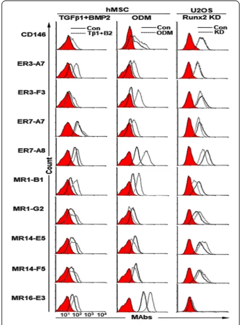
subjected to flow cytometric analysis. CD146, a marker of self-renewing osteoprogenitors in human bone mar-row [37], was increased during differentiation, and all target antigens of the selected MAbs were also increased (Fig. 1, the first column). hMSCs were also cultured for
12 days in ODM [38]. Calcium deposition was readily
detected by Alizarin Red S staining (Additional file 1: Figure S4A). After 12 days of osteogenic differentiation, CD146 was slightly increased in the ODM-differentiated hMSCs, and target antigens of 6 MAbs (A7, ER3-F3, ER7-A7, ER7-A8, MR1-B1 and MR16-E3) were also increased, while those of 3 MAbs (MR1-G2, MR14-E5 and MR14-F5) were decreased (Fig.1, Table1). Runx2 is a key transcription factor positively associated with OB phenotype and inhibits MSCs from adipogenic or
chon-drogenic differentiation [39]. Runx2 also keeps the
immature state of OB preventing the maturation of OB into osteocyte [39]. Cell surface expression of target an-tigens of 4 MAbs (ER3-F3, ER7-A7, ER7-A8, and MR1-B1) was downregulated in Runx2 knockdown U2OS cells
while cell surface expression of target antigens of 3 MAbs (MR1-G2, MR14-E5, and MR14-F5) was upregu-lated (Fig. 1, Table 1, Additional file 1: Figure S4B). Taken together, the results suggest that target antigens of 4 MAbs (ER3-F3, ER7-A7, ER7-A8 and MR1-B1) are upregulated upon osteogenic differentiation of hMSCs, while target antigens of 3 MAbs (MR1-G2, MR14-E5, and MR14-F5) are downregulated.
Identification of target antigens of MR14-E5, A7, ER7-A8, and MR1-B1
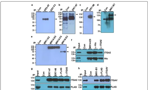
To identify the target antigens recognized by selected MAbs, biotin-labeled A549 cell lysates were cipitated with the selected MAbs, and the immunopre-cipitates were analyzed by streptavidin-HRP in Western blot analysis. Seven MAbs were able to immunoprecipi-tate surface molecules with various molecular weights (Fig. 2a–e). Mass spectrometry revealed that MR14-E5
and MR1-B1 recognized integrin α2 (ITGA2) and
integ-rin αV (ITGAV), respectively, while both ER7-A7 and
ER7-A8 recognized integrinα3 (ITGA3) (Additional file1: Figure S5, S6, S7). To demonstrate that MR14-E5
recog-nizes integrin α2, the His-tagged ITGA2 expression
plasmid was transfected into HEK293T cells, and the cell lysates were subjected to immunoprecipitation with MR14-E5, polyclonal anti-integrin α2 or anti-His antibodies, and the immunoprecipitates were detected with anti-integrinα2 or anti-His antibodies. All immunoprecipitates were de-tected with anti-integrinα2 or anti-His antibodies (Fig.2f), indicating that the MR14-E5 antigen is integrinα2 indeed. To demonstrate that both ER7-A7 and ER7-A8 recognize integrin α3, the FLAG-tagged ITGA3 expression plasmid was transfected into HEK293T cells, and the cell lysates were subjected to immunoprecipitation with A7,
ER7-Fig. 1Expression changes of target antigens of selected MAbs upon osteogenic differentiation of hMSCs. hMSCs were treated for 14 days with TGF-β1 and BMP-2 and incubated for 12 days with ODM. Expression changes of target antigens of the selected MAbs were analyzed by flow cytometry after detachment of the cells. Runx2 knockdown U2OS cells were also analyzed by flow cytometry with the same MAbs. Red-filled histograms represent the isotype controls
Table 1Expression changes of target antigens of selected MAbs in hMSCs stimulated with osteogenic inducers and in Runx2 knockdown U2OS cells
Clone Isotype hMSCs Runx2
knockdown U2OS TGF-β1 + BMP-2 ODM
CD146 – ↑ ↑ ─
ER3-A7 IgG1,κ ↑↑ ↑↑ ─
ER3-F3 IgG2a,κ ↑ ↑↑ ↓
ER7-A7 IgG2a,κ ↑ ↑↑ ↓
ER7-A8 IgG1,κ ↑ ↑↑↑ ↓↓
MR1-B1 IgG2a,κ ↑↑ ↑↑↑ ↓↓
MR1-G2 IgG1,κ ↑ ↓↓ ↑
MR14-E5 IgG2a,κ ↑ ↓↓ ↑
MR14-F5 IgG2a,κ ↑ ↓ ↑
MR16-E3 IgG2a,κ ↑ ↑↑↑ ─
A8, polyclonal anti-integrinα3, and anti-FLAG antibodies, and immunoprecipitates were detected with anti-integrin α3 or anti-FLAG antibodies. All immunoprecipitates were detected with anti-integrin α3 or anti-FLAG antibodies (Fig.2g), indicating that both ER7-A7 and ER7-A8 antigens are integrin α3 indeed. To demonstrate that MR1-B1 rec-ognizes integrin αV, the FLAG-tagged ITGAV expression plasmid was transfected into HEK293T cells, and the cell lysates were subjected to immunoprecipitation with MR1-B1, polyclonal anti-integrinαV, and anti-FLAG antibodies, and the immunoprecipitates were detected with anti-integrin αV or anti-FLAG antibodies. All immunoprecipi-tates were detected with anti-integrin αV or anti-FLAG antibodies (Fig.2h), indicating that the MR1-B1 antigen is integrinαV indeed. Shown in Fig.1, the results also suggest that integrinα3 recognized with ER7-A7 and ER7-A8 and
integrin αV recognized with MR1-B1 are upregulated
during osteogenic differentiation of hMSCs, while integrin
α2 recognized with MR14-E4 is downregulated during
osteogenic differentiation of hMSCs.
Integrinα2 is decreased during osteogenic differentiation of hMSCs while integrinα3 andαV are increased
Next, we examined expression pattern of integrinα2,α3,
and αV during differentiation of hMSCs. hMSCs were
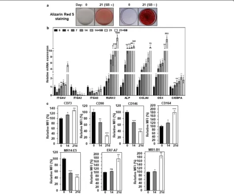
cultured for 21 days under conditions that promote osteogenic differentiation. SB431542, a specific inhibitor
of TGF-β receptor kinase, was added to ODM after 7
days of osteogenic differentiation because it enhances osteogenic differentiation of some somatic stem cells [40, 41]. Although osteogenic differentiation of hMSCs was observed in the absence of SB431542, osteogenic differentiation of hMSCs was obvious in the presence of SB-431542 at 21 days of osteogenic differentiation by
increased calcium deposition (Fig. 3a) and increased ex-pression of Runx2 (1008-fold), alkaline phosphatase (ALP, 360-fold), collagen type I (COL1A1, 15-fold), and osterix (OCX, 893-fold) (Fig. 3b). However, the adipo-genic marker C/EBPA was slightly increased (1.8-fold) at 21 days of osteogenic differentiation (Fig.3b). The obvi-ous increase of the early OB markers (Runx2, ALP, OSX) suggests that the differentiated hMSCs are mainly at the stage of early OBs [42]. Under the circumstances,
the expression level of integrin α2 mRNA (ITGA2) was
gradually decreased to 4.6-fold during osteogenic differ-entiation of hMSCs (Fig.3b). The expression level of
in-tegrin α3 mRNA (ITGA3) was initially decreased until
14 days of osteogenic differentiation of hMSCs, but it was slightly upregulated up to approximately 0.7-fold at 21 days of osteogenic differentiation (Fig. 3b).
Interest-ingly, the expression level of integrin αV mRNA
(ITGAV) was slightly increased up to approximately
1.5-Fig. 3Expression changes of integrinαV,α2,α3, and osteogenic markers during differentiation of hMSCs.aAlizarin Red S staining of OBs in differentiated hMSCs. hMSCs were incubated for 21 days with ODM (SB×). To inhibit TGF-β1 signaling that originated from the culture medium, SB431542 was also included in ODM (SB○) after 7 days of osteogenic differentiation. Calcium deposition was visualized as red color after the cells were stained with Alizarin Red S.bExpression changes of integrins and osteogenic markers during osteogenic differentiation of hMSCs. Integrins (MR14-E5, ER7-A7, MR1-B1), osteogenic markers (Runx2, ALP, COL1A1, OSX), and adipogenic marker (C/EBPA) were analyzed in differentiated hMSCs by quantitative PCR. Values are depicted as a mean fold change in gene expression (2−ΔΔCT) of differentiated hMSCs at the indicated days compared to hMSCs at day 0.△,p< 0.05;□,p< 0.01;○,p< 0.005; ns, not significant.cExpression changes of integrins and hMSC/OB surface markers during osteogenic differentiation of hMSCs. hMSCs were incubated for 21 days with ODM, and SB431542 was added to ODM after 7 days of osteogenic differentiation. Integrins (α2,α3,αV) and hMSC/OB surface markers (CD73, CD90, CD146, and CD164) were analyzed in
fold at 14 days of osteogenic differentiation of hMSCs and then increased up to approximately 5.3-fold at 21 days of osteogenic differentiation in the presence of SB431542 (Fig.3b). The results suggest that the increased expression of integrinαV is needed for the maturation of OBs.
Next, we examined the cell surface expression of
integrin α2, α3 and αV by flow cytometry during
osteogenic differentiation of hMSCs. CD73, CD90, CD146, and CD164 expression were also examined be-cause they are also known as hMSC and/or
osteopro-genitor markers [37, 43–46]. CD90 and CD146 were
drastically downregulated during osteogenic differenti-ation of hMSCs (Fig. 3c and Additional file 1: Figure S8). Interestingly, CD73 and CD164 were upregulated
during osteogenic differentiation of hMSCs (Fig. 3c
and Additional file 1: Figure S8). Under the
circum-stances, the cell surface expression of integrinα2 (rec-ognized by MR14-E5) was drastically downregulated
during osteogenic differentiation of hMSCs (Fig. 3c
and Additional file 1: Figure S8). However, the cell
surface expression of integrin α3 (recognized by ER7-A7) was slightly (106%) upregulated at 14 days of osteogenic differentiation and then further upregulated up to approximately 176% at 21 days of osteogenic dif-ferentiation (Fig. 3c and Additional file 1: Figure S8). The cell surface expression of integrinαV (recognized by MR1-B1) was also upregulated up to approximately 187% at 21 days of osteogenic differentiation (Fig. 3c and Additional file 1: Figure S8), which is also
consist-ent with increased ITGAV mRNA expression (Fig.3b).
However, integrins, including integrinα3 andαV, were drastically downregulated during adipogenic differenti-ation of hMSCs like the surface markers of hMSC/
osteoprogenitors (Additional file 1 : Figure S9),
suggesting that increased expression of integrinα3 and
αV is an osteogenic-specific property of hMSCs. To
further investigate whether increased expression of α3 andαV is due to the effects of SB431542 on osteogenic differentiation of hMSCs, hMSCs were also analyzed in the absence and presence of SB431542 during osteo-genic differentiation of hMSCs. Osteoosteo-genic differenti-ation of hMSCs was observed even without SB-431542 at 21 days of osteogenic differentiation by increased calcium deposition (Additional file1: Figure S10a). Under the same circumstances, the surface expression of hMSC/ OB markers did not show a significant difference between the presence and absence of SB431542 (Additional file1: Figure S10b). The surface expression of integrin α3 and αV also showed similar expression profiles between the
presence and absence of SB431542 (Additional file 1:
Figure S10b). The results suggest that increased expres-sion of integrin α3 andαV is the outcomes of osteogenic differentiation of hMSCs rather than the specific effects of SB431542 treatment.
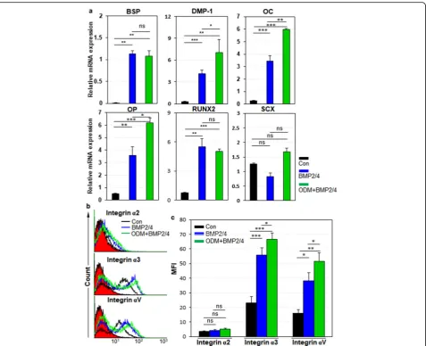
Integrinα3 andαV are upregulated in hPDLCs and hDPCs upon osteogenic differentiation
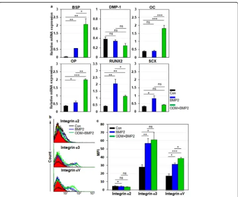
Next, we examined the expression dynamics of integrin α2,α3, andαV in hPDLCs during osteogenic differenti-ation of hPDLCs. For osteoblastic differentidifferenti-ation of hPDLCs, cells were treated with BMP-2 or ODM-plus
BMP-2 as described previously [30]. Relative mRNA
expression of bone sialoprotein (BSP), osteocalcin (OC), osteopontin (OP), and Runx2 were increased in BMP-2 and ODM-plus BMP-2 treatment, although relative mRNA expression of ligamentogenic marker scleraxis (SCX) was not altered in ODM-plus BMP-2 treatment (Fig. 4a). Under the circumstances, integrin α2 expres-sion was slightly downregulated in BMP-2 treatment but not altered in ODM-plus BMP-2 treatment (Fig. 4b, c). However, integrinα3 andαV expression were drastically upregulated in BMP-2 treatment and further increased
in ODM-plus BMP-2 treatment (Fig. 4b, c). The same
experiments were also done with hDPCs. For osteo-blastic and odontoosteo-blastic differentiation of hDPCs, cells were treated with BMP-2/4 or ODM-plus BMP-2/4 as described previously [31]. Relative mRNA expression of BSP, dentin matrix protein-1 (DMP-1), OC, OP, and
Runx2 were increased in BMP-2/4 treatment (Fig. 5a).
Relative mRNA expression of DMP-1, OC, and OP were further increased in ODM-plus BMP-2/4 treatment, while relative mRNA expression of BSP and Runx2 were just maintained in ODM-plus BMP-2/4 treatment (Fig. 5a). SCX was not altered in both culture conditions (Fig. 5a). Under the circumstances, Integrinα2 expression was not altered in both BMP2/4 and ODM-plus BMP-2/4 treatment (Fig.5b, c). Again, integrinα3 andαV expression were drastically upregulated in BMP2/4 treatment and fur-ther increased in ODM-plus BMP-2/4 treatment (Fig. 5b, c). Taken together, the results suggest that the cell surface expression of integrinα3 andαV is upregulated in hPDLCs and hDPCs upon osteogenic differentiation of them. The results also suggest that integrin α3 andαV expression is commonly upregulated in hMSCs, hPDLCs, and hDPCs upon osteogenic differentiation of them.
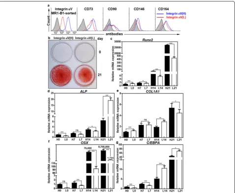
IntegrinαV-high hMSCs have a greater osteogenic potential than integrinαV-low hMSCs
Next, we examined the osteogenic potential of integrin
αV-high and -low hMSCs after MR1-B1-based magnetic
Runx2 and OSX were drastically upregulated to approxi-mately 2.7 × 10⁴- and 8.7 × 10⁶-fold, respectively, during osteogenic differentiation, and they were approximately 62-fold and 5023-fold higher, respectively, in integrin α V-high hMSCs than in integrinαV-low hMSCs at 21 days of osteogenic differentiation (Fig. 6c, f). The expression level of COL1A1, a late OB marker, was gradually upregulated and was also higher at 21 days of osteogenic differentiation
in integrin αV-high hMSCs than in integrin αV-low
hMSCs, although the expression level of ALP was down-regulated in integrinαV-high hMSCs than in integrinα V-low hMSCs (Fig. 6d, e). The expression level of C/EBPA, an adipogenesis marker, was also gradually upregulated but significantly lower at 21 days of osteogenic differentiation
in integrin αV-high hMSCs than in integrin αV-low
hMSCs (Fig. 6d, g). Thus, the expression levels of key
osteogenic transcription factors (Runx2, OSX) and
COL1A1 were upregulated in integrin αV-high hMSCs
whereas the expression level of adipogenic transcription
factor C/EBPA was downregulated in integrin αV-high
hMSCs upon osteogenic differentiation of hMSCs. The re-sults suggest that integrinαV-high hMSCs have a greater osteogenic potential than integrinαV-low hMSCs.
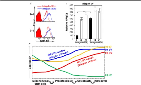
IntegrinαV induction is a good indicator of OB differentiation
Interestingly, all the osteogenic markers were also grad-ually upregulated even in integrinαV-low hMSCs during
osteogenic differentiation, and ALP was even threefold higher at 21 days of osteogenic differentiation in integrin
αV-low hMSCs than in integrin αV-high hMSCs
(Fig. 6c–g). Therefore, we compared the expression
changes of integrin αV between integrin αV-high and
αV-low hMSCs by flow cytometry. Integrin αV
expres-sion was drastically recovered (approximately sevenfold increase) at 21 days of osteogenic differentiation in integ-rin αV-low hMSCs (Fig. 7a, b). As shown in Fig. 6, the osteogenic early and late markers Runx2, ALP, OSX, and COL1A1 were also highly increased at 21 days of osteogenic differentiation in integrin αV-low hMSCs, as compared to day 0, suggesting that the increased integ-rin αV expression is closely associated with osteogenic induction. Consistent with this notion, a substantial
amount of ARS staining was observed in integrin α
V-low hMSCs at 21 days of osteogenic differentiation (Fig. 6b). Taken together, the results suggest that integ-rinαV induction is a good indicator of the early and late
stages of OB differentiation even in integrin αV-low
hMSCs.
Discussion
Osteogenesis is driven by a sequential biological pro-cesses initiated by the recruitment of MSCs to bone formation sites, subsequent proliferation, lineage com-mitment into pre-osteoblast and OB, expression of lineage-specific markers, extracellular matrix secretion,
and mineralization [47]. There are many progressive
changes in osteogenic lineage cells during osteogenic
differentiation of MSCs but there are no definitive sur-face markers for OBs except for alkaline phosphatase [42]. We postulated that some of the cell surface mole-cules on TGF-β1-treated cancer cells closely resemble
the surface molecules on OBs because TGF-β1
treat-ment enriches the population of osteogenic progenitors and activates the early differentiation of OBs by inhibit-ing late differentiation of OBs [9, 10]. To identify novel cell surface molecules on EMT-phenotypic cancer cells, in the beginning, we generated a panel of MAbs specific to the cell surface-expressed epitopes on TGF-β1-treated cancer cells by immunizing TGF-β1-treated A549 cells
in mice. Compared with conventional peptide-based ap-proaches, establishing MAbs against the surface mole-cules on intact cells expands the repertoire of MAbs against the conformation and post-translational modifi-cation of these surface molecules [48]. Therefore, we postulated that the MAbs would be useful tools to find novel surface markers or epitopes on TGF-β1-regulated OB cells. Nine MAbs were selected from the generated MAbs because they bound to hMSCs and hFOBs, and the cell surface expression of cognate antigens showed up- or downregulation during osteogenic differentiation of hMSCs (Fig. 1, Table1). The cell surface expression
Fig. 6IntegrinαV-high hMSCs have a greater osteogenic potential than integrinαV-low hMSCs.aFlow cytometric analysis of hMSCs with MR1-B1, anti-CD73, anti-CD90, anti-CD146, and anti-CD164 antibodies in integrinαV-high andαV-low hMSCs after MR1-B1-based magnetic cell sorting. bAlizarin Red S staining of OBs in integrinαV-high andαV-low hMSCs after being cultured in ODM for 21 days. To inhibit TGF-β1 signaling that originated from the culture medium, SB431542 was added to ODM after 7 days of osteogenic differentiation. Calcium deposition was visualized as red color after the cells were stained with Alizarin Red S.c–gExpression changes of osteogenic and adipogenic markers during osteogenic differentiation of hMSCs. Osteogenic markers (Runx2, ALP, COL1A1, OSX) and adipogenic marker (C/EBPA) were analyzed at 0, 7, 14, and 21 days of osteogenic differentiation in integrinαV-high (H0, H7, H14, H21) andαV-low (L0, L7, L14, L21) hMSCs by quantitative PCR. Values are depicted as a mean fold change in gene expression (2−ΔΔCT) of differentiated hMSCs at the indicated days compared to integrin
of cognate antigens was also upregulated or
downregu-lated in Runx2 knockdown osteosarcoma cells (Fig. 1,
Table1). Therefore, we thought that the MAbs were re-sponsible for physiological recognition of cognate anti-gens during osteogenic differentiation because they were generated by immunizing intact cells. Immunoprecipita-tion followed by LC-MS/MS identified the antigens of 4 MAbs (MR14-E5, ER7-A7, ER7-A8, and MR1-B1) as in-tegrin α2, α3, α3, and αV, respectively. By using
MR1-B1, we found that integrin αV-high hMSCs have a
greater osteogenic potential than integrin αV-low
hMSCs. The results also suggest that integrinαV induc-tion is a good indicator of the early and late stages of OB differentiation because the cell surface expression of
integrin αV was induced from integrin αV-low hMSCs
upon osteogenic differentiation of hMSCs. The increased
expression of integrin αV was also obvious in hPDLCs
and hDPCs during osteogenic differentiation of them. These findings suggest that integrin αV recognized by MR1-B1 can serve as a definitive cell surface marker of OB differentiation. A proposed model for expression
dynamics of integrin α2, α3, and αV upon osteogenic differentiation of hMSCs is presented (Fig.7c).
To accomplish bone formation and regeneration, it is important to investigate cell-matrix interaction mediated by various integrins of hMSC-derived OBs. Integrins are cell surface receptors which can bind extracellular matrix (ECM) components, membrane-bound cell sur-face molecules, or soluble extracellular ligands for cell migration, differentiation, survival, and proliferation [49]. The present study showed that cell surface expres-sion of integrinα2,α3, andαV was observed in undiffer-entiated hMSCs, consistent with previous studies [49] (Additional file1: Figure S3). The cell surface expression of integrin α2 was drastically downregulated after 14 days of osteogenic differentiation of hMSCs (Fig. 3c). The cell surface expression of integrin α2 was not ob-served in hPDLCs and hDPCs even in ODM (Figs.4,5). Furthermore, the drastic downregulation of integrin α2 was also observed during adipogenic differentiation of hMSCs (Additional file 1: Figure S9), suggesting that integrin α2 is a cell surface marker for undifferentiated
Fig. 7Expression changes of integrinαV during osteogenic differentiation of hMSCs after cell sorting.aFlow cytometric analysis of integrinα V-high andαV-low hMSCs after MR1-B1-based magnetic cell sorting. hMSCs were cultured in ODM for 21 days after MR1-B1-based magnetic cell sorting. To inhibit TGF-β1 signaling that originated from the culture medium, SB431542 was added to ODM after 7 days of osteogenic
differentiation. The cells were detached and analyzed at days 14 and 21 by flow cytometry.bStatistic analysis ofa. Values are depicted as relative MFIs of sorted hMSCs at the indicated days compared to sorted hMSCs cultured in normal medium. **,p< 0.01; ***,p< 0.005.cProposed model of expression dynamics of integrinα2,α3, andαV during osteogenic differentiation of hMSCs. Upon osteogenic differentiation of hMSCs, integrin
hMSCs. Consistent with this notion, integrin α2 is critical for the survival of hMSCs via collagen I binding [50, 51]. The cell surface expression of integrin α3β1
was not observed in bone marrow-derived hMSCs [52],
but integrin α3 recognized with ER7-A7 and ER7-A8
was readily detected in bone marrow-derived hMSCs (Fig. 1and Additional file1: Figure S3). The cell surface
expression of integrin α3 was downregulated during
adipogenic differentiation of hMSCs (Additional file 1: Figure S9), but it was obviously upregulated during osteogenic differentiation of hMSCs (Fig. 3), suggesting that increased expression of integrin α3 is needed for the later stage of osteogenic differentiation. Interestingly, the cell surface expression of integrinα3 was drastically induced in hPDLCs and hDPCs during osteogenic differ-entiation of them (Figs.4,5), suggesting that integrinα3 expression is needed for osteogenic differentiation of hPDLCs and hDPCs as well. Consistent with the results, integrin α3 is expressed by OBs actively synthesizing bone and some of the OB lining cells [53].
The cell surface expression of integrin αV was further increased in bone marrow-derived hMSCs during osteo-genic differentiation of bone marrow-derived hMSCs (Figs. 3, 7). The same results were also observed in
hPDLCs and hDPCs, suggesting that integrin αV is
generally increased during osteogenic differentiation of hMSCs, hPDLCs, and hDPCs. This observation drew our interest because little is known about the role of
in-tegrin αV in hMSC commitment to OB lineage. When
integrin αV-low hMSCs were subjected to osteogenic
differentiation after MR1-B1-based magnetic cell sorting, increased expression of integrinαV was obvious after 14 days of osteogenic differentiation of hMSCs (Fig. 7). It
seems that integrin αV induction is needed for the
hMSC commitment into OB lineage. IntegrinαV
expres-sion drives the proliferation and survival of human adipose-derived stem cells (hASCs) through the
suppres-sion of p21 and induction of survivin and TAZ [54].
Therefore, it is possible to speculate that integrin αV may drive the survival, proliferation, and differentiation of OBs during osteogenic differentiation of hMSCs. This phenomenon was not observed during adipogenic differ-entiation of hMSCs (Additional file1: Figure S9). It may be that this observation is due to differences between hMSCs and hASCs, which remain elusive.
Increased surface expression of integrin αV indicated the hMSC commitment into OB lineage (Figs.3, 4,5,6,
7). The results may indicate that the expression and secre-tion of ECM, especially integrinαV ligands, such as fibro-nectin, vitrofibro-nectin, and osteopontin, may be important for the survival, proliferation, and differentiation of OB during osteogenic differentiation. A previous study showed that the interaction between integrin αVβ1 and connective tissue growth factor (CTGF/CCN2) stimulates OB growth
and differentiation via integrin-mediated activation of ERK signaling and an increase in Runx2 binding to the osteocalcin promoter [55]. Integrin αVβ3 is also impli-cated in osteogenesis of bone marrow-derived hMSCs cul-tured on fibronectin coated stiff substrates [56]. By using a αVβ3 blocking antibody, another study also confirmed that integrinαVβ3 is responsible for the OB commitment from dental bud stem cells through its interaction with vitronectin and osteopontin [57]. Thus, the present studies including the previous studies indicate that increased expression of integrin αV is needed for the commitment of hMSCs to OB differentiation and maturation.
Conclusion
In this study, we generated 4 MAbs specific to integrin α2,α3, orαV on bone marrow-derived hMSCs. By using the MAbs, we found that the cell surface expression of integrins fluctuated during osteogenic differentiation of
hMSCs depending on the integrin type. Integrin α2
recognized by MR14-E5 was downregulated on hMSCs during osteogenic differentiation, while integrin α3 and
αV, recognized by ER7-A7 and MR1-B1, respectively,
were upregulated on hMSCs in accordance with upregu-lation of osteogenic markers. Subsequent studies showed that integrinα3 andαV are also upregulated in hPDLCs and hDPCs during osteogenic differentiation, suggesting that integrinα3 andαV induction is a good indicator of OB differentiation. Cell sorting revealed that key osteo-genic induction factors (Runx2, OSX, and COL1A1) and surface markers (CD73, CD90, CD146, and CD164) are higher in integrin αV-high hMSCs than in integrin α V-low hMSCs upon osteogenic differentiation of hMSCs, suggesting that integrin αV-high hMSCs have a greater osteogenic potential than integrin αV-low hMSCs. Cell sorting further revealed that integrin αV expression is
more dramatically induced in integrin αV-low hMSCs
during osteogenic differentiation of hMSCs. The results suggest that integrinαV induction is a good indicator of the early and late stages of OB differentiation.
Supplementary information
Supplementary informationaccompanies this paper athttps://doi.org/10. 1186/s13287-020-01714-7.
was analyzed in A549 and TGF-β1-treated A549 cells by flow cytometry with the indicated MAbs. The expression of E-cadherin (E-cad) and N-cadherin (N-cad) was used as controls. Red-filled histograms represent the isotype controls.Figure S3.Cell surface expression of target antigens of selected MAbs in mesenchymal stem and progenitor cells with osteo-genic potential. Cell surface expression of target antigens of selected MAbs was examined in two human osteogenic progenitor cells (hMSC and hFOB) and two human osteoblastic cancer cell lines (U2OS and SAOS-2) by flow cytometry with the indicated MAbs. Red-filled histo-grams represent the isotype controls.Figure S4.Alizarin Red S staining assay and knockdown efficiency of Runx2 in U2OS cells. (a) Alizarin Red S staining of hMSCs stimulated with ODM. hMSCs were incubated for 12 days with ODM, and calcium deposition and bone nodule were visualized as red color after the cells were stained with Alizarin Red S. The scale bar is 200μm. (b) Knockdown efficiency of Runx2 in U2OS cells. After trans-fection of control siRNA or Runx2 siRNA, the expression of Runx2 gene was evaluated by RT-PCR (left panels) and by Western blotting (right panels). GAPDH orβ-actin was used as a loading control.Figure S5.
Mass spectrometric identification of MR14-E5 antigen after immunopre-cipitation with ME14-E5. The approximately 150-kDa band from A549 cell lysates was treated with trypsin, and the resulting peptides were analyzed by mass spectrometry. Ten tryptic peptides (underlined) originating from the 150-kDa protein matched the integrinα2 precursor.Figure S6.Mass spectrometric identification of ER7-A7 and ER7-A8 antigen after immuno-precipitation with ER7-A8. The approximately 130-kDa band from A549 cell lysates was treated with trypsin, and the resulting peptides were ana-lyzed by mass spectrometry. Five tryptic peptides (underlined) originating from the 130-kDa protein matched the integrinα3 preproprotein.Figure S7.Mass spectrometric identification of MR1-B1 antigen after immuno-precipitation with MR1-B1. The approximately 130-kDa band from A549 cell lysates was treated with trypsin, and the resulting peptides were ana-lyzed by mass spectrometry. Five tryptic peptides (underlined) originating from the 130-kDa protein matched the integrinαV isoform 1 prepropro-tein.Figure S8.Expression changes of integrins and hMSC/OB surface markers upon osteogenic differentiation of hMSCs. hMSCs were incu-bated for 14, 21 days with ODM, and SB431542 was added to ODM after 7 days of the osteogenic differentiation. Integrins (α2,α3,αV), hMSC/OB surface markers (CD73, CD90, CD146 and CD164) were analyzed in undif-ferentiated (normal growth medium) and difundif-ferentiated hMSCs (ODM) by flow cytometry. Red-filled histograms represent isotype controls.Figure S9.Expression changes of integrinαV,α2,α3 and osteogenic markers during adipogenic differentiation of hMSCs. (a) Oil Red O staining of adi-pocytes in differentiated hMSCs. hMSCs were incubated for 21 days with ADM. Lipid content was visualized as red color after the cells were stained with Oil Red O. (b) Expression changes of integrins and hMSC/OB surface markers during adipogenic differentiation of hMSCs. Integrins (MR14-E5, ER7-A7, MR1-B1) and hMSC/OB surface markers (CD73, CD90, CD146 and CD164) were analyzed in differentiated hMSCs by flow cytom-etry. Values are depicted as a relative MFI of differentiated hMSCs at the indicated days compared to hMSCs at day 0. **,p< 0.01; ***,p< 0.005.
Figure S10.Expression changes of integrins and hMSC/OB surface markers upon osteogenic differentiation of hMSCs in the absence and presence of SB431542. (a) Alizarin Red S staining of hMSCs stimulated with ODM. hMSCs were incubated for 21 days with ODM in the absence and presence of SB431542. SB431542 was included in ODM after 14 days of osteogenic differentiation and calcium deposition and bone nodule were visualized as red color after the cells were stained with Alizarin Red S. (b) Flow cytometric analysis of hMSCs cultured in ODM in the absence and presence of SB431542. hMSCs were incubated for 7, 14, 21 days with ODM, and SB431542 was added to ODM after 14 days of osteogenic dif-ferentiation. Integrins (α2,α3,αV), hMSC/OB surface markers (CD73, CD90, CD146 and CD164) were analyzed by flow cytometry. Red-filled histo-grams represent isotype controls.
Abbreviations
hMSC:Human mesenchymal stem cell; MAb: Monoclonal antibody; OB: Osteoblast; TGF-β1: Transforming growth factor-β1; BMP: Bone morphogenetic protein; ODM: Osteogenic differentiation medium; hPDLCs: Human periodontal ligament cells; hDPCs: Human dental pulp cells; qPCR: Quantitative polymerase chain reaction; ARS: Alizarin Red S;
Runx2: Runt-related transcription factor 2; EMT: Epithelial-mesenchymal transition; NSCLC: Non-small cell lung carcinoma cell; bFGF: Basic fibroblast growth factor; FBS: Fetal bovine serum; PBMC: Peripheral blood mononuclear cell; FITC: Fluorescein isothiocyanate; PE: Phycoerythrin; PI: Propidium iodide; IP: Immunoprecipitation; SDS-PAGE: Sodium dodecyl sulfate-polyacrylamide gel electrophoresis; SA-HRP: Streptavidin-horse radish peroxidase; TBST: Tris-buffered saline with Tween-20; ADM: Adipogenic differentiation medium; RT-PCR: Reverse transcription-polymerase chain reaction; ALP: Alkaline phosphatase; COL1A1: Collagen type I; OCX: Osterix; ITGA: Integrinα; BSP: Bone sialoprotein; OC: Osteocalcin; OP: Osteopontin; SCX: Scleraxis; DMP-1: Dentin matrix protein-1; ECM: Extracellular matrix
Acknowledgements
We thank Dr. Hee Chul Lee for manuscript proofreading.
Authors’contributions
HML contributed to the collection of data; S-RS contributed to the collection of data; JK contributed to the collection of data; MKK contributed to the collection of data; HS contributed to the collection of data; KSK contributed to the collection of data; Y-JJ contributed to the collection of data and gave financial support; CJR contributed to the conception and design, gave financial support, and contributed to the manuscript writing. All authors approved the final manuscript.
Authors’information
H.M. Lee and S-R. Lee contributed equally to this work.
Funding
This research was supported by the National Research Foundation (NRF) of Korea (2016M3A9C6918220 and 2018M3A9H1023139).
Availability of data and materials
The datasets used and analyzed during the current study are available from the corresponding author on reasonable request.
Ethics approval and consent to participate
All human specimens’and cells’researches were reviewed and approved by the Institutional Review Board of under guidelines approved by the Dankook Dental Hospital and Dankook University (DKU NON2019-004), and the informed consent for all experiments using extracted teeth was obtained from all the participants. All animal experiments were also approved by the Institutional Animal Care and Use Committee at Sejong University (SJ-21051104).
Consent for publication Not applicable.
Competing interests
The authors declare that they have no competing interests.
Author details
1Department of Integrative Bioscience and Biotechnology, Institute of Anticancer Medicine Development, Sejong University, 209 Neungdong-ro, Gwangjin-gu, Seoul 05006, Korea.2Department of Clinical Pharmacology and Therapeutics, Kyung Hee University School of Medicine, Seoul 02447, Korea. 3Department of Nanobiomedical Science, BK21 PLUS NBM Global Research Center for Regenerative Medicine, College of Dentistry, Dankook University, Cheonan 330-714, Korea.
Received: 17 January 2020 Revised: 6 April 2020 Accepted: 7 May 2020
References
1. Pittenger MF, Mackay AM, Beck SC, et al. Multilineage potential of adult human mesenchymal stem cells. Science.1999;284:143–7.
2. James AW. Review of signaling pathways governing MSC osteogenic and adipogenic differentiation. Scientifica (Cairo). 2013;2013:684736. 3. Chen Q, Shou P, Zheng C, et al. Fate decision of mesenchymal stem cells:
4. Pino AM, Rosen CJ, Rodriguez JP. In osteoporosis, differentiation of mesenchymal stem cells (MSCs) improves bone marrow adipogenesis. Biol Res. 2012;45:279–87.
5. Bonewald LF. The amazing osteocyte. J Bone Miner Res. 2011;26:229–38. 6. Peltzer J, Montespan F, Thepenier C, et al. Heterogeneous functions of
perinatal mesenchymal stromal cells require a preselection before their banking for clinical use. Stem Cells Dev. 2015;24:329–44.
7. Phinney DG, Kopen G, Righter W, et al. Donor variation in the growth properties and osteogenic potential of human marrow stromal cells. J Cell Biochem. 1999;75:424–36.
8. Gordon KJ, Blobe GC. Role of transforming growth factor-beta superfamily signaling pathways in human disease. Biochim Biophys Acta. 2008;1782: 197–228.
9. Wu M, Chen G, Li YP. TGF-beta and BMP signaling in osteoblast, skeletal development, and bone formation, homeostasis and disease. Bone Res. 2016;4:16009.
10. Tang SY, Alliston T. Regulation of postnatal bone homeostasis by TGFbeta. Bonekey Rep. 2013;2:255.
11. Tachi K, Takami M, Sato H, et al. Enhancement of bone morphogenetic protein-2-induced ectopic bone formation by transforming growth factor-beta1. Tissue Eng Part A. 2011;17:597–606.
12. Grafe I, Alexander S, Peterson JR, et al. TGF-beta family signaling in Mesenchymal differentiation. Cold Spring Harb Perspect Biol. 2018;10: a022202.
13. Komori T. Regulation of bone development and extracellular matrix protein genes by RUNX2. Cell Tissue Res. 2010;339:189–95.
14. Niu DF, Kondo T, Nakazawa T, et al. Transcription factor Runx2 is a regulator of epithelial-mesenchymal transition and invasion in thyroid carcinomas. Lab Investig. 2012;92:1181–90.
15. Ishiwata T. Cancer stem cells and epithelial-mesenchymal transition: novel therapeutic targets for cancer. Pathol Int. 2016;66:601–8.
16. Kalluri R, Weinberg RA. The basics of epithelial-mesenchymal transition. J Clin Invest. 2009;119:1420–8.
17. Lamouille S, Xu J, Derynck R. Molecular mechanisms of epithelial-mesenchymal transition. Nat Rev Mol Cell Biol. 2014;15:178–96.
18. Gordon KJ, Kirkbride KC, How T, et al. Bone morphogenetic proteins induce pancreatic cancer cell invasiveness through a Smad1-dependent mechanism that involves matrix metalloproteinase-2. Carcinogenesis.2009; 30:238–48.
19. Zeisberg M, Shah AA, Kalluri R. Bone morphogenic protein-7 induces mesenchymal to epithelial transition in adult renal fibroblasts and facilitates regeneration of injured kidney. J Biol Chem. 2005;280:8094–100.
20. Kasai H, Allen JT, Mason RM, et al. TGF-beta1 induces human alveolar epithelial to mesenchymal cell transition (EMT). Respir Res. 2005;6:56. 21. Lee HM, Joh JW, Seo SR, et al. Cell-surface major vault protein promotes
cancer progression through harboring mesenchymal and intermediate circulating tumor cells in hepatocellular carcinomas. Sci Rep. 2017;7:13201. 22. Choi HS, Lee HM, Kim WT, et al. Detection of mycoplasma infection in
circulating tumor cells in patients with hepatocellular carcinoma. Biochem Biophys Res Commun. 2014;446:620–5.
23. Hyun SY, Mun S, Kang KJ, et al. Amelogenic transcriptome profiling in ameloblast-like cells derived from adult gingival epithelial cells. Sci Rep. 2019;9:3736.
24. Lee JH, Nam H, Um S, et al. Upregulation of GM-CSF by TGF-beta1 in epithelial mesenchymal transition of human HERS/ERM cells. In Vitro Cell Dev Biol Anim. 2014;50:399–405.
25. Akimoto T, Fujiwara N, Kagiya T, et al. Establishment of Hertwig's epithelial root sheath cell line from cells involved in epithelial-mesenchymal transition. Biochem Biophys Res Commun. 2011;404:308–12.
26. Yang Z, Hai B, Qin L, et al. Cessation of epithelial Bmp signaling switches the differentiation of crown epithelia to the root lineage in a beta-catenin-dependent manner. Mol Cell Biol. 2013;33:4732–44.
27. Saito K, Takahashi K, Huang B, et al. Loss of Stemness, EMT, and supernumerary tooth formation in Cebpb(−/−)Runx2(+/−) murine incisors. Sci Rep. 2018;8:5169.
28. Choi HS, Kim H, Won A, et al. Development of a decoy immunization strategy to identify cell-surface molecules expressed on undifferentiated human embryonic stem cells. Cell Tissue Res. 2008;333:197–206.
29. Marasini S, Chang DY, Jung JH, et al. Effects of adenoviral gene transduction on the stemness of human bone marrow mesenchymal stem cells. Mol Cells. 2017;40:598–605.
30. Hyun SY, Lee JH, Kang KJ, et al. Effect of FGF-2, TGF-beta-1, and BMPs on teno/ligamentogenesis and osteo/cementogenesis of human periodontal ligament stem cells. Mol Cells.2017;40:550–7.
31. Kang KJ, Ryu CJ, Jang YJ. Identification of dentinogenic cell-specific surface antigens in odontoblast-like cells derived from adult dental pulp. Stem Cell Res Ther. 2019;10:128.
32. Kim JH, Jang YS, Eom KS, et al. Transforming growth factor beta1 induces epithelial-to-mesenchymal transition of A549 cells. J Korean Med Sci. 2007; 22:898–904.
33. Miura M, Gronthos S, Zhao M, et al. SHED: stem cells from human exfoliated deciduous teeth. Proc Natl Acad Sci U S A. 2003;100:5807–12.
34. Yen ML, Chien CC, Chiu IM, et al. Multilineage differentiation and characterization of the human fetal osteoblastic 1.19 cell line: a possible in vitro model of human mesenchymal progenitors. Stem Cells. 2007;25: 125–31.
35. Bilbe G, Roberts E, Birch M, et al. PCR phenotyping of cytokines, growth factors and their receptors and bone matrix proteins in human osteoblast-like cell lines. Bone.1996;19:437–45.
36. Burmester A, Luthringer B, Willumeit R, et al. Comparison of the reaction of bone-derived cells to enhanced MgCl2-salt concentrations. Biomatter.2014; 4:e967616.
37. Sacchetti B, Funari A, Michienzi S, et al. Self-renewing osteoprogenitors in bone marrow sinusoids can organize a hematopoietic microenvironment. Cell.2007;131:324–36.
38. Birmingham E, Niebur GL, McHugh PE, et al. Osteogenic differentiation of mesenchymal stem cells is regulated by osteocyte and osteoblast cells in a simplified bone niche. Eur Cell Mater.2012;23:13–27.
39. Liu TM, Lee EH. Transcriptional regulatory cascades in Runx2-dependent bone development. Tissue Eng Part B Rev. 2013;19:254–63.
40. Hedayati S, Parvaneh Tafreshi A, Moradi N, et al. Inhibition of transforming growth factor-beta signaling pathway enhances the osteogenic differentiation of unrestricted somatic stem cells. J Cell Biochem. 2018;119: 9327–33.
41. Kawahara T, Yamashita M, Ikegami K, et al. TGF-Beta negatively regulates the BMP2-dependent early commitment of periodontal ligament cells into hard tissue forming cells. PLoS One. 2015;10:e0125590.
42. Rutkovskiy A, Stenslokken KO, Vaage IJ. Osteoblast differentiation at a glance. Med Sci Monit Basic Res. 2016;22:95–106.
43. Ode A, Schoon J, Kurtz A, et al. CD73/5′-ecto-nucleotidase acts as a regulatory factor in osteo−/chondrogenic differentiation of mechanically stimulated mesenchymal stromal cells. Eur Cell Mater. 2013;25:37–47. 44. Chen XD, Qian HY, Neff L, et al. Thy-1 antigen expression by cells in the
osteoblast lineage. J Bone Miner Res. 1999;14:362–75.
45. Chan CKF, Gulati GS, Sinha R, et al. Identification of the human skeletal stem cell. Cell.2018;175:43–56. e21.
46. Liu JQ, Li QW, Tan Z. New insights on properties and spatial distributions of skeletal stem cells. Stem Cells Int. 2019;2019:9026729.
47. Granero-Molto F, Weis JA, Miga MI, et al. Regenerative effects of transplanted mesenchymal stem cells in fracture healing. Stem Cells. 2009; 27:1887–98.
48. Choi HS, Kim WT, Ryu CJ. Antibody approaches to prepare clinically transplantable cells from human embryonic stem cells: identification of human embryonic stem cell surface markers by monoclonal antibodies. Biotechnol J. 2014;9:915–20.
49. Prowse AB, Chong F, Gray PP, et al. Stem cell integrins: implications for ex-vivo culture and cellular therapies. Stem Cell Res. 2011;6:1–12.
50. Popov C, Radic T, Haasters F, et al. Integrins alpha2beta1 and alpha11beta1 regulate the survival of mesenchymal stem cells on collagen I. Cell Death Dis. 2011;2:e186.
51. Barczyk M, Carracedo S, Gullberg D. Integrins. Cell Tissue Res. 2010;339:269–80. 52. Gronthos S, Simmons PJ, Graves SE, et al. Integrin-mediated interactions
between human bone marrow stromal precursor cells and the extracellular matrix. Bone.2001;28:174–81.
53. Clover J, Dodds RA, Gowen M. Integrin subunit expression by human osteoblasts and osteoclasts in situ and in culture. J Cell Sci. 1992;103(Pt 1): 267–71.
54. Morandi EM, Verstappen R, Zwierzina ME, et al. ITGAV and ITGA5 diversely regulate proliferation and adipogenic differentiation of human adipose derived stem cells. Sci Rep. 2016;6:28889.
cytoskeleton reorganization and cell differentiation. PLoS One. 2015;10: e0115325.
56. Lee J, Abdeen AA, Tang X, et al. Geometric guidance of integrin mediated traction stress during stem cell differentiation. Biomaterials.2015;69:174–83. 57. Di Benedetto A, Brunetti G, Posa F, et al. Osteogenic differentiation of
mesenchymal stem cells from dental bud: role of integrins and cadherins. Stem Cell Res. 2015;15:618–28.
Publisher’s Note