ORIGINAL ARTICLE
Diagnostic performance of 3D SPACE for comprehensive
knee joint assessment at 3 T
Pieter Van Dyck&Jan L. Gielen&
Filip M. Vanhoenacker&Eline De Smet&
Kristien Wouters&Lieven Dossche&Paul M. Parizel
Received: 19 July 2012 / Revised: 28 September 2012 / Accepted: 3 October 2012 / Published online: 26 October 2012 #The Author(s) 2012. This article is published with open access at Springerlink.com
Abstract
ObjectiveTo assess the diagnostic performance of 3D sam-pling perfection with application-optimised contrasts using variable flip-angle evolution (SPACE) turbo spin-echo (TSE) sequences compared to 2D TSE for comprehensive knee assessment at 3 T.
Methods From January to July 2011, isotropic 3D SPACE was added to a 2D knee protocol at 3 T. Forty patients underwent subsequent arthroscopy. Three readers indepen-dently assessed MR images for meniscus, anterior cruciate ligament (ACL) and cartilage lesions. Readers 1 and 2 evaluated 3D and 2D data at separate sittings; reader 3 interpreted the complete exam including 3D and 2D sequen-ces. Accuracies were calculated using arthroscopy as
reference standard. McNemar’s test (p<0.05) was used to
compare 3D and 2D techniques.
Results The highest diagnostic yield was obtained by reader
3 (accuracies ≥88 %). For the medial meniscus, readers
performed better with the 2D technique than with 3D
SPACE (accuracies 85–88 % vs. 78–80 %, respectively)
(p>0.05). For the lateral meniscus and ACL, 3D and 2D
techniques had similar performance (accuracies≥93 %). For
cartilage lesions, 3D SPACE had significantly lower
speci-ficity (p00.0156) than the 2D protocol for one reader.
Conclusion The conventional 2D TSE acquisition is more reliable than 3D SPACE for comprehensive assessment of the knee at 3.0 T.
Main Messages
• 3D SPACE is a valuable component of a knee MR proto-col at 3 T.
• 3D SPACE cannot be used as a single sequence in the MR evaluation of the knee at 3 T.
• Knee MR protocols at 3 T should include both 2D and 3D TSE sequences.
Keywords SPACE . Knee . 3 T . Cartilage . Meniscus
Introduction
Three-dimensional (3D) turbo spin-echo (TSE) sequen-ces with isotropic resolution have recently been devel-oped and are now commercially available on many magnetic resonance (MR) vendor platforms [1]. These sequences include 3D fast spin-echo (FSE) Cube (GE Healthcare), 3D Fourier Transform (FT, Philips Medical Systems) and sampling perfection with application-optimised contrasts using different flip-angle evolutions
(SPACE, Siemens Medical Systems) [1–3]. The
advan-tage of the new 3D TSE acquisitions is their capability P. Van Dyck (*)
:
J. L. Gielen:
F. M. Vanhoenacker:
E. De Smet
:
P. M. ParizelDepartment of Radiology, University Hospital Antwerp, Wilrijkstraat 10,
2650 Edegem, Belgium e-mail: [email protected] F. M. Vanhoenacker
Department of Radiology, University of Ghent, De Pintelaan 185,
9000 Ghent, Belgium F. M. Vanhoenacker
Department of Radiology, AZ St-Maarten Duffel/Mechelen, Rooienberg 25,
2570 Duffel, Belgium K. Wouters
Department of Biostatistics, University Hospital Antwerp, Wilrijkstraat 10,
2650 Edegem, Belgium L. Dossche
Department of Orthopedics, University Hospital Antwerp, Wilrijkstraat 10,
of mimicking the contrast properties of conventional two-dimensional (2D) TSE proton-density weighted acquisitions [4]. In addition, high-quality multiplanar reformatted (MPR) images may be created in any ori-entation from the volumetric source data. However, uncertainty remains as to whether a single 3D TSE acquisition has potential for replacing the multiple con-ventional 2D acquisitions currently used. Although the first clinical results on the diagnostic performance of 3D isotropic resolution TSE sequences were encouraging [3, 5], the most recently published studies have described potential limitations of these sequences for evaluating
the knee joint [6, 7]. Thus, additional studies are needed
to determine the diagnostic usefulness of 3D TSE in future knee MR protocols at 3 T. The purpose of this retrospective study was to assess the diagnostic perfor-mance of the 3D TSE SPACE sequence, as compared to routine 2D TSE sequences, for evaluating the menisci, anterior cruciate ligament (ACL) and cartilage of the knee joint in symptomatic patients at 3 T.
Materials and methods
Patient selection and medical record review
This retrospective study correlating MR imaging with ar-throscopy findings was performed with a waiver of in-formed consent from the institutional review board. All MR examinations of the knee performed at our institution with a single 3-T MR system from January to July 2011 were reviewed. Patients included in our study met the fol-lowing criteria: (1) they had undergone a 3-T MR of the knee consisting of 2D TSE sequences and the 3D SPACE sequence; (2) they had an available medical record with the relevant clinical history; (3) they had no prior history of knee surgery; (4) they had subsequent knee arthroscopy. After elimination of patients on the basis of these criteria, we identified a group of 40 patients (25 male, 15 female; average age 43 years, range 18-78 years) eligible for this study. The mean time interval between MR and arthroscopy
was 46 days (range 3–112 days).
MR imaging protocol
All MR knee examinations were performed with a single 3-T system (3-Trio 3-TIM Magnetom; Siemens Healthcare, Erlan-gen, Germany) and an eight-channel phased-array knee coil with the same imaging protocol. The imaging protocol con-sisted of standard 2D TSE acquisitions and a SPACE 3D TSE acquisition with the imaging parameters of all
sequen-ces summarised in Table1. The 2D protocol consisted of a
coronal fat-suppressed (FS) TSE intermediate-weighted
(IM-w) acquisition, an axial FS TSE IM-w acquisition and a coronal SE T1-weighted acquisition. The 3D pro-tocol consisted of a single 3D TSE acquisition in the sagittal plane with the commercially available SPACE sequence. The SPACE isotropic source data were post-processed on a high-performance workstation (Leonardo, Siemens Healthcare) to create sagittal, coronal and axial MPR images with 1-mm slice thickness. Moreover, read-ers were free to use volumetric data to create MPRs in any orientation and slice thickness.
MR image analysis
Three radiologists who had between 10 and 25 years of experience in musculoskeletal radiology and who were blinded to clinical and arthroscopic results at the time of review retrospectively and independently assessed MR images for meniscus, ACL and cartilage lesions. Readers 1 and 2 evaluated 3D and 2D data sets at separate sittings with a 4-week interval to minimise recall bias. During the first review, they used the SPACE sequence with MPR images to detect meniscal, ACL and cartilage lesions within the knee joint. During the second review, the readers used the 2D sequences to detect these joint abnormalities. Reader 3 interpreted the complete MR exam including the 3D and 2D sequences at one sitting. The diagnostic criterion for meniscal tear was abnormal signal intensity within the me-niscus that definitely extended to the meniscal articular surface on one or more sections or abnormal morphology of the meniscus [8]. If a meniscal tear was diagnosed on MR, the observers localised tears in the anterior horn, body or posterior horn of the meniscus in order to make sure that
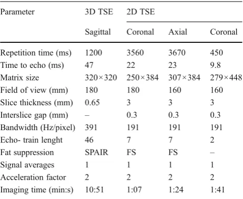
Table 1 Parameters for MR imaging sequences Parameter 3D TSE 2D TSE
Sagittal Coronal Axial Coronal
Repetition time (ms) 1200 3560 3670 450 Time to echo (ms) 47 22 23 9.8 Matrix size 320×320 250×384 307×384 279×448 Field of view (mm) 180 180 160 160 Slice thickness (mm) 0.65 3 3 3 Interslice gap (mm) – 0.3 0.3 0.3 Bandwidth (Hz/pixel) 391 191 191 191 Echo- train lenght 46 7 7 2 Fat suppression SPAIR FS FS –
Signal averages 1 1 1 1
Acceleration factor 2 2 2 2 Imaging time (min:s) 10:51 1:07 1:24 1:41
the tear identified at MR imaging was the same as that identified at arthroscopy. ACL tears were diagnosed on MR imaging on the basis of the presence of increased signal intensity in the ligament. If ligament margins were intact, the tear was termed partial. If fibre disruption could clearly be detected in the anteromedial (AM) or posterolateral bun-dle (PL) of the ACL, an isolated bunbun-dle tear was reported [9]. If margins were not identified or there was ligament retraction and no identifiable central ligament was present, the tear was termed complete [10]. Cartilage abnormalities were graded using the Noyes classification system [11]. Only cartilage lesions grade 3 and 4 were recorded for the purpose of this study. Six cartilage compartments (medial femoral, medial tibial, lateral femoral, lateral tibial, patellar and femoral trochlea) were assessed separately.
Arthroscopic knee surgery
All arthroscopic procedures were performed at our institu-tion by one of three experienced orthopaedic surgeons who specialised in sports medicine and knee surgery and who had between 10 and 25 years of clinical experience. Stan-dard anteromedial and anterolateral portals were used with blunt probing of both menisci and the ACL to evaluate their integrity. Once identified, the location of a meniscal tear was recorded (anterior horn, body and/or posterior horn). The ACL was classified as either normal, partially torn or com-pletely torn. If possible, partial discontinuity of ACL fibres was located in the AM or PL bundle [9]. All articular surfaces of the knee joint were graded at arthroscopy by using the Noyes classification system [11]. The ortho-paedic surgeons were aware of the prospective interpre-tations of the MR imaging studies in all patients at the time of arthroscopy.
Statistical analysis
For each reader and each imaging series, the sensitivity, specificity and accuracy of MR, with corresponding 95 % confidence intervals, were calculated using arthroscopy as
the standard of reference. McNemar’s test was used to
identify differences between 3D and 2D TSE sequences for the diagnosis of meniscal and ACL tears as well as cartilage lesions. Differences were considered to be
signif-icant if thep-value was less than 0.05. For the assessment of
interobserver agreement, kappa (к) coefficients were calcu-lated. According to the recommendations of Landis and
Koch [12], к -values were interpreted as slight (к00.0–
0.20), fair (к00.21–0.40), moderate (к00.41–0.60),
sub-stantial (к00.61–0.80) or excellent (к00.81–1.0). All
anal-yses were performed using the Statistical Package for Social Sciences for Windows (version 17.0; SPSS Inc., Chicago, IL, USA).
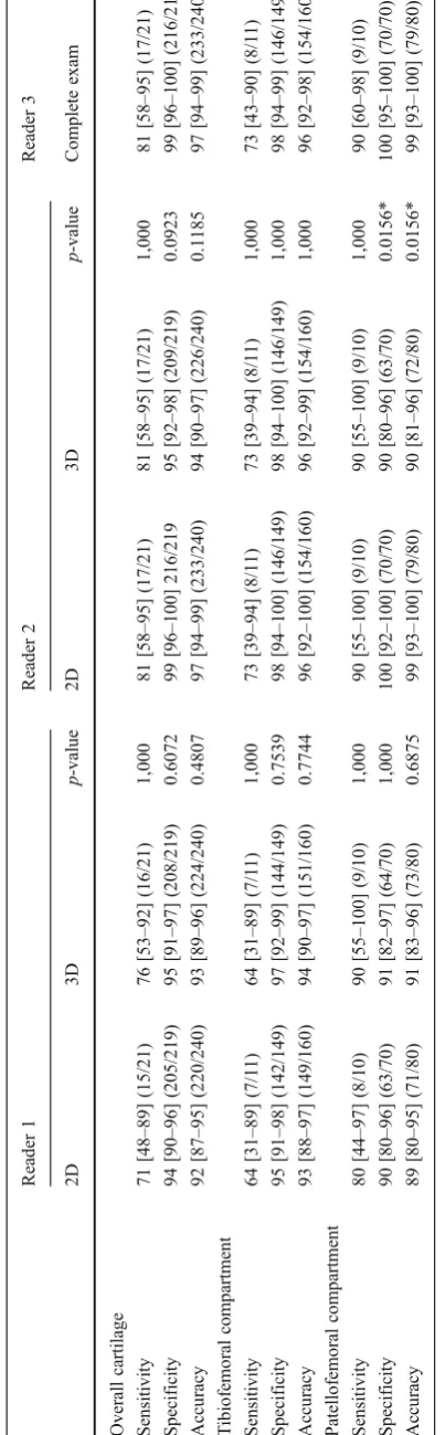
Results
Arthroscopy revealed 24 tears of the medial meniscus, 8 tears of the lateral meniscus, 10 ACL (9 complete and 1
isolated PL bundle) tears and 21 grade 3–4 cartilage lesions
(medial tibia, n01; medial femur, n05; lateral tibia,n03;
lateral femur, n02; patella, n07; trochlea, n03). Tables 2
and 3 show the sensitivities, specificities and accuracies,
with corresponding 95 % confidence intervals, of the 3D SPACE and 2D TSE sequences for the MR diagnoses ren-dered by the three readers. The highest diagnostic yield was
obtained by reader 3 (accuracies≥88 % for all lesions). For
the medial meniscus, both readers 1 and 2 performed better with routine 2D acquisition than with 3D SPACE
acquisi-tion (accuracies 85–88 % and 78–80 %, respectively). This
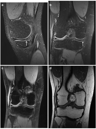
difference was not statistically significant. There were five false-positive MR interpretations of medial meniscal tear for both reader 1 and 2 using 3D SPACE (specificity 69 %)
(Fig.1). In the detection of eight lateral meniscal tears, the
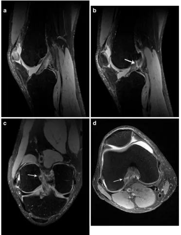
3D and 2D techniques had similar performance (accuracy 95 %). Both reader 1 and 2 missed one tear in the posterior horn of the lateral meniscus using only 2D sequences. In the detection of ten ACL tears, readers 1 and 2 had similar performance with the 3D and 2D techniques (accuracies
93–100 %). One partial (PL bundle) ACL tear was correctly
identified by all readers using both 3D and 2D sequences
(Fig.2). There were three false-positive MR interpretations of
(partial) ACL tear for reader 1 using 2D sequences. For detecting cartilage lesions within the knee joint, reader 1 had similar performances with the 3D and 2D acquisition
meth-ods. However, we found significantly lower specificity (p0
0.0156) for reader 2 using 3D SPACE compared to 2D sequences for evaluating the patellofemoral compartment
[90 % (63/70) and 100 % (70/70), respectively] (Fig.3). Both
the 3D and 2D sequences had excellent interobserver agree-ment for meniscus and ACL lesions (к>0.81), and moderate
interobserver agreement for cartilage lesions (к00.60).
Discussion
Current knee MR protocols typically consist of 2D TSE sequences repeated in multiple planes. These sequences have excellent tissue contrast and high in-plane spatial res-olution [1]. However, they have relatively thick slices and small gaps between slices that can obscure pathology
sec-ondary to partial volume averaging [1, 2].
Three-dimensional sequences can reduce partial volume averaging
by acquiring thin, continuous slices through joints [2, 4].
lack of contrast between abnormal and normal tissue [1,2, 13,14]. Recently, 3D TSE sequences have become available allowing for comprehensive knee joint assessment because of its high tissue contrast and slice resolution [1]. These acquisitions, typically used at 3 T, entail a variable flip angle refocussing pulse and allow extremely large turbo factors [2, 4]. Few studies have directly compared 3D and 2D TSE
sequences for comprehensive knee joint assessment [3,5,7,
13]. Therefore, we undertook this study to determine the diagnostic value of 3D SPACE as compared to routine 2D TSE sequences for the assessment of internal derangements of the knee joint at 3 T.
In our study, readers performed better with conventional 2D acquisition than with 3D SPACE acquisition for evaluation of the menisci of the knee with 3.0-T MR imaging. Although the differences did not reach statistical significance, we found that a conventional 2D TSE protocol was more accurate than an isotropic 3D SPACE protocol for the evaluation of the medial meniscus. The false-positive MR interpretations of medial
meniscal tear (n05 for both readers) using the 3D SPACE
protocol were related to poor image contrast and blurring on SPACE images. Our study results are in concordance with the findings of Kijowski et al. [6] and Subhas et al. [7] who also found the 2D TSE technique to be more accurate in the
evaluation of the knee meniscus as compared to 3D TSE. Also, Ristow et al. [15] found a decreased visualisation of low contrast structures such as bone marrow and menisci because of a higher amount of image blurring and indistinctness of the structural edges on 3D TSE images.
The 3D SPACE sequence may have advantages over 2D sequences for evaluating the knee ligaments. The thin, con-tinuous sections of 3D SPACE minimise the effect of partial-volume averaging, which can be a source of diag-nostic error when evaluating the ACL of the knee [3]. However, in our study, 3D SPACE had similar sensitivity, specificity and accuracy as the routine MR imaging protocol in the detection of nine complete and one partial ACL tear.
Our study findings are in line with prior studies dem-onstrating high diagnostic accuracy of 3D SPACE for evaluating the articular cartilage of the knee joint (overall
accuracies ≥93 %) [3, 13, 16, 17]. However, 3D SPACE
had significantly lower specificity (p00.0156) than the
routine MR protocol for detecting patellofemoral cartilage lesions for one radiologist (reader 2). This lower speci-ficity of 3D SPACE is most likely related to decreased in-plane spatial resolution and image blurring due to acquisition of high spatial frequencies late in the echo train. This may cause a normal articular surface to appear indistinct and ill defined, simulating the appearance of cartilage degeneration [13].
Recently, in a study by Notohamiprodjo et al. [18], the SPACE sequence was further optimised and used in combination with a 15-channel knee coil for 3D imaging of the knee at 3 T. These authors found a considerable refinement of image quality of this opti-mised 3D SPACE sequence with increased in-plane resolution and reduction of image blurring as com-pared to the non-optimised version of 3D SPACE with the eight-channel coil.
Our study had several limitations. First, an important limitation was the small patient population. However, chance fluctuations causing differences in MR accuracy can occur even with sample sizes as large as 100 [19]. In this era of limited resources and cost savings in health care, a study including more than 100 patients would not be possible in our busy clinical practice. We believe that, without reaching statistical significance, our study found clinically significant results demonstrating that 3D SPACE is a good but not superior sequence compared to currently used 2D sequences for detecting cartilage and ACL lesions and that 3D SPACE is less accurate compared to routine MR for detecting meniscal lesions. Second, we did not obtain 2D sagittal images because of time constraints. However, accurate assess-ment of the knee joint could be made using coronal and axial 2D sequences, as indicated by the high accuracy rates obtained by reader 1 and 2 for all lesions. More-over, replacing the sagittal 2D sequence by the sagittally oriented 3D SPACE sequence did not decrease the ac-curacy of our knee MR protocol at 3 T. This is evidenced by the accuracy rates obtained by reader 3, all being well within the range of previously reported accuracy rates at 3 T. Third, we compared sequences with different slice thicknesses and spatial resolutions. However, we did not want to reconstruct the 3D SPACE images with a larger slice thickness as we wanted to assess the full potential of the small slice thickness. Moreover, in an earlier study by Notohamiprodjo et al. [3], it was found that SPACE 1-mm MPRs were supe-rior to SPACE 2-mm MPRs for visualisation of anatom-ic structures. Fourth, we only assessed grade 3 and grade 4 cartilage lesions according to the Noyes classi-fication system. However, low-grade cartilage lesions are diagnosed with less accuracy during arthroscopy, making it an imperfect gold standard for identification of these lesions [20]. Fifth, the arthroscopy findings could have been biased by the availability of the MR reports introducing surgical bias and limiting the refer-ence standard. Sixth, selection bias was introduced, as our study group consisted of only a proportion of all patients undergoing MR of the knee at our institution.
Given our results, we believe the present study adds to the increasing pool of data suggesting that 3D SPACE may be a valuable component of a knee MR protocol at 3 T. However, the 3D SPACE sequence needs further optimisa-tion regarding image quality and further acceleraoptimisa-tion of acquisition for improving time efficiency and patient com-fort. Until this goal is achieved, 3D SPACE cannot be used as a single sequence in the MR evaluation of the knee at 3 T. In conclusion, conventional 2D acquisition is more reli-able compared to 3D SPACE for comprehensive assessment of the knee joint at 3.0 T.
Conflict of interest The authors declare that they have no conflict of interest.
Open Access This article is distributed under the terms of the Crea-tive Commons Attribution License which permits any use, distribution, and reproduction in any medium, provided the original author(s) and the source are credited.
References
1. Kijowski R, Gold GE (2011) Routine three-dimensional magnetic resonance imaging of joints. J Magn Reson Imaging 33(4):758–771 2. Gold GE, Busse RF, Beehler C et al (2007) Isotropic MRI of the knee with 3D fast spin-echo extended echo-train acquisition (XETA): initial experience. AJR Am J Roentgenol 188(5):1287–1293 3. Notohamiprodjo M, Horng A, Pietschmann MF et al (2009) MRI
of the knee at 3T: first clinical results with an isotropic PDfs-weighted 3D-TSE-sequence. Investig Radiol 44(9):585–597 4. Yao L, Pitts JT, Thomasson D (2007) Isotropic 3D fast spin-echo
with proton-density-like contrast: a comprehensive approach to musculoskeletal MRI. AJR Am J Roentgenol 188:W199–W201 5. Jung JY, Yoon YC, Kwon JW, Ahn JH, Choe BK (2009) Diagnosis of
internal derangement of the knee at 3T MR imaging: 3D isotropic intermediate-weighted versus 2D sequences. Radiology 253(3):780–787 6. Kijowski R, Davis KW, Blankenbaker DG, Woods MA, Del Rio AM, De Smet AA (2012) Evaluation of the menisci of the knee joint using three-dimensional isotropic resolution fast spin-echo imaging: diagnostic performance in 250 patients with surgical correlation. Skeletal Radiol 41(2):169–178
7. Subhas N, Kao A, Freire M, Polster JM, Obuchowski NA, Winalski CS (2011) MRI of the knee ligaments and menisci: comparison of isotropic resolution 3D and conventional 2D fast spin-echo sequences at 3T. AJR Am J Roentgenol 197:442–450 8. Stoller DW, Martin C, Crues JV 3rd, Kaplan L, Mink JH (1987)
Meniscal tears: pathologic correlation with MR imaging. Radiology 163(3):731–735
9. DeFranco MJ, Bach BR Jr (2009) A comprehensive review of partial anterior cruciate ligament tears. J Bone Joint Surg Am 91 (1):198–208
10. Robertson PL, Schweitzer ME, Bartolozzi AR, Ugoni A (1994) Anterior cruciate ligament tears: evaluation of multiple signs with MR imaging. Radiology 193(3):829–834
11. Noyes FR, Stabler CL (1989) A system for grading articular cartilage lesions at arthroscopy. Am J Sports Med 17(4):505–513 12. Landis JR, Koch GG (1977) The measurement of observer
agree-ment for categorical data. Biometrics 33(1):159–174
13. Kijowski R, Davis KW, Woods MA et al (2009) Knee joint: comprehensive assessment with 3D isotropic resolution fast spin-echo MR imaging—diagnostic performance compared with that of conventional MR imaging at 3T. Radiology 252(2):486–495 14. Yoon YC, Kim SS, Chung HW, Choe B-K, Ahn JH (2007)
Diagnostic efficiency in knee MRI comparing conventional tech-nique and multiplanar reconstruction with one-millimeter FSE PDW images. Acta Radiol 48(8):869–874
15. Ristow O, Steinbach L, Sabo G et al (2009) Isotropic 3D fast spin-echo imaging versus standard 2D imaging at 3T of the knee— image quality and diagnostic performance. Eur Radiol 19(5):1263– 1272
16. Milewski MD, Smitaman E, Moukaddam H et al (2012) Comparison of 3D vs 2D fast spin-echo imaging for evaluation of articular cartilage in the knee on a 3T system scientific research. Eur J Radiol 81(7):1637–1643
17. Friedrich KM, Reiter G, Kaiser B et al (2011) High-resolution cartilage imaging of the knee at 3T: basic evalu-ation of modern isotropic 3D MR-sequences. Eur J Radiol 78(3):398–405
18. Notohamiprodjo M, Horng A, Kuschel B et al (2012) 3D-imaging of the knee with an optimized 3D-FSE-sequence and a 15-channel knee-coil. Eur J Radiol 81(11):3441–3449
19. De Smet AA, Norris MA, Yandow DR, Graf BK, Keene JS (1993) Diagnosis of meniscal tears of the knee with MR imaging: effect of observer variation and sample size on sensitivity and specificity. AJR Am J Roentgenol 160(3):555–559