R E S E A R C H
Open Access
Predictive value of C-reactive protein and
carotid intimal medial thickness in acute
ischemic stroke
Mahmoud Elbelkimy, Naglaa ELkhayat, Ahmed ElSadek
*, Alia Mansour and Mariam Aboutaleb
Abstract
Background:Elevated CRP and increased CCA-IMT are both associated with the occurrence of stroke. CRP and IMT are closely associated; the higher the CRP, the greater the carotid atherosclerosis as measured by carotid IMT. Objectives:To study the relationship between elevated C-reactive protein as a blood biomarker and increased intimal media thickness of carotid artery, and its relation to infarct size and its impact on prognosis.
Materials and methods:This study is an analytical observational study, in which 73 patients who have recently suffered first-ever acute ischemic stroke in the anterior circulation within 72 h were recruited. Only 64 of them were able to continue the study with follow-up during the 1 month and 3 months durations. Magnetic resonance imaging for the brain was done and the infarct volume was measured. All patients had quantitative Serum CRP level within 72 h from stroke onset and carotid duplex with assessment of carotid intimal media thickness (IMT). Results:The results showed there is a significant positive correlation between highly sensitive C-reactive protein (hs-CRP) and MRS after 1 month yet no significant correlation was found between hs-CRP and IMT.
Conclusion:Highly sensitive C-reactive protein (hs-CRP) could serve as prognostic blood biomarker in long-term follow-up of stroke patients. Non-significant correlation was found in our study between increased hs-CRP and increased intima-media thickness (CCA-IMT).
Keywords:C-reactive protein, Carotid intima-media thickness, Acute ischemic stroke
Introduction
Increased intima-media thickness is considered the earli-est sign of carotid atherosclerosis. IMT is measured on the far wall of distal common carotid arteries (CCA). There is an association between brain infarction (BI) and common carotid arteries-intima media thickness (CCA-IMT); this association was present in the main BI sub-types [1]. Major anterior circulation ischemic strokes, caused by occlusion of the distal internal carotid artery or proximal middle cerebral artery or both, accounts for about one-third of ischemic strokes with mostly poor outcomes. The pathophysiology of stroke is complex and involves excitotoxicity mechanisms, inflammatory pathways, oxidative damage, ionic imbalances, apoptosis, angiogenesis, and neuroprotection [2, 3]. As a reflector of systemic atherosclerosis, increased carotid artery IMT
has been associated with a higher risk for stroke. Each 1 standard deviation (SD) change in IMT increased the in-cidence of future stroke by approximately 30%, inde-pendent of traditional cardiovascular risk factors [4, 5]. Elevated CRP and increased CCA-IMT are both associ-ated with the occurrence of stroke. CRP and IMT are closely associated; the higher the CRP, the greater the carotid atherosclerosis as measured by carotid IMT [6]. C-reactive protein is an acute-phase reactant with half-life 19-h, synthesized by liver within 6 h of an inflamma-tory stimulus, e.g., acute ischemic stroke, ischemic heart disease, and autoimmune disorders. It is also suggested that C-reactive protein is a marker of inflammatory process and has an important value in coronary artery disease and also important prognostic factor ischemic stroke patients [7]. The development of atherosclerosis is now considered to be due in part to an inflammatory response. Several investigators have examined a variety
© The Author(s). 2019Open AccessThis article is distributed under the terms of the Creative Commons Attribution 4.0 International License (http://creativecommons.org/licenses/by/4.0/), which permits unrestricted use, distribution, and reproduction in any medium, provided you give appropriate credit to the original author(s) and the source, provide a link to the Creative Commons license, and indicate if changes were made.
* Correspondence:[email protected]
of circulating markers of inflammation as potential pre-dictors of the presence of cardiovascular disease (CVD) and the risk of future CVD events. Of the variety of cir-culating markers C-reactive protein has the most con-sistent relations to the risk of CVD events in a variety of clinical settings. High-sensitivity CRP predicts CVD risk in healthy men and women, in selected high-risk pa-tients with traditional risk factors, and among papa-tients with CVD. The ability of hs-CRP to predict CVD risk is independent of the effects of traditional risk factors. Earlier carotid atherosclerosis, without direct threat to the brain, has been linked to an elevated risk for stroke. The linkage is supported by an association between ca-rotid and systemic atherosclerosis [8].
Aim of the work
To study the correlation between C-reactive protein as a blood biomarker and intimal media thickness of the ca-rotid artery, and its relation to infarct size and its impact on prognosis.
Materials and methods
This study is an analytical observational study, in which 73 patients who have recently suffered acute ischemic within 72 h, were recruited from Ain-Shams University Hospitals from October 2014 and May 2015. Only 64 of them were able to continue the study with follow-up during the 1 month and 3 months durations.
Inclusion criteria and subject selection
Stroke patients who were included in the study had the following criteria: first ischemic stroke, and admitted to the hospital within 72 h of onset of stroke of anterior circulation origin.
Exclusion criteria
Transient ischemic attack, recurrent stroke or second stroke, intracerebral hemorrhage, subarachnoid hemo rrhage, posterior circulation stroke, lack of baseline data, if they received intravenous or intra-arterial thrombolytic therapy; interventional treatment,, ongoing acute coronary syndrome. Patients with clinical symptoms and signs of active infection including fever, cough, burning mictur-ition, asymptomatic subjects with evidence of infection on investigations such as leukocytosis on peripheral smear, infiltrates on chest radiograph, patients with a history of prior inflammatory diseases, like rheumatoid arthritis and systemic lupus erythematosus (SLE), and those on steroids or immunomodulatory drugs were also excluded, evidence of cardioembolic stroke and patients refused to participate in the study protocol.
Ethical consideration
The study was approved by Ain-Shams University Eth-ical Committee. All patients or relatives are informed with written consent was taken about the study and its possible benefits.
A total of 64 patients of acute ischemic stroke within 24 h from stroke onset were enrolled. All patients had a detailed neurologic examination and relevant investigation to determine the mechanism of stroke and also the risk factors. Patient’s neurological status was assessed via National Institute of Health and Stroke Scale (NIHSS) which was done on admission and after 1 month and modified Rankin Scale (mRS) after 3 months. All patients had quantitative serum CRP level using a turbid metric assay, the samples were collected within 24 h from stroke onset and the cut points of low risk is (< 1.0 mg/L), average risk (1–3 mg/L), and high risk (> 3 mg/L).
Magnetic resonance imaging of the brain (MRI) was
done for all patients. The infarct volume can be reliably approximated using the formula ABC/2, where A, B, and C are the longest three orthogonal distances of the lesion as measured on the MR console [9]. According to infarction volume, small vessel disease was considered that of less than 508 mm3,while large vessel disease exceeds 508 mm3[3]. Carotid duplex with assessment of carotid intimal media thickness which was measured at the distal part of CCA before carotid bulb manually (using General Electric Logic 5, USA machine with linear probe“5–13 M hertz”) and it was considered abnormal when it exceeds 0.8 mm.
Statistical analysis
The collected data of the present study was revised, coded, tabulated, and statistically analyzed using Statis-tical Package for Social Sciences (SPSSR); software pro-gram version 20.0. Data was presented and suitable analysis was done according to the type of data obtained for each parameter.
Descriptive statistics
Mean, standard deviation, and minimum and maximum of the range for numerical data.
Analytical statistics
Student’sttest was used to assess the statistical significance of the difference between the two study group means.
linear relationship between two variables. The level of
significance was considered significant at P value of
≤0.05, otherwise was non-significant at higher P values. The P value is a statistical measure for the probability that the results observed in a study could have occurred by chance. Chi-square test was used to examine the rela-tionship between two qualitative variables.
Results
Analysis of the demographic data showed that this study included 36 males (56.3%) and 28 females (43.8%), and their mean age was 62.47 ± 10.03 ranging from 45 to 89 as shown in (Table1).
Highly sensitive C-reactive protein (Hs-CRP) among patients showed mean value 0.99 mg/dL ± 0.91 ranging from 0.1 mg/dL to 3.8 mg/dL.
Vascular risk factors (e.g., dyslipidemia, diabetes, hypertension, and ischemic heart disease) were assessed in our study as follows:
Low-density lipoprotein (LDL) showed mean value 117.45 mg/dL ± 40.65 ranging from 36 mg/dL to 257 mg/ dL. High-density lipoprotein (HDL) showed mean value 45.92 mg/dL ± 12.68 ranging from 27 mg/dL to 81 mg/dL.
Glycated hemoglobin (HbA1c) showed a mean value 6.99% ± 1.75 ranging from 4.5 to 13.5%.
Troponin showed with value 0.1 ng/mL ± 0.1 ranging from 0 ng/mL to 0.04 ng/mL.
On the measurement of blood pressure, it was found that mean systolic blood pressure (SBP) on admission was 152.19 mmHg ± 24.33 ranging from 100 to 220 mmHg, mean diastolic blood pressure (DBP) was 89.69 mmHg ± 9.59 ranging from 60 to 110 mmHg as shown in Tables2and3.
In this study, we correlate hs-CRP values with values of LDL (as an indicator for dyslipidemia). It was found that 78% of patient with high level of LDL had high
hs-CRP value. While only 21% of people with high serum LDL had a normal value of hs-CRP.
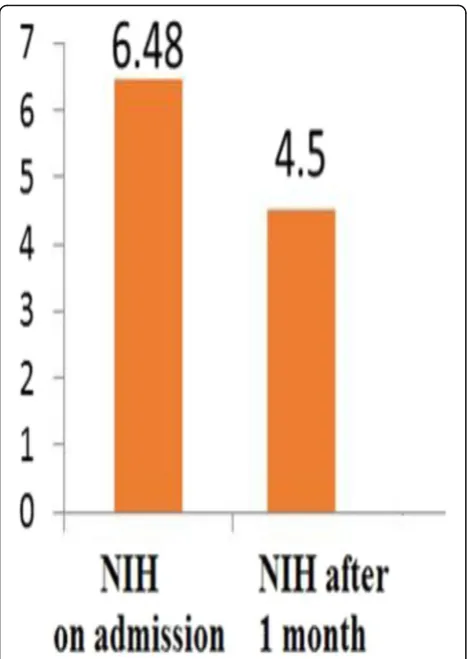
Patients were assessed using NIH stroke scale on admission, after 1 month and by modified Rankin scale after 3 months, mean value of NIH stroke scale on admission was 6.48 ± 2.97 ranging from 1 to 16, mean value of NIH stroke scale after 1 month was 4.5 ± 2.48 ranging from 0 to 9, mean value of modi-fied Rankin scale after 3 months was 2.13 ± 1.09 ran-ging from 0 to 4.
NIH on admission, after 1 month and MRS after 3
months (Table4) (Fig.1)
In this study, the mean value of common carotid artery intimal medial thickness was 1.25 as shown in Table5
Common carotid artery intimal medial thickness
Large versus small vessel stroke according to TOAST criteria among patients:
According to TOAST classification, percentage of large vessel stroke was 21.9 and small vessel stroke was
78.1 as shown in Table6
Correlation between hs-CRP and IMT
There was non significant correlation between
Hs-CRP and IMT as shown in Table7
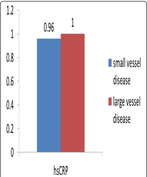
Comparison between patients with large and small
volumes regarding Hs-CRP value (Table8) (Fig.2)
Table 2Mean systolic and diastolic blood pressure on admission
Mean SD Range
Systolic blood pressure (SBP) on admission mmHg
152.19 24.33 100–220
Diastolic blood pressure (DBP) on admission mmHg
89.69 9.59 60–110
Table 1Age and gender of patients recruited in this study
Gender MaleN(%) 36 (56.3%)
FemaleN(%) 28 (43.8%)
Age, mean ± SD 62.47 ± 10.03
Table 3Mean value of common carotid artery intimal medial thickness
Mean SD
Carotid duplex IMT 1.25 0.58
Table 4Mean value of Hs-CRP, LDL, HbA1c, HDL, and troponin among patients
Mean SD Range
Hs-CRP mg/dL
.99 .91 0.1–3.8
LDL mg/dL
117.45 40.65 36–257
HbA1c % 6.99 1.75 4.5–13.5
HDL mg/dL
45.92 12.68 27–81
Troponin ng/mL
There was non significant difference between patients with large and small volumes regarding Hs-CRP value as
shown in Table8
Correlation between the volume of infarction and
the intimal medial thickness:
There was non significant difference between stroke volume and IMT on the symptomatic side and on the
asymptomatic side as shown in Table9
Correlations between Hs-CRP and NIH on
admis-sion, NIH after 1 month and MRS
There was significant correlation between Hs-CRP and MRS after 1 month yet non significant correl-ation with NIH on admission and after 1 month as
shown in Table 10.
Discussion
This study is considered pilot study aimed to correlate between C-reactive protein as a blood biomarker and in-timal media thickness of carotid artery, and its relation to infarct size and its impact on prognosis using clinical scales (NIHSS and MRS) which show significantly posi-tive correlation between highly sensiposi-tive C-reacposi-tive pro-tein with carotid intimal media thickness and also with MRS after 1 month. Elevated CRP and increased com-mon carotid artery-intimal medial thickness (CIMT) are both associated with the occurrence of stroke. CRP and IMT are closely associated; the higher the CRP, the greater the carotid atherosclerosis as measured by
ca-rotid IMT [6]. Many studies demonstrated that high
levels of inflammatory mediator such as plasma IL-6, TNF, IL-1, and CRP could predict stroke severity and poor long-term outcome have. CRP is one of the most extensively studied markers, which was associated with all stages of atherosclerosis, from the formation of the plaque to the occurrence and the progression of acute ischemic stroke [10].
In the current study, we correlate hs-CRP and C-IMT which was non-significant may be due to high percentage of stroke patients with Small vessel occlu-sion subtype in comparison to other subtypes of stroke in our sample. This result is different from meta-analysis done by Baldassarre and his colleagues [2, 11], who studied in their meta-analysis the rela-tionship between plasma levels of hs-CRP and C-IMT of 65 groups within 54 studies. A significant univari-ate correlation/association was found in 39 groups
Table 5Mean value of NIH on admission after 1 month and MRS after 3 months
Mean SD Range
NIH on admission 6.48 2.97 1–16
NIH after 1 month 4.50 2.48 0–9
MRS 2.13 1.09 0–4
Fig. 1Mean value of NIH on admission, after 1 month and MRS after 3 months. Patients were assessed using NIH stroke scale on admission, after 1 month and by modified Rankin scale after 3 months, mean value of NIH stroke scale on admission was 6.48 ±
2.97 ranging from 1–16, mean value of NIH stroke scale after one
month was 4.5 ± 2.48 ranging from 0 to 9
Table 6Percentage of large versus small vessel stroke according to TOAST criteria among patients
Number (N) Percentage (%)
Large vessel stroke 14 21.9
Small vessel stroke 50 78.1
Table 7Correlation between hs-CRP and IMT
IMT
Hs-CRP rs 0.234
(60%) within 29 studies. Whereas fewer than 4 (5%) were expected by chance if there were no relationship between the two variables and assuming no publica-tion bias.
Correlations between hs-CRP and NIH on admis-sion, NIH after 1 month and MRS were done, it showed there was a positive significant correlation be-tween Hs-CRP and MRS while no significant correla-tions were found as regards hs-CRP and NIH on admission. However, in a study done by Shoeab and his colleagues [9] where 50 patients with a first-ever acute stroke and they assessed stroke severity by the National Institute of Health Stroke Scale (NIHSS),
Hs-CRP level assay and CT brain were done to all patients on admission. Further assessment was done 7 days later, and the outcome was evaluated by a modified Rankin Scale (mRS). Results revealed that serum Hs-CRP level on admission was predictive of stroke severity (positively correlated with NIHSS, as well as outcome positively correlated with mRS, which was consistent with the current study yet they had shorter follow-up and smaller sample size.
While comparing large vessel stroke with small ves-sel stroke, no significant correlation was found be-tween high value of CRP and large vessel and small vessel disease stroke. This may be due to the small sample size of our study in comparison to other stud-ies that show significant correlation between level of hs-CRP and type of stroke. In contrast to a study
done by Terruzzi and his colleagues [12], where
pa-tients were included after a first-ever CT/MR docu-mented cerebral infarction. Hs-CRP was measured from blood samples. The study included 648 stroke patients. CRP levels were mostly increased in large vessel stroke, followed by small vessel stroke. They had larger sample size than the current study which may account to the non-significant results.
In the current study results, there was non-significant correlation between IMT with MRI stroke volume. This could be explained by the fact that IMT assesses the common carotid artery which is considered a large ar-tery, yet not enough information about the status of small vessels is indicated by carotid duplex IMT. The re-sult was in favor with previous study done by Nagai and his colleagues [13] which did not find significant differ-ence between CCA-IMT in large vessel disease (LVD) and small vessel disease (SVD).
However, one of the limitations of our study is the small sample size. So, this study can be considered as a pilot study.
Table 8Comparison between patients with large and small volumes regarding hs-CRP value
Volume
Small vessel stroke (14)
Large vessel stroke (50) t
test
Mean SD Mean SD Pvalue Sig
Hs-CRP 0.96 0.93 1.00 0.91 0.89 Non-significant (NS)
Fig. 2Comparison between patients with large and small volumes regarding hs-CRP value. Comparison between Hs-CRP value and large vessel stroke and small vessel stroke was done, and it was found that mean CRP was slightly higher in large vessel stroke with mean 1 mg/dL ± 0.91 while 0.96 mg/dL ± 0.93 yet it was insignificant (Pvalue = 0.89)
Table 10Correlation between hs-CRP and NIH on admission, NIH after 1 month and MRS
NIH on admission
NIH after 1 month
MRS NIH percent of change
Hs-CRP rs
0.23 0.21 0.28 −0.179
P value
0.06 NS 0.09 NS 0.02 Significant (S)
0.156 NS Table 9Correlation between stroke volume and IMT on the symptomatic side and on the asymptomatic side
IMT
MRI stroke volume rs −0.11
Conclusion
At the end of the study, it was concluded that highly sensitive C-reactive protein (hs-CRP) could serve as prognostic blood biomarker in long-term follow-up of stroke patients. Non-significant correlation was found in our study between increased hs-CRP and increased intima-media thickness (CCA-IMT).
Abbreviations
BI:Brain infarction; CCA: Common carotid arteries; CCA-IMT: Common carotid
arteries–intima-media thickness; CVD: Cardiovascular disease; DBP: Diastolic blood pressure; HbA1c: Glycated hemoglobin; HDL: High-density lipoprotein; Hs-CRP: Highly sensitive C-reactive protein; IMT: Intima media thickness; LDL: Low-density lipoprotein; mRS: Magnetic resonance imaging (MRI), modified Rankin Scale; N: Number; NIHSS: National Institute of Health and Stroke Scale; NS: Non-significant; S: Significant; SBP: Systolic blood pressure; SD: Standard deviation; SLE: Systemic lupus erythematosus; SPSSR: Statistical Package for Social Sciences
Acknowledgements
Not applicable.
Authors’contributions
ME and AE conceived of the study and participated in its design and coordination and helped to draft the manuscript (FG). AM and NE participated in the sequence alignment (MT). MA participated in the design of the study and performed the statistical analysis (ES). All authors read and approved the final manuscript.
Funding
Not applicable.
Availability of data and materials
Dataset is available as a master sheet in Excel format and publicly available in Neurology Department, Ain-Shams University through communicating corresponding authors.
Ethics approval and consent to participate
The study was approved by Ain-Shams University Ethical Committee in Octo-ber 2014. Written informed consent was obtained from the patients partici-pating in the study.
Consent for publication
Not applicable.
Competing interests
The authors declare that they have no competing interests.
Received: 23 February 2018 Accepted: 3 September 2019
References
1. Bathala L, Mehndiratta MM, Sharma VK. Cerebrovascular ultrasonography: technique and common pitfalls. Ann Indian Acad Neurol. 2013;16(1):121–7.
2. Deb P, Sharma S, Hassan KM. Pathophysiologic mechanisms of acute
ischemic stroke: an overview with emphasis on therapeutic significance beyond thrombolysis. Pathophysiology. 2010;17(3):197–218.
3. Hakimelahi R, Vachha BA, Copen WA, et al. Time and diffusion lesion size in major anterior circulation ischemic strokes. Stroke. 2014.https://doi.org/10. 1161/STROKEAHA.114.005644.
4. Martinic-Popovic I, Simundic AM, Dukic L, et al. The association of inflammatory markers with cerebral vasoreactivity and carotid atherosclerosis in transient ischaemic attack. Clin Biochem. 2014;47(16-17):182–6.
5. Wang TJ, Nam BH, Wilson PW, et al. Association of C-reactive protein with carotid atherosclerosis in men and women: the Framingham Heart Study. Arterioscler Thromb Vasc Biol. 2002;22:1662–7.
6. Ock SY, Cho KI, Kim HJ, et al. The impacts of C-reactive protein and atrial fibrillation on carotid atherosclerosis and ischemic stroke in patients with
suspected ischemic cerebrovascular disease: a single-center retrospective observational cohort study. Korean Circ J. 2013;43(12):796–803. 7. Sadreddini SA, Abolfathi AA, Khandagi R. C-reactive protein, fibrinogen,
lipoprotein (a), and lipid profile levels and platelet counts in ischemic stroke patients. Neurosciences (Riyadh). 2007;12(3):202–6.
8. Salisbury D, Bronas U. Inflammation and immune system contribution to
the etiology of atherosclerosis: mechanisms and methods of assessment. Nurs Res. 2014;63(5):375–85.
9. Shoeab MA, Shehata MA, Taema KM, Hammouda MA. CRP in cerebrovascular
stroke: prognostic implications. Egypt J Crit Care Med. 2014;2(1):43–52. 10. Zeng L, He X, Liu J, et al. Differences of circulating inflammatory markers
between large- and small vessel disease in patients with acute ischemic stroke. Int J Med Sci. 2013;10(10):1399–405.
11. Baldassarre D, De Jong A, Amato M, et al. Carotid intima-media thickness and markers of inflammation, endothelial damage and hemostasis. Ann Med. 2008;40:21–44.
12. Terruzzi A, Valente L, Mariani R, et al. C-reactive protein and aetiological subtypes of cerebral infarction. Neurol Sci. 2008;29:245–9.
13. Nagai Y, Kitagawa K, Yamagami H, et al. Carotid artery intima-media thickness and plaque score for the risk assessment of stroke subtypes. Ultrasound Med Biol. 2002;28:1239–43.
Publisher’s Note