imprinting interactions as well as unique expression patterns
Ben-Yehudah
et al.
Introduction
To identify the mechanisms underlying pluripotency, a number of studies have been carried out, and these have been recently summarized [1]. Th e fi rst two studies that characterized the ‘stemness gene’ list [2,3] identifi ed about 250 putative genes involved in mouse embryonic stem cell (mESC) pluripotency, and many other genes are being studied today [4-7]. While these experiments identifi ed many genes involved in maintenance of
pluri-potency, such as Oct-4, Nanog and Sox-2, they also
usually showed that human ESCs (hESCs) are quite diff erent from each other [4,8-10]. A more comprehensive
study showed that although closely related, the 59 ESC lines showed heterogeneity in gene expression [11]. Interestingly, variations in gene expression were found not only for genes correlated with the pluripotent state or diff erentiation, but also for housekeeping genes [12]. Th erefore, interactions among many genes likely form an active network that allows the pluripotent state to be main tained [13]. In addition, due to this variation between lines, better models need to be established to unders tand the true underlying mechanisms of pluripotency.
While gene regulatory networks that enhance our knowledge of pluripotency will help our understanding of stem cell biology, there are additional implications. As described below, ESCs are derived from the inner cell mass of a blastocyst [14,15]. Th erefore, the ESCs are closely related to the inner cell mass, from which, via post-implantation development, the embryo and fetus form. Th us, diff erentiation of ESCs recapitulates the earliest stages of human development, and understanding Abstract
The study of pluripotent stem cells has generated much interest in both biology and medicine. Understanding the fundamentals of biological decisions, including what permits a cell to maintain pluripotency, that is, its ability to self-renew and thereby remain immortal, or to diff erentiate into multiple types of cells, is of profound importance. For clinical applications, pluripotent cells, including both embryonic stem cells and adult stem cells, have been proposed for cell replacement therapy for a number of human diseases and disorders, including Alzheimer’s, Parkinson’s, spinal cord injury and diabetes. One challenge in their usage for such therapies is understanding the mechanisms that allow the maintenance of pluripotency and controlling the specifi c diff erentiation into required functional target cells. Because of regulatory restrictions and biological feasibilities, there are many crucial investigations that are just impossible to perform using pluripotent stem cells (PSCs) from humans (for example, direct comparisons among panels of inbred embryonic stem cells from prime embryos obtained from pedigreed and fertile donors; genomic analysis of parent versus progeny PSCs and their identical diff erentiated tissues; intraspecifi c chimera analyses for pluripotency testing; and so on). However, PSCs from nonhuman primates are being investigated to bridge these knowledge gaps between discoveries in mice and vital information necessary for appropriate clinical evaluations. In this review, we consider the mRNAs and novel genes with unique expression and imprinting patterns that were discovered using systems biology approaches with primate pluripotent stem and germ cells.
© 2010 BioMed Central Ltd
Systems biology discoveries using non-human
primate pluripotent stem and germ cells: novel
gene and genomic imprinting interactions as well
as unique expression patterns
Ahmi Ben-Yehudah
1,2, Charles A Easley IV
1,2,
Brian P Hermann
1,2, Carlos Castro
1,2, Calvin Simerly
1,2, Kyle E Orwig
1,2,
Shoukhrat Mitalipov
3and Gerald Schatten
1,2*
R E V I E W
*Correspondence: [email protected]
2Departments of Ob/Gyn and Reproductive Sciences, University of Pittsburgh
School of Medicine, Pittsburgh, PA 15213, USA
Full list of author information is available at the end of the article
the gene regulatory networks in these cells will enhance our knowledge of the regulation of the earliest stages of development.
Currently, we lack much information on specifi c stages in development. As described, all tissues of the embryo arise from the same cells; however, they are diff erent from each other, not only in their morphology and function, but also in their total DNA content. While somatic cells are diploid, gametes are only haploid. We have almost no knowledge on how these initial cells are selected and diff erentiated.
Another area that would benefi t from the delineation of pluripotent gene networks is the better understanding of reproductive mechanisms. While the sperm meets the egg to form the zygote, what gene expression in the egg allows the fi rst stages of development to proceed? Since in vitro fertilization (IVF) and other assisted reproductive technologies are so commonly used, can better under-standing of these fi rst stages increase the effi ciency of these technologies?
However, while ESC lines contribute to a chimera - which is when cells injected into a new blastocyst contri-bute to all tissues in the newborn - epiblast lines do not. We have no understanding of the mechanisms that underline the diff erence between these two types of cells.
ESCs have been studied not only for cell replacement therapy or basic stem cell biology, but also as a tool for development of better and safer drugs. Since the ESC lines resemble, in many aspects, the developing fetus, they can be used as a fi rst and quick tool for drug screen-ing without exposscreen-ing pregnant mothers and their babies to harmful drugs. Th erefore, better understanding of gene regulatory networks that control these cells or allow them to diff erentiate under specifi c signals will allow the development of new therapies. Th ese therapies will be based on the fi nding of new targets and hence the development of treatments specifi cally for those targets and the processes they control.
Certain cancers have been suggested to have a stem cell origin. Treatment today is usually directed to the amplifying cell rather than to the source of the cancer. Hence, understanding of the gene networks that have changed from those in stem cells and have led to cancer will allow the development of new treatments for the cancer, and as described before, the development of specifi cally targeted molecules for these pathologies.
To summarize, understanding of the gene regulatory networks that enable a cell to maintain its pluripotent phenotype are of great interest today. Better understand-ing of these networks will lead to better understandunderstand-ing of basic biology questions, the control of the specifi c diff erentiation of stem cells into target cells for cell replacement therapy, and the development of new drugs and treatments for cancer, among other diseases. Th is
review summarizes our current knowledge of gene
expres sion networks in non-human primates, which
resem bles the human model and also has greater
advantages.
Embryonic stem cells
ESCs are defi ned as a population of cells capable of self-renewing while maintaining their pluripotency. ESCs diff erentiate and give rise to cells from all three germ layers: ectoderm, mesoderm and endoderm, including the pancreas [16]. Th ese cells are derived from the inner cell mass of mammalian blastocyst stage embryos [17,18]. While mESCs were cloned over three decades ago, we have only recently celebrated our fi rst decade of hESC derivation. Hence, our knowledge on hESCs is more limited than that on mESCs. While the two types of ESC share many features, such as the expression of the pluri-potent marker OCT-4, they also diff er from each other, such as the dependency of mESCs on leukemia inhibitory factor (LIF) to maintain pluripotency. Presumably, therefore, pathways that contribute to both pluripotency as well as specifi c diff erentiation might diff er between
mESCs and hESCs. Th us, our knowledge on gene
expression in mESCs as well as their utilization for therapy must be verifi ed in hESCs.
Nuclear transfer
While the best known successful NT was Dolly the Sheep, derived by Sir Ian Wilmut [20], NT has been successful in many species, including dog, cat, mouse, cow, goat and others [21-23]. Recently, primate cells have been cloned, though with low effi ciency [24]. Th ese results support the contention that human cells could also be generated by NT, though very signifi cant
bioethical challenges remain. Th e reasons for the low effi ciency of primate cloning are not clear, and better methods for cloning are being investigated.
hurdles of this process: the lack of suffi cient numbers of donor eggs. However, the control experiments in this study were lacking. While the authors were able to demon strate that there was no development in the cloned intraspecies cells, they did not show their ability to clone human cells, raising questions about the effi ciency of cloning in this study [25].
ESCs and NT-ESCs have been proposed as possible treatments for diseases such as Parkinson’s, Alzheimer’s, juvenile diabetes and others. Specifi cally, NT-ESCs as cell replacement therapy - that is, using NT to derive ESCs - have a great advantage when compared to fertilized ESCs. Patients could provide their own fi broblasts for the derivation of NT-ESCs, and the resulting line would be patient-specifi c. However, since this process uses donated eggs that contain mitochondrial DNA, these cells are not identical to the donor patient [26]. Th erefore, the newly derived ESCs are mostly genetically similar to the donor, and hence might be rejected when fully diff eren tiated cells are transplanted. In contrast, many patients could not (for gender reasons) and/or should not (for ethical reasons) [26,27] donate eggs for the derivation of fertilized ESCs, and even then the newly derived cells would only be similar but not identical to the fi broblast from which they were derived, while fertilized ESCs can be closely related to the donor only if the egg donor is the patient herself or a fi rst degree relative.
NT-ESCs and fertilized ESCs have been shown to have many similar properties, including the ability to diff eren-tiate into cells from all three germ layers [28]. Since most diff erentiation procedures used for NT-ESCs will follow the same methods from fertilized ESCs, much emphasis is placed on generat ing NT-ESCs more effi ciently, and other studies are carried out to improve the diff eren-tiation procedures. As proof of principle, a number of publications have shown that NT-ESCs can be used for therapy. Th is was achieved in a Parkinson’s model [29] as well as a diabetic model in which NT-ESCs were diff erentiated into beta cells [30], thereby bridging the gap between the two routes of research. Th e method of diff er entiation for these NT-ESCs was based on protocols described previously [30]. Th ese cells were able to main-tain normal glucose levels after transplantion into a diabetic mouse [30]; however, an increase in blood glucose levels was seen 8 weeks after transplantation, presumably because the cells were not fully diff erentiated beta cells.
Induced pluripotent stem cells
One major goal of stem cell research is the generation of patient-specifi c stem cells. While successful in mice [31], the derivation of genetically matched, patient-specifi c human ESCs using somatic cell NT (SCNT) has not yet been accomplished. Furthermore, the use of donor oocytes or pre-implantation embryos to derive patient-specifi c
stem cells using techniques such as SCNT, cell fusion and parthenogenesis elicits ethical concerns. Th e recent advance in reprogramming adult somatic cells into ES-like cells, termed induced pluripotent stem (iPS) cells, provides another avenue for generating patient-specifi c stem cells without the ethical concerns of other methodologies. Th us, iPS cell derivation is the latest innovation for generating large pools of patient-specifi c stem cells that can be used to treat a wide range of human diseases.
In 2006, Takahashi and Yamanaka [32] demonstrated that mouse embryonic fi broblasts and adult tip fi bro-blasts could be reprogrammed into a pluripotent, ES-like state by transducing these cells with four transcription factors (Oct4, Sox2, Klf4 and c-Myc) along with a knock-in Fbx15 neomycknock-in-resistant reporter gene. After 2 weeks of culture, these mouse iPS cells exhibited similar characteristics to ESCs, such as alkaline phosphatase activity, expression of SSEA-1 and Nanog (two pluri-potency markers), and the ability to diff erentiate into all three germ layers (endoderm, ectoderm and mesoderm) via in vitro diff erentiation or teratoma formation in
immuno defi cient mice. While this ground-breaking
research provided a proof-of-principle for the repro-gram ming of adult somatic cells to ES-like cells, these iPS cells diff ered from ESCs in the genomic expression of several genes and the inability of the iPS cells to fully chimerize with donor mouse embryos. Shortly after the publication of this work, three groups showed that dispens ing with the reactivation of the Fbx15 reporter gene generated iPS cells that yielded fully chimerized pups following blastocyst injection and could contribute to germ cell transmission [33-35].
Recently, this work has been extended to human cells, as three groups originally demonstrated that human iPS cells could be generated from embryonic, neonatal and adult fi broblasts [36-38]. Like ESCs, these human iPS cells exhibited alkaline phosphatase activity, expressed SSEA-3, TRA-1-60, Oct4 and Nanog (human pluri-potency markers) from endogenous loci, and exhibited a genomic profi le more similar to ESCs than the originating fi broblasts. Interestingly, two diff erent combinations of retrovirally introduced transcription factors were used: Oct4, Sox2, Klf4 and c-Myc were utilized by two groups [36,37], while Yu and colleagues [38] generated human iPS cells using Oct4, Sox2, Nanog and Lin28, an RNA binding protein that regulates synthesis of the let-7 family of microRNAs (miRNAs). Currently, this work has been repeated in several other labs and has included repro-gramming of more terminally diff erentiated cells, such as pancreatic beta cells [39-42].
anemia model by diff erentiating iPS cells into hemato-poietic stem cells and re-introducing these cells back into the animal model [43]. Similarly, human iPS cells have been directly diff erentiated into motor neurons and insulin-secreting islet-like clusters in vitro [44,45]. While these results highlight the great promise for iPS cells in a clinical setting, transplanting iPS cells or diff erentiated iPS cells into humans carries a high risk. Around 20% of chimeric mice generated from iPS cells developed tumors within a 2- to 10-month time frame [34]. Th is fi nding is most likely due to the reactivation of c-Myc in iPS cells. While dispensing with c-Myc in iPS cell formation reduces the overall effi ciency of obtaining iPS cell colonies, chimeric mice derived from these cells appear to be free of tumors [46,47]. However, aberrant expres-sion of Oct4, Sox2, Klf4 and/or Nanog has been observed in a number of human malignancies [48-50]. Likewise, the random integration of the retroviruses could induce tumorigenesis by activating other oncogenic factors [51]. Because of these concerns, current research has been targeted at deriving iPS cells in a more clinically-friendly manner. Such protocols include using adenoviruses instead of retroviruses, and using miRNAs or a combi-nation of chemical and genetic modifi cations to induce reprogramming [52-54]. While iPS cell derivation is years away from being utilized in a clinical setting, the proof-of-principal results shown thus far indicate that iPS cells have the promise of treating a wide range of human disorders without the concern of immuno-rejection. Also, without requiring donor oocytes or pre-implan ta-tion embryos, iPS cell technology reduces the ethical concerns about generating patient-specifi c, pluripotent stem cells (PSCs).
Germ cells
Another potential source of stem cells is derived from precursor germ cells. In early embryonic development, a subset of pluripotent cells diff erentiate into primordial germ cells (PGCs) [55,56]. Th ese cells migrate, proliferate and colonize the genital ridge and represent a population of cells that will eventually further diff erentiate to form gametes. Initially discovered in mice, failure of PGCs to mitotically arrest following colonization leads to the formation of teratomas, tumors that contain cells repre-senting all three germ layers: ectoderm, mesoderm, and endoderm [57]. Th e fi rst isolations and cultures of these proliferating PGCs yielded a multipotent cell line termed embryonal carcinoma cells. Th ese cells are capable of being diff erentiated in culture into various cell types, including neurons and cardiomyocytes [58,59]. It was also shown that culture of isolated PGCs prior to genital ridge colonization resulted in germ cell colonies that express numerous pluripotency markers akin to those of ESCs, such as OCT-4 [60,61]. Th ese unique cells, termed
embryonic germ cells (EGCs) were shown to be highly pluripotent. EGCs have previously been an interesting cell source for studying gametogenesis in vitro because mouse EGCs appear to follow similar diff erentiation patterns as observed in in vivo gametogenesis [62]. However, ethical concerns about obtaining human EGCs have tamed interest in this fi eld.
Several groups have shown the ability of mouse, non-human primate and non-human ESCs to diff erentiate into germ cell lineages, specifi cally in vitro-derived PGCs (invPGCs) [63-79]. However, three groups in particular have demonstrated three diff erent methodologies for faithfully deriving invPGCs from ESCs at higher effi ci-encies [63-65]. Yamauchi and colleagues [64] successfully diff erentiated cynomolgus monkey ESCs into germ cells by forming embryoid bodies (EBs) with retinoic acid and culturing these EBs for 28 days. At day 28, germ cells could be identifi ed by positive immunostaining for SSEA1, VASA or DAZL. Furthermore, these researchers showed up-regulation of germ cell gene expression for CXCR4, NANOS1, NANOS2, NANOS3, VASA, PIWIL1 and TEKT1 upon EB formation with retinoic acid for 28 days. Likewise, this group was able to demonstrate that day 28 EBs grown in retinoic acid or bone morphogenetic protein (BMP)-4 elevated expression of the meiotic marker SCP1 but not SCP3. Kee and colleagues [63] showed that adherent diff erentiation with a BMP cocktail (BMP4, BMP7 and BMP8b) induced diff eren tiation of hESCs into invPGCs in 7 to 14 days. Using a green fl uorescent protein (GFP) transgene driven by the VASA promoter, these researchers showed that
diff eren tiation medium supplemented with BMPs
resulted in increased expres sion of two PGC markers in
diff eren tiating hESCs: VASA and DAZL. Kee and
colleagues also demonstrated that VASA-GFP+ cells could be isolated and cultured on mouse embryonic fi broblasts for 7 days to form invPGC colonies. Th ese cultured cells also exhibited hypomethylation of the H19 locus, suggesting that these cells, like in vivo PGCs, undergo de-methy lation prior to gametogenic progres-sion [63]. More importantly, Kee and colleagues
demon-strated that over expression of DAZ family members
(DAZ, DAZL and BOULE) in cultured invPGCs induces
meiotic progres sion as determined by immunofl
uor-escence staining for SCP3 and γH2AX [63]. Even more striking, they demonstrated haploid formation by over-expression of the DAZL family members by the appear-ance of a small 1N peak in their propidium iodide FACS analysis and the expression of acrosin in a small fraction of cells. Th is remarkable discovery highlights the poten-tial of driving gametogenesis in vitro from PSCs [63].
showed that diff erentiation of hESCs on human fetal gonadal stromal cells signifi cantly improved germ cell diff erentiation. Strikingly, these researchers showed that c-kit+/SSEA1+/VASA+ invPGCs (5% of the total popu la-tion of cells) could be isolated from diff erentiated hESCs as early as 3 days of culture on human fetal gonadal stromal cells. Similarly to Kee and colleagues, Park and colleagues demonstrated that invPGCs exhibit imprint erasures and show expression of a wide range of germ cell markers [63,65]. Th e work of Park and colleagues demon-strates progress towards a highly effi cient methodology for generating PGCs from ESCs in vitro [65].
Further-more, Park and colleagues are the fi rst group to
diff erentiate human iPS cells into early germ cell lineages. Th ese exciting results combined with the work of Kee and colleagues [63] and Yamauchi and colleagues [64] high light the similarities between in vivo PGCs and invPGCs illustrate the possibility of treating infertility by diff erentiating patient-matched ESCs into gametes or male germline stem cells for transplantation.
Th e ability to generate transplantable male germline stem cells or haploid gametes in culture has signifi cant therapeutic implications for couples with infertility [80,81]. Th e appeal of these approaches is enhanced by iPS cell and NT technologies, which would theoretically enable men to derive germline stem cells or sperm from their own skin cells in vitro. Th us, it is hypothetically possible for a man who is rendered infertile by toxic treatment for cancer (chemotherapy or radiation), and who did not cryopreserve semen prior to treatment, to father his own genetic children from germ cells derived from NT-ESCs or iPS cells. Th is potential can only be realized after extensive feasibility and safety studies are conducted, ideally in nonhuman primate models that are relevant to human physiology. Th ere is a lack of consen-sus among species regarding the potential of PGCs to undergo spermatogenesis when introduced into semini-ferous tubules (mouse PGCs can [82] and rat PGCs cannot (K Orwig, unpublished)). However, there is con sen sus in rodents and several large animal species that gono cytes and spermatogonia from neonate, pup and adult testes
undergo spermatogenesis when trans planted into the
testes of infertile recipients [82-87]. Human PSCs can be diff erentiated into PGCs in the context of EBs [70,88] or adherent diff erentiation cultures [63,65,67]. Similarly, two groups have reported macaque PSC to PGC diff eren tiation in EBs [64,89].Th ere are no reports of PSC to spermato-gonial stem cell (SSC) diff erentiation, but several studies have reported PSC diff erentiation to haploid germ cells [63,77,78], suggesting a transient transition through an SSC-like intermediate. Th us, direct diff erentiation of PSCs to SSCs would provide a source of transplantable cells that could be used to ask important questions about the safety and effi cacy of PSC-derived cells.
Interestingly, the postnatal mammalian testis itself may provide an alternative source of PSCs that bypasses the need for an embryonic intermediate or genetic manipu-lation. Several groups have shown the ability of germ cells in the mouse postnatal testis to produce PSCs in vitro [90-96]. Several recent studies have also provided evidence for PSCs derived from the adult human testis [97-100]. Th ese cells arise in vitro from spermatogonia and can give rise to tissues of all three embryonic germ layers. Given that germ cells are responsible for initiating embryogenesis, it seems possible that germ cell factors could infl uence their ability to become pluripotent (for example, including expression of genes associated with pluripotency). Among the genes that are thought to form a core regulatory network in ESCs (OCT4, SOX2, and NANOG) [101], only OCT-4 is expressed by a few postnatal germ cells or cultured SSCs. Several reports have described a relatively small group of normal mouse spermatogonia that express OCT-4, including those in the adult testis, which could potentially be those that have the capacity to produce PSCs in vitro [102-107]. In human spermatogonia, though, only a few postnatal spermatogonia retain embryonic-expressed OCT-4, and this expression is lost after the fi rst few months of infant life except in pathological conditions [108,109]. In cultured SSCs, Oct-4 mRNA and protein can be detected, albeit at substantially lower levels than in ESCs [90,95,110,111], and this feature may be required for long-term survival of SSCs in culture [112]. Th us, the mechanisms that predispose spermatogonia (presumably SSCs) to acquire a pluripotent phenotype in a culture dish are unclear, but may involve similar gene expression features with other pluripotent cells (ESCs).
line, including genes expressed, and compared them to the literature on gene and mRNA expression of human ESCs.
Using the newly derived nhpESC lines, we examined their gene expression [9] and found that they are very similar (>97%) to each other. We next compared the gene expression of these lines to that of two types of fi broblast: skin fi broblasts from the parents from which gametes were taken to derive the lines; and fi broblasts derived from teratomas generated by the injection of nhpESCs into severe combined immunodefi ciency (SCID) mice [1]. We found a unique set of genes that is diff erentially expressed between the nhpESCs and the two types of
fi broblast. Interestingly, many of these genes were
membrane-bound proteins and receptors [1] (Table 1). We have also shown that there are chromosomes that show an overabundance of over-expressed genes, such as chromosomes 16, 19 and X, which correlate to human chromosomes 17, 20 and X, respectively.
Our second study [114] has shown that there is indeed a unique set of genes in nhpESCs that maintains pluripotency, and that these diff erentially expressed genes are involved in many pathways. As expected, both studies revealed the over-expression of the ‘classic’ stem cell factors OCT-4, Nanog and Sox-2 (Table 1). Interest-ingly, when imprinting was examined, the nhpESCs were found to have aberrant imprinting, the implications of which are unknown [121].
When the two studies are compared, as depicted in Table 1, many of the top diff erentially expressed genes are similar. Taking into account that these were two separate studies, the resemblance between the two gene lists is striking. While Ben-Yehudah and colleagues [1] com-pared the gene expression of nhpESCs to fi broblasts and generated a list of genes over-expressed in ESCs, Mitalipov and colleagues [117] generated a list of genes that were highly expressed in a number of nhpESC lines. Out of the top 25 genes over-expressed in nhpESCs in both studies, 6 (24%) were found on both lists. As expected, genes known to be involved in maintenance, such as OCT-4 and Nanog, can be found on both lists. Additionally, PTPRZ1 is found on both lists; this gene has been shown to be expressed by hESCs and is down-regulated upon diff erentiation. Depletion of PTPRZ1 resulted in decreased colony formation and lower recovery of hESCs. However, the lists include genes that have yet to be associated with pluripotency, such as TACSTD1. TACSTD1, also called Ep-CAM, is an epi-thelial adhesion molecule that was originally identifi ed as a marker of carcinomas and is also expressed by rat SSCs [122-124]. We found this gene to be the most diff erentially expressed gene between stem cells and fi broblasts, indicating that it might have other functions in signaling rather than solely adhesion. In addition, it
should be pointed out that 40% of the genes on the list in Ben-Yehudah and colleagues [1] are hypo thetical; these genes may also play a signifi cant role in pluripotency.
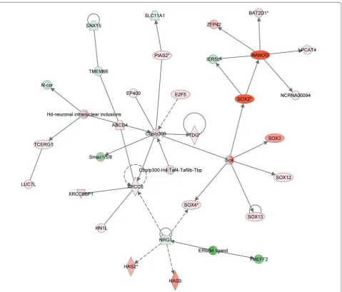
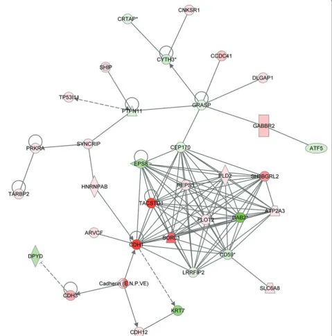
When we compiled the data in Ben-Yehudah and colleagues [1] using Ingenuity software to identify system networks responsible for the regulation of the pluripotent state in nhpESCs, we were able to create many gene networks. Some of these networks contained anticipated candidate genes, including SOX2, OCT-4 and NANOG, as we have shown previously [1]. In addition we could identify networks that have been shown to be diff eren tially expressed between stem cells and fi broblasts, as depicted in Table 1. Th ese genes participate in networks that have yet to be associated with pluripotency. Although most of the genes depicted in Figure 1 and Table 1 are unidentifi ed or have not been associated with pluripotency, some were found to play roles in regulating the transition from pluripotency to diff erentiation; for example, the gene TACSTD1 is included in both Figure 1 and Table 1.
Since ESCs can serve as a method of studying development [125], much research has been carried out to understand the mechanisms that underlie regulation of this specifi c process, such as the gene regulatory networks that control pluripotency. Th ese regulatory networks have been studied in mice [126] and have revealed the importance of key regulators of the pluri-potent state, including OCT-4, Sox-2 and Nanog. A comprehensive review described similar fi ndings in humans [127,128] and has also been discussed by us [1]. It should be pointed out that although many genes have been implicated in the networks controlling pluripotency, little is known about the networks controlling this process. An exception is the OCT-4/Sox-2/Nanog net-work, which has been shown to be invaluable for main-taining pluripotency. In our hands, we could identify the pluripotent genes and networks [1,24], but could not fi t all the diff erentially expressed genes into these networks or form new ones.
Imprinted genes in nhpESCs
While genes involved in pluripotency can be identifi ed and even gene regulatory networks can be described, other mechanisms controlling the expression of genes in pluripotent cells can be established - for example, epi-genetic mechanisms that control gene expression in pluri-potent cells. One such epigenetic mechanism is DNA methylation, which is considered a key factor in the formation of cellular memory and identity [129]. A comprehensive review summarized the key features of the regulatory mechanisms that control the trans crip tional regulatory features in hESCs [130,131], which complemented their work with ChIP-chip in mESCs [132].
can help answer questions on the epigenetic state of cells that undergo reprogramming - for example, whether they are closer to ESCs or to the somatic cell from which they originate. Th e answer to this question might shed light on why it is very diffi cult for primate cells to undergo NT compared to mice and other animals. Th is could lead to improvements in primate NT.
We have recently compared DNA methylation in native ESCs, fi broblasts, and ESCs generated by SCNT [129]. We wished to examine if the SCNT cells undergo changes in methylation state that would resemble a stem cell rather than a somatic cell. We have identifi ed and com-pared epigenome programming and reprogramming. Based on our previous knowledge, we have characterized hundreds of regions that are hyper- or hypomethylated in fi broblasts compared to native ESCs. We found that these regions are conserved in human cells and tissues. When ESCs were compared to the SCNT cells, we found to our surprise that the vast majority of these regions were
reprogrammed in SCNT ESCs. Th e meaning of these
phenomena is that these cells do indeed undergo repro-gramming of their DNA methylation during SCNT. Th is reprogramming leads to an almost perfect corre lation between the epigenomic profi les of the native (ESC) and reprogrammed (NTSC) lines.
We also found that at least 58% of these changes are correlated in cis to transcription changes, Polycomb repressive complex-2 occupancy, or binding by the CTCF insulator [129].
As expected, since the process of adding or removing a methyl group from the DNA must be a complex process, we found that while epigenomic repro gram-ming is extensive and globally accurate, the effi ciency of adding and stripping DNA methylation during reprogramming is regionally variable. In several cases, this variability results in regions that remain methylated in a fi broblast-like pattern even after reprogramming [129].
Table 1. Twenty-fi ve genes over-expressed in Ben-Yehudah and colleagues [1] and Mitalipov and colleagues [117]
Top 25 over-expressed genes in [1] Top 25 over-expressed genes in [117]
Aff ymetrix ProbeSet ID Gene symbol Aff ymetrix ProbeSet ID Gene symbol
1 MmuSTS.2870.1.S1_at TACSTD1 MmuSTS.3741.1.S1_at PTPRZ1
2 MmugDNA.35532.1.S1_at LOC697750 MmugDNA.32128.1.S1_at NANOG
3 MmuSTS.4178.1.S1_at CTSL2 MmugDNA.33796.1.S1_s_at FLJ16517
4 MmugDNA.17159.1.S1_at NFE2L3 MmugDNA.12465.1.S1_at LIN28
5 MmugDNA.20158.1.S1_at NELL2 MmuSTS.1454.1.S1_at MAL2
6 MmugDNA.11043.1.S1_at LOC705355 MmuSTS.2862.1.S1_at SPP1
7 MmunewRS.431.1.S1_at NPY1R MmuSTS.3364.1.S1_at PDZK1
8 MmuSTS.2285.1.S1_at POU5F1 MmugDNA.37987.1.S1_at SALL1
9 MmunewRS.475.1.S1_at LOC703107 MmuSTS.1929.1.S1_at MYCN
10 MmugDNA.24757.1.S1_at LOC702395 MmugDNA.20158.1.S1_at NELL2
11 MmuSTS.3573.1.S1_at PCDH8 MmuSTS.2870.1.S1_at TACSTD1
12 MmuSTS.3621.1.S1_at CHGB MmugDNA.17017.1.S1_at OTX2
13 MmuSTS.4813.1.S1_at GABRB3 MmugDNA.24774.1.S1_s_at APOA1
14 MmugDNA.38382.1.S1_at LOC696162 MmuSTS.1037.1.S1_at SH3GL3
15 MmugDNA.41477.1.S1_at NLGN4X MmugDNA.11977.1.S1_at MBD2
16 MmugDNA.17159.1.S1_s_at NFE2L3 MmugDNA.33242.1.S1_at PODXL
17 MmugDNA.19721.1.S1_at LOC696085 MmugDNA.6117.1.S1_at CECR2
18 MmuSTS.3827.1.S1_at LOC696132 MmuSTS.4090.1.S1_at EBAF
19 MmugDNA.32128.1.S1_at Nanog MmugDNA.36148.1.S1_at CYP26A1
20 MmugDNA.27729.1.S1_at SOX2 MmuSTS.2285.1.S1_at POU5F1
21 MmuSTS.3741.1.S1_at PTPRZ1 MmugDNA.3748.1.S1_at LOC112868
22 MmugDNA.7641.1.S1_at LOC712710 MmugDNA.32848.1.S1_at ST8SIA4
23 MmugDNA.33796.1.S1_s_at LOC696130 MmuSTS.214.1.S1_at ZIC3
24 MmugDNA.26523.1.S1_s_at NFE2L3 MmuSTS.1436.1.S1_at LCK
25 MmugDNA.31842.1.S1_s_at LOC696002 MmuSTS.4824.1.S1_at GDF3
Small RNAs and other RNAs
We have carried out many systems analyses using Ingenuity, as described in Figure 1. Th ese usually identi-fi ed networks not directly associated with pluripotency. However, we could occasionally identify a connection between genes associated with pluripotency and unique genes. One example is depicted in Figure 2. While Sox2 and Nanog are expressed in pluripotent cells (red), they
are also associated with the gene NCRNA00094. Th is
gene has been shown previously to be a non-coding RNA with unknown activity that is expressed in ESCs [134].
NCRNA00094 is an example of a large number of non-coding RNAs that might play a role in maintaining
a connection between the ‘stem cell factors’ and miRNAs that inhibit them [145]. However, more work has to be done to identify their specifi c targets and actions, such as the crosstalk between stem cell factors and miRNAs, as in the case of Lin-28 and Let7, for example [139].
A comprehensive comparison of gene and RNA profi les of mouse fertilized and SCNT lines has been carried out recently [146]. Th ey found that the two types of ESCs have similar miRNA and protein expression profi les. Th ey conclude that this phenomenon is consistent with their similar developmental potentials and might result from their similar transcriptional profi les.
While much research has been conducted on miRNA involvement in pluripotency in hESCs and mESCs, this has been little studied in the monkey. A recent study by some of us [137] has computationally searched the rhesus genome to identify novel miRNAs involved in
pluri-potency by homology to human miRNAs. Th is study
identifi ed 383 novel miRNAs: 173 have 100% homology to human miRNAs and 281 have >90% homology in the
seed sequence of the miRNAs [137]. Th is study also
identifi ed miRNAs that are involved in human ESC pluripotency, such as miR302, as described above.
Conclusions
In this review we have summarized our and other results from the past decade on the generation of nhpESCs, SCNT, the generation of iPS cells and our work on primordial germ cells. All these fi elds of research cumu-latively enhance our understanding of the early stages of human development. Th ese exciting results, together with our results on gene expression in rhesus macaques and other primates, open the possibility of studying the gene expres sion and its control by miRNAs that results in the undiff erentiated state of ESCs. Moreover, these studies
may lead to better understanding of the mecha nisms
behind processes such as induced pluripotency, the knowledge of which could be used to test cellular therapies in nonhuman primates before introduction in humans.
Abbreviations
BMP = bone morphogenetic protein; EB = embryoid body; EGC = embryonic germ cell; ESC = embryonic stem cell; hESC = human embryonic stem cell; invPGC = in vitro-derived primordial germ cell; iPS = induced pluripotent stem; IVF = in vitro fertilization; mESC = mouse embryonic stem cell; miRNA = microRNA; nhpESC = non-human primate embryonic stem cell; NT = nuclear transfer; PGC = primordial germ cell; PSC = pluripotent stem cell; SCNT = somatic cell nuclear transfer; SSC = spermatogonial stem cell.
Competing interests
The authors declare that they have no competing interests.
Acknowledgements
The support of this research by the NIH is acknowledged gratefully.
Author details
1Pittsburgh Development Center, 204 Craft Avenue, Pittsburgh, PA 15213, USA. 2Departments of Ob/Gyn and Reproductive Sciences, University of Pittsburgh
School of Medicine, Pittsburgh, PA 15213, USA. 3Oregon Stem Cell Center,
Oregon Health and Science University, Beaverton, Oregon 97006, USA.
Published: 5 August 2010
References
1. Ben-Yehudah A, Navara CS, Redinger CJ, Mich-Basso JD, Castro CA, Oliver S, Chensny LJ, Richards TJ, Kaminski N, Schatten G: Pluripotency genes overexpressed in primate embryonic stem cells are localized on homologues of human chromosomes 16, 17, 19, and X. Stem Cell Res 2010, 4:25-37.
2. Ivanova NB, Dimos JT, Schaniel C, Hackney JA, Moore KA, Lemischka IR: A stem cell molecular signature. Science 2002, 298:601-604.
3. Ramalho-Santos M, Yoon S, Matsuzaki Y, Mulligan RC, Melton DA: “Stemness”: transcriptional profi ling of embryonic and adult stem cells. Science 2002, 298:597-600.
4. Allegrucci C, Young LE: Diff erences between human embryonic stem cell lines. Hum Reprod Update 2007, 13:103-120.
5. Bhattacharya B, Cai J, Luo Y, Miura T, Mejido J, Brimble SN, Zeng X, Schulz TC, Rao MS, Puri RK: Comparison of the gene expression profi le of undiff erentiated human embryonic stem cell lines and diff erentiating embryoid bodies. BMC Dev Biol 2005, 5:22.
6. Bhattacharya B, Miura T, Brandenberger R, Mejido J, Luo Y, Yang AX, Joshi BH, Ginis I, Thies RS, Amit M, Lyons I, Condie BG, Itskovitz-Eldor J, Rao MS, Puri RK: Gene expression in human embryonic stem cell lines: unique molecular signature. Blood 2004, 103:2956-2964.
7. Cai J, Chen J, Liu Y, Miura T, Luo Y, Loring JF, Freed WJ, Rao MS, Zeng X: Assessing self-renewal and diff erentiation in human embryonic stem cell lines. Stem Cells 2006, 24:516-530.
8. Abeyta MJ, Clark AT, Rodriguez RT, Bodnar MS, Pera RA, Firpo MT: Unique gene expression signatures of independently-derived human embryonic stem cell lines. Hum Mol Genet 2004, 13:601-608.
9. Navara CS, Mich-Basso JD, Redinger CJ, Ben-Yehudah A, Jacoby E, Kovkarova-Naumovski E, Sukhwani M, Orwig K, Kaminski N, Castro CA, Simerly CR, Schatten G: Pedigreed primate embryonic stem cells express homogeneous familial gene profi les.Stem Cells 2007, 25:2695-2704. 10. Rao RR, Calhoun JD, Qin X, Rekaya R, Clark JK, Stice SL: Comparative
transcriptional profi ling of two human embryonic stem cell lines.
Biotechnol Bioeng 2004, 88:273-286.
11. Adewumi O, Afl atoonian B, Ahrlund-Richter L, Amit M, Andrews PW, Beighton G, Bello PA, Benvenisty N, Berry LS, Bevan S, Blum B, Brooking J, Chen KG, Choo AB, Churchill GA, Corbel M, Damjanov I, Draper JS, Dvorak P, Emanuelsson K, Fleck RA, Ford A, Gertow K, Gertsenstein M, Gokhale PJ, Hamilton RS, Hampl A, Healy LE, Hovatta O, Hyllner J, et al.: Characterization of human embryonic stem cell lines by the International Stem Cell Initiative. Nat Biotechnol 2007, 25:803-816.
12. Synnergren J, Giesler TL, Adak S, Tandon R, Noaksson K, Lindahl A, Nilsson P, Nelson D, Olsson B, Englund MC, Abbot S, Sartipy P: Diff erentiating human embryonic stem cells express a unique housekeeping gene signature.
Stem Cells 2007, 25:473-480.
13. Vogel G: Stem cells.‘Stemness’ genes still elusive.Science 2003, 302:371. 14. Navara CS, Redinger C, Mich-Basso J, Oliver S, Ben-Yehudah A, Castro C,
Simerly C: Derivation and characterization of nonhuman primate embryonic stem cells.Curr Protoc Stem Cell Biol 2007, Chapter 1:Unit 1A 1. 15. Thomson JA, Itskovitz-Eldor J, Shapiro SS, Waknitz MA, Swiergiel JJ, Marshall
VS, Jones JM: Embryonic stem cell lines derived from human blastocysts.
Science 1998, 282:1145-1147.
16. Keller G: Embryonic stem cell diff erentiation: emergence of a new era in biology and medicine. Genes Dev 2005, 19:1129-1155.
17. Evans MJ, Kaufman MH: Establishment in culture of pluripotential cells from mouse embryos. Nature 1981, 292:154-156.
18. Martin GR: Isolation of a pluripotent cell line from early mouse embryos cultured in medium conditioned by teratocarcinoma stem cells.Proc Natl Acad Sci U S A 1981, 78:7634-7638.
19. Yang X, Smith SL, Tian XC, Lewin HA, Renard JP, Wakayama T: Nuclear reprogramming of cloned embryos and its implications for therapeutic cloning.Nat Genet 2007, 39:295-302.
20. Wilmut I, Taylor J: Stem cells: primates join the club. Nature 2007, 450:485-486.
21. Markoulaki S, Meissner A, Jaenisch R: Somatic cell nuclear transfer and derivation of embryonic stem cells in the mouse. Methods 2008, 45:101-114.
23. Wilmut I, Beaujean N, de Sousa PA, Dinnyes A, King TJ, Paterson LA, Wells DN, Young LE: Somatic cell nuclear transfer. Nature 2002, 419:583-586. 24. Byrne JA, Pedersen DA, Clepper LL, Nelson M, Sanger WG, Gokhale S, Wolf DP,
Mitalipov SM: Producing primate embryonic stem cells by somatic cell nuclear transfer. Nature 2007, 450:497-502.
25. Chung Y, Bishop CE, Treff NR, Walker SJ, Sandler VM, Becker S, Klimanskaya I, Wun WS, Dunn R, Hall RM, Su J, Lu SJ, Maserati M, Choi YH, Scott R, Atala A, Dittman R, Lanza R: Reprogramming of human somatic cells using human and animal oocytes. Cloning Stem Cells 2009, 11:213-223.
26. Condic ML, Rao M: Regulatory issues for personalized pluripotent cells.
Stem Cells 2008, 26:2753-2758.
27. Rao M, Condic ML: Alternative sources of pluripotent stem cells: scientifi c solutions to an ethical dilemma. Stem Cells Dev 2008, 17:1-10.
28. Wakayama S, Jakt ML, Suzuki M, Araki R, Hikichi T, Kishigami S, Ohta H, Van Thuan N, Mizutani E, Sakaide Y, Senda S, Tanaka S, Okada M, Miyake M, Abe M, Nishikawa S, Shiota K, Wakayama T: Equivalency of nuclear transfer-derived embryonic stem cells to those derived from fertilized mouse blastocysts.
Stem Cells 2006, 24:2023-2033.
29. Tabar V, Tomishima M, Panagiotakos G, Wakayama S, Menon J, Chan B, Mizutani E, Al-Shamy G, Ohta H, Wakayama T, Studer L: Therapeutic cloning in individual parkinsonian mice. Nat Med 2008, 14:379-381.
30. Jiang W, Shi Y, Zhao D, Chen S, Yong J, Zhang J, Qing T, Sun X, Zhang P, Ding M, Li D, Deng H: In vitroderivation of functional insulin-producing cells from human embryonic stem cells.Cell Res 2007, 17:333-344.
31. Rideout WM 3rd, Hochedlinger K, Kyba M, Daley GQ, Jaenisch R: Correction of a genetic defect by nuclear transplantation and combined cell and gene therapy. Cell 2002, 109:17-27.
32. Takahashi K, Yamanaka S: Induction of pluripotent stem cells from mouse embryonic and adult fi broblast cultures by defi ned factors. Cell 2006, 126:663-676.
33. Maherali N, Sridharan R, Xie W, Utikal J, Eminli S, Arnold K, Stadtfeld M, Yachechko R, Tchieu J, Jaenisch R, Plath K, Hochedlinger K: Directly reprogrammed fi broblasts show global epigenetic remodeling and widespread tissue contribution.Cell Stem Cell 2007, 1:55-70.
34. Okita K, Ichisaka T, Yamanaka S: Generation of germline-competent induced pluripotent stem cells. Nature 2007, 448:313-317.
35. Wernig M, Meissner A, Foreman R, Brambrink T, Ku M, Hochedlinger K, Bernstein BE, Jaenisch R: In vitro reprogramming of fi broblasts into a pluripotent ES-cell-like state. Nature 2007, 448:318-324.
36. Park IH, Zhao R, West JA, Yabuuchi A, Huo H, Ince TA, Lerou PH, Lensch MW, Daley GQ: Reprogramming of human somatic cells to pluripotency with defi ned factors.Nature 2008, 451:141-146.
37. Takahashi K, Tanabe K, Ohnuki M, Narita M, Ichisaka T, Tomoda K, Yamanaka S: Induction of pluripotent stem cells from adult human fi broblasts by defi ned factors. Cell 2007, 131:861-872.
38. Yu J, Vodyanik MA, Smuga-Otto K, Antosiewicz-Bourget J, Frane JL, Tian S, Nie J, Jonsdottir GA, Ruotti V, Stewart R, Slukvin, II, Thomson JA: Induced pluripotent stem cell lines derived from human somatic cells. Science 2007, 318:1917-1920.
39. Lowry WE, Richter L, Yachechko R, Pyle AD, Tchieu J, Sridharan R, Clark AT, Plath K: Generation of human induced pluripotent stem cells from dermal fi broblasts. Proc Natl Acad Sci U S A 2008, 105:2883-2888.
40. Hanna J, Markoulaki S, Schorderet P, Carey BW, Beard C, Wernig M, Creyghton MP, Steine EJ, Cassady JP, Foreman R, Lengner CJ, Dausman JA, Jaenisch R: Direct reprogramming of terminally diff erentiated mature B lymphocytes to pluripotency.Cell 2008, 133:250-264.
41. Mali P, Ye Z, Hommond HH, Yu X, Lin J, Chen G, Zou J, Cheng L: Improved effi ciency and pace of generating induced pluripotent stem cells from human adult and fetal fi broblasts. Stem Cells 2008, 26:1998-2005. 42. Stadtfeld M, Brennand K, Hochedlinger K: Reprogramming of pancreatic
beta cells into induced pluripotent stem cells. Curr Biol 2008, 18:890-894. 43. Hanna J, Wernig M, Markoulaki S, Sun CW, Meissner A, Cassady JP, Beard C, Brambrink T, Wu LC, Townes TM, Jaenisch R: Treatment of sickle cell anemia mouse model with iPS cells generated from autologous skin. Science 2007, 318:1920-1923.
44. Dimos JT, Rodolfa KT, Niakan KK, Weisenthal LM, Mitsumoto H, Chung W, Croft GF, Saphier G, Leibel R, Goland R, Wichterle H, Henderson CE, Eggan K: Induced pluripotent stem cells generated from patients with ALS can be diff erentiated into motor neurons. Science 2008, 321:1218-1221. 45. Tateishi K, He J, Taranova O, Liang G, D’Alessio AC, Zhang Y: Generation of
insulin-secreting islet-like clusters from human skin fi broblasts. J Biol Chem
2008, 283:31601-31607.
46. Nakagawa M, Koyanagi M, Tanabe K, Takahashi K, Ichisaka T, Aoi T, Okita K, Mochiduki Y, Takizawa N, Yamanaka S: Generation of induced pluripotent stem cells without Myc from mouse and human fi broblasts. Nat Biotechnol
2008, 26:101-106.
47. Wernig M, Meissner A, Cassady JP, Jaenisch R: c-Myc is dispensable for direct reprogramming of mouse fi broblasts. Cell Stem Cell 2008, 2:10-12.
48. Clark AT:The stem cell identity of testicular cancer. Stem Cell Rev 2007, 3:49-59.
49. Gu G, Yuan J, Wills M, Kasper S: Prostate cancer cells with stem cell characteristics reconstitute the original human tumor in vivo. Cancer Res
2007, 67:4807-4815.
50. Rowland BD, Peeper DS: KLF4, p21 and context-dependent opposing forces in cancer. Nat Rev Cancer 2006, 6:11-23.
51. Zhao R, Daley GQ: From fi broblasts to iPS cells: induced pluripotency by defi ned factors. J Cell Biochem 2008, 105:949-955.
52. Li W, Wei W, Zhu S, Zhu J, Shi Y, Lin T, Hao E, Hayek A, Deng H, Ding S: Generation of rat and human induced pluripotent stem cells by combining genetic reprogramming and chemical inhibitors. Cell Stem Cell
2009, 4:16-19.
53. Lin SL, Chang DC, Chang-Lin S, Lin CH, Wu DT, Chen DT, Ying SY: Mir-302 reprograms human skin cancer cells into a pluripotent ES-cell-like state.
RNA 2008, 14:2115-2124.
54. Stadtfeld M, Nagaya M, Utikal J, Weir G, Hochedlinger K: Induced pluripotent stem cells generated without viral integration. Science 2008, 322:945-949. 55. Saitou M, Payer B, Lange UC, Erhardt S, Barton SC, Surani MA: Specifi cation of
germ cell fate in mice. Philos Trans R Soc Lond B Biol Sci 2003, 358:1363-1370. 56. Surani MA: Germ cells: the eternal link between generations.C R Biol 2007,
330:474-478.
57. Stevens LC: The biology of teratomas. Adv Morphog 1967, 6:1-31.
58. Kleinsmith LJ, Pierce GB Jr: Multipotentiality of single embryonal carcinoma cells.Cancer Res 1964, 24:1544-1551.
59. McBurney MW, Jones-Villeneuve EM, Edwards MK, Anderson PJ: Control of muscle and neuronal diff erentiation in a cultured embryonal carcinoma cell line. Nature 1982, 299:165-167.
60. Matsui Y, Zsebo K, Hogan BL: Derivation of pluripotential embryonic stem cells from murine primordial germ cells in culture.Cell 1992, 70:841-847. 61. Resnick JL, Bixler LS, Cheng L, Donovan PJ: Long-term proliferation of mouse
primordial germ cells in culture. Nature 1992, 359:550-551. 62. Donovan PJ, Stott D, Cairns LA, Heasman J, Wylie CC: Migratory and
postmigratory mouse primordial germ cells behave diff erently in culture.
Cell 1986, 44:831-838.
63. Kee K, Angeles VT, Flores M, Nguyen HN, Reijo Pera RA: Human DAZL, DAZ and BOULE genes modulate primordial germ-cell and haploid gamete formation.Nature 2009, 462:222-225.
64. Yamauchi K, Hasegawa K, Chuma S, Nakatsuji N, Suemori H: In vitro germ cell diff erentiation from cynomolgus monkey embryonic stem cells. PLoS One
2009, 4:e5338.
65. Park TS, Galic Z, Conway AE, Lindgren A, van Handel BJ, Magnusson M, Richter L, Teitell MA, Mikkola HK, Lowry WE, Plath K, Clark AT: Derivation of primordial germ cells from human embryonic and induced pluripotent stem cells is signifi cantly improved by coculture with human fetal gonadal cells. Stem Cells 2009, 27:783-795.
66. Bucay N, Yebra M, Cirulli V, Afrikanova I, Kaido T, Hayek A, Montgomery AM: A novel approach for the derivation of putative primordial germ cells and sertoli cells from human embryonic stem cells. Stem Cells 2009, 27:68-77. 67. Tilgner K, Atkinson SP, Golebiewska A, Stojkovic M, Lako M, Armstrong L:
Isolation of primordial germ cells from diff erentiating human embryonic stem cells. Stem Cells 2008, 26:3075-3085.
68. Salvador LM, Silva CP, Kostetskii I, Radice GL, Strauss JF 3rd: The promoter of the oocyte-specifi c gene, Gdf9, is active in population of cultured mouse embryonic stem cells with an oocyte-like phenotype. Methods 2008, 45:172-181.
69. Ohinata Y, Sano M, Shigeta M, Yamanaka K, Saitou M: A comprehensive, non-invasive visualization of primordial germ cell development in mice by the Prdm1-mVenus and Dppa3-ECFP double transgenic reporter.
Reproduction 2008, 136:503-514.
70. Kee K, Gonsalves JM, Clark AT, Pera RA: Bone morphogenetic proteins induce germ cell diff erentiation from human embryonic stem cells. Stem Cells Dev 2006, 15:831-837.
Yang YS, Ho HN: Derivation, characterization and diff erentiation of human embryonic stem cells: comparing serum-containing versus serum-free media and evidence of germ cell diff erentiation. Hum Reprod 2007, 22:567-577.
72. Mikkola M, Olsson C, Palgi J, Ustinov J, Palomaki T, Horelli-Kuitunen N, Knuutila S, Lundin K, Otonkoski T, Tuuri T: Distinct diff erentiation
characteristics of individual human embryonic stem cell lines. BMC Dev Biol
2006, 6:40.
73. Diaconu M, Tangat Y, Bohm D, Kuhn H, Michelmann HW, Schreiber G, Haidl G, Glander HJ, Engel W, Nayernia K: Failure of phospholipid hydroperoxide glutathione peroxidase expression in oligoasthenozoospermia and mutations in the PHGPx gene. Andrologia 2006, 38:152-157.
74. Payer B, Chuva de Sousa Lopes SM, Barton SC, Lee C, Saitou M, Surani MA: Generation of stella-GFP transgenic mice: a novel tool to study germ cell development. Genesis 2006, 44:75-83.
75. Lacham-Kaplan O, Chy H, Trounson A: Testicular cell conditioned medium supports diff erentiation of embryonic stem cells into ovarian structures containing oocytes. Stem Cells 2006, 24:266-273.
76. Clark AT, Rodriguez RT, Bodnar MS, Abeyta MJ, Cedars MI, Turek PJ, Firpo MT, Reijo Pera RA: Human STELLAR, NANOG, and GDF3 genes are expressed in pluripotent cells and map to chromosome 12p13, a hotspot for teratocarcinoma. Stem Cells 2004, 22:169-179.
77. Geijsen N, Horoschak M, Kim K, Gribnau J, Eggan K, Daley GQ: Derivation of embryonic germ cells and male gametes from embryonic stem cells.
Nature 2004, 427:148-154.
78. Toyooka Y, Tsunekawa N, Akasu R, Noce T: Embryonic stem cells can form germ cells in vitro. Proc Natl Acad Sci U S A 2003, 100:11457-11462. 79. Hubner K, Fuhrmann G, Christenson LK, Kehler J, Reinbold R, De La Fuente R,
Wood J, Strauss JF, 3rd, Boiani M, Scholer HR: Derivation of oocytes from mouse embryonic stem cells. Science 2003, 300:1251-1256.
80. Orwig KE, Schlatt S: Cryopreservation and transplantation of
spermatogonia and testicular tissue for preservation of male fertility. J Natl Cancer Inst Monogr 2005:51-56.
81. Brinster RL: Male germline stem cells: from mice to men. Science 2007, 316:404-405.
82. Chuma S, Kanatsu-Shinohara M, Inoue K, Ogonuki N, Miki H, Toyokuni S, Hosokawa M, Nakatsuji N, Ogura A, Shinohara T: Spermatogenesis from epiblast and primordial germ cells following transplantation into postnatal mouse testis. Development 2005, 132:117-122. 83. Shinohara T, Orwig KE, Avarbock MR, Brinster RL: Remodeling of the
postnatal mouse testis is accompanied by dramatic changes in stem cell number and niche accessibility. Proc Natl Acad Sci U S A 2001, 98:6186-6191. 84. Ryu BY, Orwig KE, Avarbock MR, Brinster RL: Stem cell and niche
development in the postnatal rat testis. Dev Biol 2003, 263:253-263. 85. Honaramooz A, Megee SO, Dobrinski I: Germ cell transplantation in pigs.
Biol Reprod 2002, 66:21-28.
86. Honaramooz A, Behboodi E, Megee SO, Overton SA, Galantino-Homer H, Echelard Y, Dobrinski I: Fertility and germline transmission of donor haplotype following germ cell transplantation in immunocompetent goats. Biol Reprod 2003, 69:1260-1264.
87. Kim Y, Turner D, Nelson J, Dobrinski I, McEntee M, Travis AJ: Production of donor-derived sperm after spermatogonial stem cell transplantation in the dog. Reproduction 2008, 136:823-831.
88. Clark AT, Bodnar MS, Fox M, Rodriquez RT, Abeyta MJ, Firpo MT, Pera RA: Spontaneous diff erentiation of germ cells from human embryonic stem cells in vitro. Hum Mol Genet 2004, 13:727-739.
89. Teramura T, Takehara T, Kawata N, Fujinami N, Mitani T, Takenoshita M, Matsumoto K, Saeki K, Iritani A, Sagawa N, Hosoi Y: Primate embryonic stem cells proceed to early gametogenesis in vitro. Cloning Stem Cells 2007, 9:144-156.
90. Kanatsu-Shinohara M, Inoue K, Lee J, Yoshimoto M, Ogonuki N, Miki H, Baba S, Kato T, Kazuki Y, Toyokuni S, Toyoshima M, Niwa O, Oshimura M, Heike T, Nakahata T, Ishino F, Ogura A, Shinohara T: Generation of pluripotent stem cells from neonatal mouse testis. Cell 2004, 119:1001-1012.
91. Kanatsu-Shinohara M, Lee J, Inoue K, Ogonuki N, Miki H, Toyokuni S, Ikawa M, Nakamura T, Ogura A, Shinohara T: Pluripotency of a single spermatogonial stem cell in mice. Biol Reprod 2008, 78:681-687.
92. Guan K, Nayernia K, Maier LS, Wagner S, Dressel R, Lee JH, Nolte J, Wolf F, Li M, Engel W, Hasenfuss G: Pluripotency of spermatogonial stem cells from adult mouse testis. Nature 2006, 440:1199-1203.
93. Huang YH, Chin CC, Ho HN, Chou CK, Shen CN, Kuo HC, Wu TJ, Wu YC, Hung
YC, Chang CC, Ling TY: Pluripotency of mouse spermatogonial stem cells maintained by IGF-1- dependent pathway. FASEB J 2009, 23:2076-2087. 94. Izadyar F, Pau F, Marh J, Slepko N, Wang T, Gonzalez R, Ramos T, Howerton K,
Sayre C, Silva F: Generation of multipotent cell lines from a distinct population of male germ line stem cells.Reproduction 2008, 135:771-784. 95. Seandel M, James D, Shmelkov SV, Falciatori I, Kim J, Chavala S, Scherr DS,
Zhang F, Torres R, Gale NW, Yancopoulos GD, Murphy A, Valenzuela DM, Hobbs RM, Pandolfi PP, Rafi i S: Generation of functional multipotent adult stem cells from GPR125+ germline progenitors. Nature 2007, 449:346-350. 96. Ko K, Tapia N, Wu G, Kim JB, Bravo MJ, Sasse P, Glaser T, Ruau D, Han DW,
Greber B, Hausdorfer K, Sebastiano V, Stehling M, Fleischmann BK, Brustle O, Zenke M, Scholer HR: Induction of pluripotency in adult unipotent germline stem cells. Cell Stem Cell 2009, 5:87-96.
97. Conrad S, Renninger M, Hennenlotter J, Wiesner T, Just L, Bonin M, Aicher W, Buhring HJ, Mattheus U, Mack A, Wagner HJ, Minger S, Matzkies M, Reppel M, Hescheler J, Sievert KD, Stenzl A, Skutella T: Generation of pluripotent stem cells from adult human testis. Nature 2008, 456:344-349.
98. Kossack N, Meneses J, Shefi S, Nguyen HN, Chavez S, Nicholas C, Gromoll J, Turek PJ, Reijo-Pera RA: Isolation and characterization of pluripotent human spermatogonial stem cell-derived cells. Stem Cells 2009, 27:138-149. 99. Golestaneh N, Kokkinaki M, Pant D, Jiang J, DeStefano D, Fernandez-Bueno C,
Rone JD, Haddad BR, Gallicano GI, Dym M: Pluripotent stem cells derived from adult human testes. Stem Cells Dev 2009, 18:1115-1126.
100. Mizrak SC, Chikhovskaya JV, Sadri-Ardekani H, van Daalen S, Korver CM, Hovingh SE, Roepers-Gajadien HL, Raya A, Fluiter K, de Reijke TM, de la Rosette JJ, Knegt AC, Belmonte JC, van der Veen F, de Rooij DG, Repping S, van Pelt AM: Embryonic stem cell-like cells derived from adult human testis. Hum Reprod 2010, 25:158-167.
101. Boyer LA, Lee TI, Cole MF, Johnstone SE, Levine SS, Zucker JP, Guenther MG, Kumar RM, Murray HL, Jenner RG, Giff ord DK, Melton DA, Jaenisch R, Young RA: Core transcriptional regulatory circuitry in human embryonic stem cells. Cell 2005, 122:947-956.
102. Pesce M, Wang X, Wolgemuth DJ, Scholer H: Diff erential expression of the Oct-4 transcription factor during mouse germ cell diff erentiation. Mech Dev 1998, 71:89-98.
103. Yoshimizu T, Sugiyama N, De Felice M, Yeom YI, Ohbo K, Masuko K, Obinata M, Abe K, Scholer HR, Matsui Y: Germline-specifi c expression of the Oct-4/ green fl uorescent protein (GFP) transgene in mice. Dev Growth Diff er 1999, 41:675-684.
104. Ohbo K, Yoshida S, Ohmura M, Ohneda O, Ogawa T, Tsuchiya H, Kuwana T, Kehler J, Abe K, Scholer HR, Suda T: Identifi cation and characterization of stem cells in prepubertal spermatogenesis in mice small star, fi lled. Dev Biol 2003, 258:209-225.
105. Ohmura M, Yoshida S, Ide Y, Nagamatsu G, Suda T, Ohbo K: Spatial analysis of germ stem cell development in Oct-4/EGFP transgenic mice. Arch Histol Cytol 2004, 67:285-296.
106. Buaas FW, Kirsh AL, Sharma M, McLean DJ, Morris JL, Griswold MD, de Rooij DG, Braun RE: Plzf is required in adult male germ cells for stem cell self-renewal. Nat Genet 2004, 36:647-652.
107. Tokuda M, Kadokawa Y, Kurahashi H, Marunouchi T: CDH1 is a specifi c marker for undiff erentiated spermatogonia in mouse testes. Biol Reprod
2007, 76:130-141.
108. Looijenga LH, Stoop H, de Leeuw HP, de Gouveia Brazao CA, Gillis AJ, van Roozendaal KE, van Zoelen EJ, Weber RF, Wolff enbuttel KP, van Dekken H, Honecker F, Bokemeyer C, Perlman EJ, Schneider DT, Kononen J, Sauter G, Oosterhuis JW: POU5F1 (OCT3/4) identifi es cells with pluripotent potential in human germ cell tumors. Cancer Res 2003, 63:2244-2250.
109. Rajpert-De Meyts E, Hanstein R, Jorgensen N, Graem N, Vogt PH, Skakkebaek NE: Developmental expression of POU5F1 (OCT-3/4) in normal and dysgenetic human gonads. Hum Reprod 2004, 19:1338-1344.
110. Kanatsu-Shinohara M, Ogonuki N, Iwano T, Lee J, Kazuki Y, Inoue K, Miki H, Takehashi M, Toyokuni S, Shinkai Y, Oshimura M, Ishino F, Ogura A, Shinohara T: Genetic and epigenetic properties of mouse male germline stem cells during long-term culture. Development 2005, 132:4155-4163.
111. Imamura M, Miura K, Iwabuchi K, Ichisaka T, Nakagawa M, Lee J, Kanatsu-Shinohara M, Kanatsu-Shinohara T, Yamanaka S: Transcriptional repression and DNA hypermethylation of a small set of ES cell marker genes in male germline stem cells. BMC Dev Biol 2006, 6:34.
2008, 26:2928-2937.
113. Simerly CR, Navara CS, Castro CA, Turpin JC, Redinger CJ, Mich-Basso JD, Jacoby ES, Grund KJ, McFarland DA, Oliver SL, Ben-Yehudah A, Carlisle DL, Frost P, Penedo C, Hewitson L, Schatten G: Establishment and characterization of baboon embryonic stem cell lines: An Old World Primate model for regeneration and transplantation research. Stem Cell Res
2009 [Epub ahead of print].
114. Byrne JA, Mitalipov SM, Clepper L, Wolf DP: Transcriptional profi ling of rhesus monkey embryonic stem cells. Biol Reprod 2006, 75:908-915. 115. Byrne JA, Mitalipov SM, Wolf DP: Current progress with primate embryonic
stem cells. Curr Stem Cell Res Ther 2006, 1:127-138.
116. Dighe V, Clepper L, Pedersen D, Byrne J, Ferguson B, Gokhale S, Penedo MC, Wolf D, Mitalipov S: Heterozygous embryonic stem cell lines derived from nonhuman primate parthenotes. Stem Cells 2008, 26:756-766.
117. Mitalipov S, Kuo HC, Byrne J, Clepper L, Meisner L, Johnson J, Zeier R, Wolf D: Isolation and characterization of novel rhesus monkey embryonic stem cell lines. Stem Cells 2006, 24:2177-2186.
118. Mitalipov SM, Wolf DP: Nuclear transfer in nonhuman primates. Methods Mol Biol 2006, 348:151-168.
119. Muller T, Fleischmann G, Eildermann K, Matz-Rensing K, Horn PA, Sasaki E, Behr R: A novel embryonic stem cell line derived from the common marmoset monkey (Callithrix jacchus) exhibiting germ cell-like characteristics. Hum Reprod 2009, 24:1359-1372.
120. Eiges R, Urbach A, Malcov M, Frumkin T, Schwartz T, Amit A, Yaron Y, Eden A, Yanuka O, Benvenisty N, Ben-Yosef D: Developmental study of fragile X syndrome using human embryonic stem cells derived from preimplantation genetically diagnosed embryos. Cell Stem Cell 2007, 1:568-577.
121. Fujimoto A, Mitalipov SM, Kuo HC, Wolf DP: Aberrant genomic imprinting in rhesus monkey embryonic stem cells. Stem Cells 2006, 24:595-603. 122. Baeuerle PA, Gires O: EpCAM (CD326) fi nding its role in cancer. Br J Cancer
2007, 96:417-423.
123. Trzpis M, McLaughlin PM, de Leij LM, Harmsen MC: Epithelial cell adhesion molecule: more than a carcinoma marker and adhesion molecule. Am J Pathol 2007, 171:386-395.
124. Ryu BY, Orwig KE, Kubota H, Avarbock MR, Brinster RL: Phenotypic and functional characteristics of spermatogonial stem cells in rats. Dev Biol
2004, 274:158-170.
125. Dvash T, Benvenisty N: Human embryonic stem cells as a model for early human development. Best Pract Res Clin Obstet Gynaecol 2004, 18:929-940. 126. Zhou Q, Chipperfi eld H, Melton DA, Wong WH: A gene regulatory network
in mouse embryonic stem cells. Proc Natl Acad Sci U S A 2007, 104:16438-16443.
127. Aiba K, Carter MG, Matoba R, Ko MS: Genomic approaches to early embryogenesis and stem cell biology. Semin Reprod Med 2006, 24:330-339. 128. Sharov AA, Masui S, Sharova LV, Piao Y, Aiba K, Matoba R, Xin L, Niwa H, Ko MS:
Identifi cation of Pou5f1, Sox2, and Nanog downstream target genes with statistical confi dence by applying a novel algorithm to time course microarray and genome-wide chromatin immunoprecipitation data.
BMC Genomics 2008, 9:269.
129. Cohen NM, Dighe V, Landan G, Reynisdottir S, Palsson A, Mitalipov S, Tanay A: DNA methylation programming and reprogramming in primate embryonic stem cells. Genome Res 2009, 19:2193-2201.
130. Cole MF, Young RA: Mapping key features of transcriptional regulatory circuitry in embryonic stem cells. Cold Spring Harb Symp Quant Biol 2008, 73:183-193.
131. Marson A, Levine SS, Cole MF, Frampton GM, Brambrink T, Johnstone S, Guenther MG, Johnston WK, Wernig M, Newman J, Calabrese JM, Dennis LM, Volkert TL, Gupta S, Love J, Hannett N, Sharp PA, Bartel DP, Jaenisch R, Young RA: Connecting microRNA genes to the core transcriptional regulatory circuitry of embryonic stem cells.Cell 2008, 134:521-533.
132. Mathur D, Danford TW, Boyer LA, Young RA, Giff ord DK, Jaenisch R: Analysis of the mouse embryonic stem cell regulatory networks obtained by ChIP-chip and ChIP-PET.Genome Biol 2008, 9:R126.
133. Mitalipov SM, Zhou Q, Byrne JA, Ji WZ, Norgren RB, Wolf DP: Reprogramming following somatic cell nuclear transfer in primates is dependent upon nuclear remodeling. Hum Reprod 2007, 22:2232-2242.
134. Darr H, Mayshar Y, Benvenisty N: Overexpression of NANOG in human ES cells enables feeder-free growth while inducing primitive ectoderm features. Development 2006, 133:1193-1201.
135. Zhang B, Pan X, Anderson TA: MicroRNA: a new player in stem cells. J Cell Physiol 2006, 209:266-269.
136. Lakshmipathy U, Hart RP: Concise review: MicroRNA expression in multipotent mesenchymal stromal cells. Stem Cells 2008, 26:356-363. 137. Yue J, Sheng Y, Orwig KE: Identifi cation of novel homologous microRNA
genes in the rhesus macaque genome. BMC Genomics 2008, 9:8. 138. Houbaviy HB, Murray MF, Sharp PA: Embryonic stem cell-specifi c
microRNAs. Dev Cell 2003, 5:351-358.
139. Viswanathan SR, Daley GQ, Gregory RI: Selective blockade of microRNA processing by Lin28. Science 2008, 320:97-100.
140. Lakshmipathy U, Love B, Goff LA, Jornsten R, Graichen R, Hart RP, Chesnut JD: MicroRNA expression pattern of undiff erentiated and diff erentiated human embryonic stem cells. Stem Cells Dev 2007, 16:1003-1016. 141. Brameier M, Wiuf C: Ab initio identifi cation of human microRNAs based on
structure motifs. BMC Bioinformatics 2007, 8:478.
142. Bar M, Wyman SK, Fritz BR, Qi J, Garg KS, Parkin RK, Kroh EM, Bendoraite A, Mitchell PS, Nelson AM, Ruzzo WL, Ware C, Radich JP, Gentleman R, Ruohola-Baker H, Tewari M: MicroRNA discovery and profi ling in human embryonic stem cells by deep sequencing of small RNA libraries. Stem Cells 2008, 26:2496-2505.
143. Suh MR, Lee Y, Kim JY, Kim SK, Moon SH, Lee JY, Cha KY, Chung HM, Yoon HS, Moon SY, Kim VN, Kim K: Human embryonic stem cells express a unique set of microRNAs. Dev Biol 2004, 270:488-498.
144. Laurent LC, Chen J, Ulitsky I, Mueller FJ, Lu C, Shamir R, Fan JB, Loring JF: Comprehensive microRNA profi ling reveals a unique human embryonic stem cell signature dominated by a single seed sequence. Stem Cells 2008, 26:1506-1516.
145. Tay Y, Zhang J, Thomson AM, Lim B, Rigoutsos: MicroRNAs to Nanog, Oct4 and Sox2 coding regions modulate embryonic stem cell diff erentiation.
Nature 2008, 455:1124-1128.
146. Ding J, Guo Y, Liu S, Yan Y, Chang G, Kou Z, Zhang Y, Jiang Y, He F, Gao S, Sang J: Embryonic stem cells derived from somatic cloned and fertilized blastocysts are post-transcriptionally indistinguishable: a microRNA and protein profi le comparison. Proteomics 2009, 9:2711-2721.
doi:10.1186/scrt24
Cite this article as: Ben-Yehudah A, et al.: Systems biology discoveries using non-human primate pluripotent stem and germ cells: novel gene and genomic imprinting interactions as well as unique expression patterns.
![Table 1. Twenty-fi ve genes over-expressed in Ben-Yehudah and colleagues [1] and Mitalipov and colleagues [117]](https://thumb-us.123doks.com/thumbv2/123dok_us/704294.2068143/9.612.64.549.99.482/table-genes-expressed-ben-yehudah-colleagues-mitalipov-colleagues.webp)