Research Article
Vascular Endothelial Growth Factor-C (VEGF-C) Expression
Can Not Predict Pelvic Lymph Node Metastases and Response
to Neo-adjuvant Chemotherapy in Bulky Cervical Cancer
Ekspresi Vascular Endothelial Growth Factor-C (VEGF-C) tidak dapat
Memprediksi Metastasis ke Kelenjar Getah Bening Pelvis dan
Respons terhadap Kemoterapi Neo Ajuvan pada Kanker Serviks Berukuran Besar
Johnson Hutapea1, Nugroho Kampono1, Bambang Sutrisna2, Emil Taufik3
1Gynecologic Oncology Division, Department of Obstetrics and Gynecology 2Department of Epidemiology
3Department of Anatomical Pathology Faculty of Medicine University of Indonesia/
Dr. Cipto Mangunkusumo Hospital Jakarta
Abstract
Objective: To assess whether VEGF-C expression can predict the re-sponse to neoadjuvant chemotherapy and pelvic lymphnode metas-tases in bulky cevical cancer.
Methods: Seventeen cervical cancer stage IB2 and IIA2 cases during
the period of July 2009 until June 2010 were collected consecutively and given neoadjuvant chemotherapy (NAC) PVB prior radical surgery. Re-sponse to treatment was evaluated based on the change of tumour size. VEGF-C expression was examined immunohistochemically at tumour bi-opsy before chemotherapy. The presence of lymphnode metastases his-topathologically were obtained from pelvic lymphnode dissection. The difference and correlation of response and metastases on VEGF-C ex-pression were analized statistically. The validity of the cut off percentage of immunopositive cells to VEGF-C to identify non responding and me-tastatic cases was calculated with the ROC. Multivariate analysis were done to determine the predictor of no response to chemotherapy.
Results: Clinical response, using the RECIST version 1.1 criteria, was found in 41.18% cases and lymphnode metastases were found in 27.27% cases. VEGF-C was expressed in all cases. Statistically, there were no significant differences and correlation in response to treat-ment and pelvic lymphnode metastases on VEGF-C expression. At the cut off ≥ 76% immunopositivity to VEGF-C, the sensitivity to identify no response and the specificity to identify response to NAC are 70.00% and 71.43% respectively (LR+ 2.45 and LR- 0.42); whereas at the cut off ≥ 75% immunopositivity to VEGF-C, the sen-sitivity to identify lymphnode metastases and the specificity to iden-tify no lymphnode metastases are 100.00% and 75.00% (LR+ 4.0 dan LR- 0). With multivariate analysis using logistic regression, the cut off ≥ 76% immunopositive cells to VEGF-C were found to have positive coefficient, largest OR and statistic score, 1.93, 6.88 (96% CI OR 0.45; 104.34) and 41 respectively, to predict non responders in a prediction score model.
Conclusion: VEGF-C expression on biopsy specimen bulky cervical cancers can not differentiate cases that respond to NAC and metas-tases to the pelvic lymphnode from that do not. The cut off ≥ 76% immunopositive cells to VEGF-C in a prediction model can be used as an alternative predictor to identify non responders.
[Indones J Obstet Gynecol 2012; 36-3: 144-9]
Keywords: bulky cervical cancer, neoadjuvant chemotherapy, re-sponse and metastases prediction, VEGF-C immunohistochemistry expression
Abstrak
Tujuan: Untuk menilai apakah ekspresi VEGF-C dapat memprediksi respons terhadap kemoterapi neoajuvan dan metastasis ke kelenjar getah bening pelvis pada kanker leher rahim lesi besar.
Metode: Sebanyak 17 kasus kanker leher rahim stadium IB2 dan IIA2
dikumpulkan secara konsekutif selama periode Juli 2009-Juni 2010 dan diberi kemoterapi neoajuvan PVB sebelum bedah radikal. Penilaian respons terhadap kemoterapi didasarkan pada perubahan ukuran tumor. Ekspresi VEGF-C diperiksa secara imunohistokimia dari biopsi tumor sebelum pemberian kemoterapi. Adanya metastasis ke kelenjar getah bening pelvis diperoleh dari diseksi kelenjar getah bening pelvis. Uji beda dan korelasi respons terapi dan metastasis tu-mor pada ekspresi VEGF-C dianalisis secara statistik. Reliabilitas cut off persentase imunopositif VEGF-C untuk mengidentifikasi kasus tidak respons dan bermetastasis ditentukan dengan ROC. Analisis multi-variat dilakukan untuk menentukan prediktor tidak adanya respons terhadap kemoterapi.
Hasil: Respon klinis, menggunakan kriteria RECIST 1.1, dijumpai pada 41,18% kasus dan metastasis kelenjar dijumpai pada 27,27% kasus. VEGF-C diekspresikan pada seluruh kasus. Secara statistik tidak di-jumpai perbedaan dan korelasi respons terapi dan metastasis kelenjar yang bermakna pada ekspresi VEGF-C. Pada cut off ≥ 76% imunopo-sitif terhadap VEGF-C, sensitivitas untuk mengidentifikasi kasus tidak respons dan spesifisitas untuk mengidentifikasi kasus respons ter-hadap kemoterapi neoajuvan masing-masing adalah 70,00% dan 71,43% (LR+ 2.45 dan LR- 0.42); sementara pada cut off ≥ 75% imu-nopositif terhadap VEGF-C, sensitivitas untuk mengidentifikasi tasis kelenjar dan spesifisitas untuk mengidentifikasi tidak ada metas-tasis kelenjar adalah 100,00% dan 75,00% (LR+ 4,0 dan LR- 0). De-ngan analisis multivariat menggunakan regressi logistik, cut off ≥ 76% imunopositif terhadap VEGF-C dijumpai mempunyai koefisien positif, OR dan skor statistik terbesar, masing-masing 1,928 dan 6,88 (96% CI OR 0,45; 104,34) dan 41, untuk memprediksi kasus tidak respons di dalam suatu model skor prediksi.
Kesimpulan: Ekspresi VEGF-C pada spesimen biopsi kanker leher rahim stadium IB2 dan IIA2 tidak dapat membedakan kasus yang
berespons terhadap kemoterapi neoajuvan maupun yang bermetasta-sis dari yang tidak. Cut off ≥ 76% imunopositif terhadap VEGF-C di dalam suatu model skor prediksi dapat digunakan sebagai alat prediktor alternatif untuk menemukan kasus tidak respons.
[Maj Obstet Ginekol Indones 2012; 36-3:144-9]
INTRODUCTION
Cervical cancer is the 3rd most common cancer in
women, and the seventh overall, with an estimated
530,000 new cases in 2008.1 In Indonesia, based
on the National Cancer Registry on 2010, the inci-dence rate of cervical cancer accounts for 27.89% of cancer in women and from 509 clinical staging done on 2009, there were 18 and 36 cases with
stage IB2 and IIA disease.2
Caused by the high incidence of pelvic lymph-nodes metastases and disease recurrence in bulky tumour, FIGO (Federation of International Gyne-cologists and Obstetricians) on 1995 divided cervi-cal cancer stage IB into 2 sub-stages, and also stage IIA on 2008 (Revised FIGO staging for cervical
can-cer). The sub-stages represents lesion size ≤ 4 cm
and > 4 cm (bulky tumour).3-4
Although chemoradiation has already been ac-cepted as standard therapy for cervical cancer pa-tients with bulky tumour size, the role of neoadju-vant chemotherapy (NAC) before radical surgery need to be explored further because it can reduce
tumour size and improve overall survival.5-6 On the
other hand, delay of definitive therapy, acceleration of tumour growth, and even metastases can be ex-pected in patients who do not shows any response to this approach. A convenient predictor that can predict response to neoadjuvant chemotherapy and metastases to the pelvic lymphnodes before choosing an available therapy modality, has
be-come an important issue.7
A biomarker is still controversial as a predictor of response to chemotherapy and in testing
metas-tases.8-9 But for Indonesia, where sophisticated
imaging techniques are not always available and considered expensive, Vascular Endothelial Growth Factor-C (VEGF-C) that has been shown to have a role in the development and progression of cancer
could be a valuable alternative predictor.10
In cervical cancer, including bulky lesion, VEGF-A on previous studies has been shown to have a role in prediction of response to neoadjuvant
che-motherapy11-13 and also VEGF-C serum level in
prediction of metastases.14 In this study we want
to know whether VEGF-C expression can predict response and metastases to pelvic lymphnodes be-fore neoadjuvant chemotherapy in order to im-prove patient management.
METHODS
From 17 cervical cancer patients stage IB2 and IIA2,
studied in Dr. Cipto Mangunkusumo Jakarta and Dr. Hasan Sadikin Bandung referral hospital (Indo-nesia) from July 2009 until June 2010 and ap-proved by the tumour board to give neoadjuvant chemotherapy, were collected data about tumour size, histopathology, tumour VEGF-C expression and pelvic lymphnode status. All subjects, that had no history of therapy and been diagnosed his-topathologically, had given written informed con-sent.
Chemotherapy in this study consists of cisplatin
50 mg/m2, vincristine 2 mg/m2 and bleomycin 15
mg at 7 days intervals for 3 cycles. After receiving complete chemotherapy, patients considered suit-able for surgery, had been performed radical
hys-terectomy and pelvic ≤ lymphnode dissection in 6
weeks. Metastases to the lymphnodes and other high risk factors were evaluated histopathologi-cally. Patients who did not respond or not suitable for surgery received radiation.
Response criteria to therapy was based on re-vised RECIST (Response Evaluation Criteria in Solid Tumours) (1.1). Measurement of the tumour diameter, was done in 4 weeks before and in 3 weeks after chemotherapy, through pelvic exami-nation using a tampon forcep measured with a ruler (cm) in 3 dimensions. If the stage of the dis-ease clinically progressed the response was consid-ered progressive. If the response was clinically complete, with no evidence of primary tumour, pel-vic lymphnode and parametrial involvement his-topathologically, the response was considered as
complete pathologic response.15-16
Cervical biopsy specimen, preserved with 10% formalin and embedded in paraffin to build tissue blocks, were processed for histologic examination with hematoxylin and eosin (H&E) staining and im-munohistochemistry analysis using avidin-biotin-peroxidase method. VEGF-C protein expression was detected with mouse anti-human monoclonal VEGF (C-1) antibody using Star Trek Universal HRP Detection System (Biocare Medical, LLC, Concord CA, USA) as a detector. Paraffin blocks were
sec-tioned 4μm thick, developed in waterbath, adhered
to object glasses and incubated at 60°C for 30 mi-nutes. The slides were deparaffinized in xylene, de-hydrated through graded alcohol concentrations,
incubated at 94°C with 0.5% methanol-H2O2 for 30
so-lution as pretreatment for antigen retrieval. The slides were then incubated using microwave at 90-100°C followed at 50-60°C for 5 minutes respec-tively. After cooling at room temperature and washed with Phosphate-Buffer Saline (PBS) at pH 7.4, to the slides were added a protein blocking agent to block background sniper. The primary an-tibody was then added and incubated for 30-45 minutes at room temperature and washed with PBS. After adding the secondary antibody, biotiny-lated secondary antibody (Trekkie Universal Link), and bound with Horseradish peroxidase (HRP) la-beled with septravidine, the antigen antibodi com-plex was visualized with diaminobenzidine that is brown stained. Positive controls originates from colon tissue that express VEGF-C and negative con-trols originates from the study cervical cancer tis-sue without giving primary antibody.
VEGF immunoreactivity was scored based on brown staining of cytoplasm of tumour cells at im-munohistochemistry slides semiquantitatively
us-ing the IRS (Immunoreactive Scorus-ing System).17
(Figure 1).
Assesments were done independently by two investigators, who were not aware of the clinical outcome, and then evaluated together until a con-sensus reached.
Statistical analysis were done using SPSS 15. Bi-variate analysis were calculated using Fisher exact, Pearson chi square and Wilcoxon signed-rank tests. Probability was considered significant if p value <0.05 with confidence interval 95%. Correlation
beween variables were evaluated with Spearman test. A ROC (Receiver Operating Characteristic) analysis and a multivariate analysis using logistic regression, were done to determine a reliable cut off point of the percentage immunopositive cells to VEGF-C and a predictive score.
RESULTS
Thirty cases were collected consecutively. As many as 13 cases (43%) had to excluded, primarily caused by delay in chemotherapy schedule.
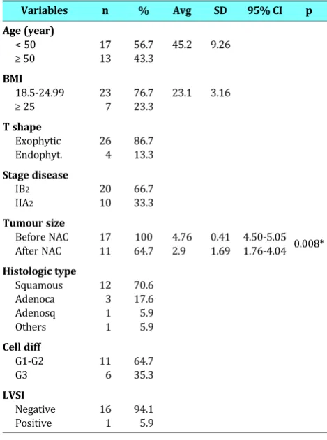
The average age of the cases is 45.20 years and cummulatively were more at the age <50 years. Other clinicopathologic characteristics are as listed in Table 1.
Table 1. Clinicopathologic characteristics
Variables n % Avg SD 95% CI p
Age (year) < 50
≥ 50
17 13
56.7 43.3
45.2 9.26
BMI 18.5-24.99
≥ 25
23 7
76.7 23.3
23.1 3.16
T shape Exophytic Endophyt.
26 4
86.7 13.3 Stage disease
IB2
IIA2
20 10
66.7 33.3 Tumour size
Before NAC After NAC
17 11
100 64.7
4.76 2.9
0.41 1.69
4.50-5.05 1.76-4.04 0.008* Histologic type
Squamous Adenoca Adenosq Others
12 3 1 1
70.6 17.6 5.9 5.9 Cell diff
G1-G2 G3
11 6
64.7 35.3 LVSI
Negative Positive
16 1
94.1 5.9
* Wilcoxon signed-rank test Avg=average
All cases expressed VEGF-C. After neoadjuvant chemotherapy there was a significant decrease in tumour size (p = 0.0087). From 7 responding cases (41.18%), 2 (11.76%) showed clinical (CR) and pathological complete response (CPR). To all cases that were considered respectable were performed radical hysterectomy and pelvic lymphnode
dissec-Figure 1. VEGF-C expression (magnified 400x) at IHC slides. A. Negative expression; B. Weak expression;
tion (10 cases, 58.82%), while cases that were con-sidered too large or disease progressed were irra-diated (7 cases, 41.18%). To one irrairra-diated case, was performed also lymphnode dissection. Tu-mour cells at surgical margins after radical surgery were found only in 1 of 10 cases (10%).
The p values of response to NAC at clinico-pathologic characteristics were not significantly different. Also at VEGF-C expression, respectively at every grade, there were no statistical differences (p = 0.65, 0.69 dan 0.67) (Table 2).
Table 2. Differences in response to NAC at VEGF-C expression
VEGF-C
expression Response ResponseNo Total p Weak 4 (23.53%) 6 (35.29%) 10 (58.82%) 0.65*
Moderate 2 (11.76%) 3 (17.65%) 5 (29.41%) 0.69*
Strong 1 ( 5.88%) 1 ( 5.88%) 2 (11.76%) 0.67*
Total 7 (41.18%) 10 (58.82%) 17 (100.0%)
* Fisher exact test
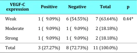
Metastases to the pelvic lymphnodes were found in 3 cases (27.27%). Metastatic tumour cells, re-spectively were found at 1 of 3 pelvic lymphnodes with infiltration to the right parametrium, at 1 of 5 obturator lymphnodes, and at 3 pelvic lymph-nodes each side of 19 lymphlymph-nodes dissected. There were no statistically significant differences in metastases to the pelvic lymphnodes at VEGF-C expression (p=0.44). (Table 3)
Table 3. Differences in lymphnode metastases at VEGF-C expression
VEGF-C
expression Positive Negative Total p
Weak 1 ( 9.09%) 6 (54.55%) 7 (63.64%) 0.44*
Moderate 1 ( 9.09%) 1 ( 9.09%) 2 (18.18%)
Strong 1 ( 9.09%) 1 ( 9.09%) 2 (18.18%)
Total 3 (27.27%) 8 (72.73%) 11 (100.0%)
* Pearson chi square
With Spearman test, a correlation could not be demonstrated between VEGF-C expression and re-sponse; neither also between VEGF-C expression and metastases (Spearman’s rho = -0.0286, 0.3858 and p = 0.9133, 0.2413 respectively).
Because bivariate analysis and Spearman test could not answer the hypothesis of this study, we
decide to determine a reliable cut off point at the percentage of immunopositive cells to VEGF-C to identify non responding and metastatic cases with the ROC analysis. The rationality to use the per-centage of immunopositive cells to VEGF-C, be-cause this continuous numeric data were not cate-gorized like the IRS that could hide the differences. On each percentage, we tested the sensitivity and specificity, positive likelyhood ratio (LR+) and negative likelyhood ratio (LR-).
The reliable cut off point to identify non
re-sponder was at ≥ 76% immunopositive cells
(sen-sitivity 70% and specificity 71.43%, correctly clas-sified 70.59%, with LR+ 2.45 and LR- 0.42), al-though not statistically different (p=0.09). The area under the curve (AUC) at the ROC analysis was 0.61 with SE 0.17 (95% CI 0.27 - 0.95). (Figure 2)
Whereas the reliable cut off point to identify
me-tastatic cases, was at ≥ 75% immunopositive cells
(sensitivity 100% and specificity 75.00%, correctly classified 81.82%, with LR+ 4.0 and LR- 0). The AUC at the ROC analysis was 0.75 with SE 0.16 (95% CI 0.43-1.00). (Figure 3)
Area under ROC curve = 0.7500
0.00 0.25 0.50 0.75 1.00
0.
00
0.
25
0.
50
0.
75
1.
00
Figure 3. ROC analysis curve of metastases at VEGF-C immunopositivity
Area under ROC curve = 0.6071
0.00 0.25 0.50 0.75 1.00
0.
00
0.
25
0.
50
0.
75
1.
00
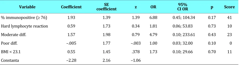
At the multivariate analysis with logistic regres-sion, the variables that included in the prediction model of no response are as listed in Table 4. A positive coefficient, highest OR (Odds ratio) and
statistical score were found at the variable ≥ 76
percentage immunopositivity to VEGF-C to predict no response, respectively 1.93, 6.88 (96% CI OR 0.45; 104.34) and 41.
If we compared the AUC from the mathematical model with the AUC from the model that has been transformed into statistical score, there were no statistical differences (p=0.3068). The statistical
score with cut off ≥ 52 can be used as a predictor
of no response (sensitivity 60%, specificity 85.71% and accuracy 70.59%).
DISCUSSION
The number of complete clinical response in this study are smaller compared with the results in Mo-darress et al and Benedetti-Panici P et al studies (respectively 16.7% and 78%); either is complete pathologic response compared with the studies of Choi CH et al (27.6%), but similar with the studies of Modarress et al and Benedetti-Panici P et al (re-spectively 10 and 13%).
In this study, despite VEGF-C was expressed in all cases, it could not differentiate responder from responder neither also metastatic from non-metastatic cases. These observations are different with that reported by Choi CH et al and Cheng WF
et al at VEGF expression,11-13 and Andrijono dan
Priyanto H at VEGF-C serum level in their studies.14
The differences of the results reported in the studies mentioned above at response and metasta-ses, is probably caused by the sample size needed to see differences in a relatif homogen percentage
of responder and non responder population (41.18% vs 58.82%) is larger, although the minimum sam-ple size to see differences in response and metas-tases at VEGF-C expression has been reached. This difference can be referred as a statistical bias by chance.
At the ROC analysis, the percentage =76 and =75 immunopositive cells to VEGF-C, respectively for no response and metastases, are reliable and the AUC of both ROC curves in this analysis (0.61 and 0.75 respectively) were considered to have fairly and good reliability. With multivariate analysis, al-though statistically not significant (p = 0.17), the
variable percentage ≥ 76 immunopositive cells to
VEGF-C can be demonstrated as the strongest pre-dictor of no response (Table 4).
The variables included in the prediction
mo-del, with cut off ≥ 52 prediction score, can
fur-ther be used as a tool to predict non responding cases; although this conclusion should be further studied with more samples. If the prediction score found for a certain cervical cancer with bulky
le-sion is ≥ 52, chemoradiaton would be more
reason-able than neoadjuvant chemotherapy followed with radical surgery as the definite therapy. Alter-natively, if the choice is radical surgery anti VEGF should be added, although this approach in cervical cancer need to be explored.
CONCLUSION
VEGF-C expression in this study can not differenti-ate non responding from responding and
metas-tatic from non-metasmetas-tatic cases in stage IB2 and
IIA2 cervical cancer given neoadjuvant
chemother-apy. The prediction score, using ≥ 76 percentage
immunopositive cells, with cut off ≥ 52 could be a
valuable alternative tool in this approach.
Table 4. Prediction model of no response to NAC with statistical score
Variable Coefficient coefficientSE z OR CI OR95% p Score
% immunopositive (≥ 76) 1.93 1.39 1.39 6.88 0.45; 104.34 0.17 41
Hard lymphocyte reaction 0.59 1.73 0.34 1.81 0.06; 53.83 0.73 10
Moderate diff. 1.57 1.98 0.79 4.79 0.10; 233.61 0.43 23
Poor diff. –.005 1.77 –.003 1.00 0.03; 32.00 0.10 0
BMI < 23.1 0.55 1.45 .378 1.73 0.10; 29.66 0.70 11
In the immunohistochemistry report of VEGF-C expression, the percentage of immunopositive cells to VEGF-C need to be included in order to have a more clearer impression of the immunopathology of the disease.
REFERENCES
1. Cancer, I.A.f.R.o., Cervical Cancer Incidence and Mortality Worldwide in 2008, in GLOBOCAN 2008. CANCER FACT SHEET. 2008, WHO
2. Andrijono. Cervical Cancer In Indonesia. Div of Oncology Dept of Obstetrics and Gynecology University of Indonesia Jakarta. 2010
3. H-S, R., Optimal Management for Cervical Cancer Stage IB2.
11th Annual Fall Symposium 2006. 38 (Suppl. 2): 27-31
4. ONCOLOGY, F.C.O.G., Revised FIGO staging for carcinoma of the vulva, cervix, and endometrium. Int J Gynecol Obstet, 2009; 105: 103-4
5. Choi YS, S.J., Kim JH. Survival Benefits of Neoadjuvant Che-motherapy Followed by Radical Surgery versus Radiother-apy in Locally Advanced Chemo resistant Cervical Cancer. J Korean Med Sci, 2006; 21: 683-9
6. Vermorken JB, E.E., Current developments in the treatment of cervical cancer. CME J Gynecol Oncol. 2006; 18: 52-60 7. Benedetti-Panici P, G.S., Colombo A, et al, Neoadjuvant
che-motherapy and radical surgery versus exclusive radiother-apy in locally advanced squamous cell cervical cancer: re-sults from the Italian multicenter randomized study. J Clin Oncol, 2002; 20: 179-88
8. Ménard S, C.M, Balsari A. Prediction of response to therapy by biomolecular markers: from the research laboratory to the clinic. Annals Oncol, 2003; 14: 178-9
9. Selman TJ, M.C., Zamora J. Diagnostic accuracy of tests for lymphnode status in primary cervical cancer: a systematic review and meta-analysis. Canad Med Associat J, 2008; 178(7)
10. Hu G, R.S., Zhu Y. Vascular endothelial growth factor modu-lates cisplatin sensitivity in human ovarian carcinoma cells. Cancer Therapy, 2003; 1: 31-7
11. Choi CH, S.S., Choi J-J. Prognostic significance of VEGF ex-pression in patients with bulky cervical carcinoma under-going neoadjuvant chemotherapy. BMC Cancer 2008; 8: 295.
12. Cheng W-F, C.C.-A., Lee C-N. Vascular Endothelial Growth Factor in Cervical Carcinoma. Am College Obstet and Gyne-col 1993; 93: 761-4.
13. Cheng WF, C.C., Lee CN. Vascular Endothelial Growth Factor and Prognosis of Cervical Carcinoma. Obstetrics & Gynecol-ogy, 2000; 96(5): 721-6.
14. Andrijono, P.H. VEGF-C level as a predictor of pelvic lymph node metastases of cervical cancer at early stage Med J In-dones, 2009; 18(4): 257-61.
15. Eisenhauer EA, T.P., Bogaerts J et al. New response evalu-ation criteria in solid Tumours: Revised RECIST guideline (version 1.1). Euro J Cancer 2009; 45: 228-47
16. Modarres M, M., Govalnaz M. Comparative study of chemoradiation and neoadjuvant chemotherapy effects be-fore radical hysterectomy in stage IB-IIB bulky cervical can-cer and with tumor diameter greater than 4 cm. Int J Gyne-col Cancer, 2005; 15: 483-8