The Role of Genital Hiatus (Gh), Perineal Body (Pb), Summation (Gh+Pb)
of POP-Q Examination in Maximum Levator Hiatal Area of Women with
Symptomatic Pelvic Organ Prolapse
Peran Hiatus Genitalis, Badan Perineum dan Penjumlahannya dari Pemeriksaan Pop-Q
pada Luas Area Hiatus Levator Maksimal pada Perempuan dengan Prolaps Organ
Panggul Simtomatik
Kukuh W Kustarto, Fernandi Moegni
Division of Urogynecology Reconstruction Departement of Obsterics and Gynecology Faculty of Medecine Universitas Indonesia Dr. Cipto Mangunkusumo General Hospital
Jakarta
Correspondence author : Kukuh Wibowo. [email protected]
Research Article
Abstract
Objective : To provide data on the correlation of levator hiatus area measurements in symptomatic POP using 3D / 4D Ultrasound with clinical examination of Gh, Pb and summation (Gh+Pb).
Methods : Secondary data analysis of 160 POP patients examined from January 2012 to April 2017 at the Urogynecology Clinic of Dr. Cipto Mangunkusumo Hospital, Jakarta, Indonesia. Patient characteristics, maximum 3D / 4D Ultrasound measurement of Levator Hiatus Area, and clinical measurement results using pelvic organ prolapse quantifi cation system (POP-Q) were recorded.
Results : There was a positive correlation between clinical examination and measurement of hiatal area area using ultrasound with r = 0.43 for Gh length, and the medium correlation on the sum of Gh and Pb with r = 0,51. No correlation for Pb length with r = 0.23. The optimal cut to differentiate degrees 2 by 3 is 7.5 cm / 29.7 cm2 and degree 3 by 4 is 8.3 cm / 32.1 cm2
Conclusions : Clinical examination by summing the lengths of Gh and Pb may be consider refl ects the examination of the hiatal area by using transperineal ultrasound to see the strain on levator ani called “ballooning” in an area with limited resources.
Keywords : genital hiatus, levator hiatus area, pelvic organ prolapse, perineal body.
Abstrak
Tujuan : Untuk memberikan data mengenai korelasi pengukuran area hiatus levator pada POP simtomatik mengunakan Ultrasonografi 3D/4D dengan pemeriksaan klinis yaitu panjang Gh, panjang Pb dan penjumlahannya.
Metode : Analisa data sekunder sebanyak 160 pasien POP yang diperiksa dari Januari 2012 hingga April 2017 di poliklinik Urogynecology Rumah Sakit Dr. Cipto Mangunkusumo (RSCM), Jakarta, Indonesia. Diambil data karakteristik pasien, pengukuran Ultrasonografi 3D/4D maksimal Area Hiatal Levator, dan hasil pengukuran secara klinis dengan menggunakan pelvic organ prolapse quantifi cation system (POP-Q)
Hasil : Terdapat korelasi positif antara pemeriksaan klinis dengan pengukuran luas area hiatal menggunakan USG dengan r = 0,43 untuk panjang Gh, dan korelasi pada penjumlahan Gh dan Pb dengan r=0,51 termasuk kategori sedang, sedangkan untuk panjang Pb dengan r = 0,23 tidak didapatkan adanya korelasi. Didapatkan titik potong optimal untuk membedakan derajat 2 dengan derajat 3 adalah 7,5 cm / 29,7 cm2 dan derajat 3 dan derajat 4 adalah 8,3 cm / 32,1 cm2
Kesimpulan : Pemeriksaan klinis dengan menjumlahkan panjang Gh dan panjang Pb dapat dipertimbangkan untuk mencerminkan pemeriksaan area hiatal dengan mengunakan USG 3 / 4 dimensi transperineal pada daerah dengan sarana terbatas untuk melihat regangan pada levator ani atau yang disebut sebagai “ballooning
maternal tissue. Avulsion levator ani occurs in 15-30% of women who deliver vaginally Avulsion is a risk factor for 'ballooning' (an abnormal hiatal area at Valsalva maneuver > 25 cm2) and is a risk factor for POP, especially in the anterior and
middle compartments.9,11
In addition to avulsion, microtrauma or traumatic over distention leads to changes in levator hiatus biometry and boils down to POP.11
Damage to pelvic fl oor muscle structure during
vaginal delivery eliminates the ability to keep the urogenital always closed, so that eventually the ligaments fail to retain the pelvic organs due to persistent intra-abdominal pressure.12
In 2005 Dietz HP et al found a signifi cant
association between pelvic organ mobility with levator hiatus area at rest and Valsalva
maneuvers.13 Their further study has suggested
that measurement of levator ani distensibility is the most basic approach for determining the biomechanical properties of the muscle, and labor increases the distensibility of the levator ani hiatus, although without signifi cant levator ani trauma. They also found that the levator hiatus area has a very strong statistical relationship with clinical symptoms of POP. Therefore, the distensibility of hiatus may be an independent
etiologic factor of POP.9 Rodrigues Jr AA et al
found that the Levator Ani Subtended Volume (LASV) demonstrated a strong association with the increase in POP levels defi ned by POP-Q.14
Punarbawa shows a correlation between the maximum of hiatal levator area and the degree of uterine prolapse, the optimal cut off point with the highest sensitivity and specifi city was 28.5
cm2.15Santoso showed the optimal cut off point
of the levator ani muscle area in distinguishing
cystocele grade I-II and III-IV was 29 cm2.The
optimal cut of point of the levator ani muscle area in distinguishing rectoceles grade I-II and III-IV was 30 cm2.16 In addition to the ultrasound
examination of the above-mentioned hiatal area, there is also a study by Khunda A et al linking the levator hiatal area with clinical examination of the sums of Gh and Pb where a 7 cm cutoff point is defi ned as an excessive strain of levator hiatus.17
Gerges B et al states that the measurements of length of Gh and Pb can clinically determine the degree of excess stretching of levator hiatus without the need for ultrasound.18 All of
these clinical examinations were conducted in
INTRODUCTION
Pelvic organ prolapse (POP) is an abnormal descent of pelvic organs such as the uterus, bladder, urethra, and rectum from the normal position into the vagina or out of the vagina due to decreased function of the pelvic organ supporting
system.1,2 This support function results from
interactions between the pelvic bone, muscles, ligaments, fascia and nerves.2 POP is part of pelvic fl oor dysfunction, strongly associated with other
pelvic fl oor disorder symptoms such as urinary
incontinence, constipation, decreased sexual quality.3,4 The incidence of pelvic organ prolapse
in a study was 30.8% at age above 50 years. An American study found that 79-year-old women had an 11.1% risk for at least one POP surgery or urinary incontinence, with a possible 29.2%
reoperation.5,6 Based on studies in the United
States, the operating costs for POP and urinary incontinence reached more than 1 billion dollars.7
An increase in fi nancing is also expected to occur, as it is estimated that over the next 30 years the number of women seeking treatment will double, as a result of age and lifestyle changes.8
The levator hiatus area is an area formed by the levator ani muscle that is strongly associated with prolapsed occurrence and is a potentially high-potential site or portal for the occurrence of POP and rectal prolapse.9 It is also a central
opening of the levator plate, which is known to be strongly associated with signs and symptoms of POP and risk for recurrence. There are several explanations that cause excess strain of levator hiatus or so-called ballooning due to congenital or acquired abnormalities. The existence of microtrauma for example over distention, due to hormonal effects on labor and because of the process of childbirth. Subsequent over distence leads to secondary avulsion of the puborectal muscle, where the muscle escapes from its insertion in the symphony bone. Prolapse of the anterior portion of the vagina or cystocele is the most common type of prolapse, and is the most persistent place and the highest incidence of recurrence.10
Caucasian races and have not been studied in the Malay race.
The use of 3 and 4-dimensional ultrasound is ideal for assessing the morphology as well as the
dimensions of the pelvic fl oor. The emergence
of ultrasound 3 and 4 dimension provides an advantage in imaging, which can imaging the three areas of the body. This allows both qualitative and quantitative assessment of the pelvic fl oor support structure, the integrity of the levator ani muscle, the avulsion and in addition to the internal and external sphincters at the same time. Data at the time of examination can be saved and transfer for analysis and interpretation in the future.19
The use of Utrasound 3 and 4 dimensions to measure the hiatal levator ani preoperative area is the goal to determine the operating technique to be performed to reduce the genitals of hiatus, whether the use of mesh or the reduction of the genitals hiatus with zakarin levatorplasty.20,21
Procurement of ultrasound 3 and 4 dimensions of course requires considerable funds and required special skills to assess the pelvic
fl oor.22 Not all hospitals in Indonesia have this
facility. Pelvic fl oor examination with POP-Q
is currently a common clinical examination to assess the degree of POP. The measurement sum of the length of the genital hiatus (Gh) and the perineal body (Pb) taken from clinical POP-Q examination can be to determine the degree of hiatal or ballooning strain that equivalent with
ultrasound examination.18However, there is a
research that shows the suitability of Gh length with the degree of weight of POP while Pb does not show a meaningful suitability.23,24 Therefore,
research needs to be conducted so that methods and techniques of clinical measurement of length of Gh, length of Pb and addition can be studied in relation to the area of hiatal levator and can be used for clinical benefi t and used by many people in the future.
METHOD
This was a cross-sectional study, where the data source was from secondary data of medical record in POP patient in Urogynecology and reconstruction subdivision department of Obstetrics and Gynecology Dr. Cipto Mangunkusumo Hospital, Jakarta, Indonesia.
Data was taken from January 2012 to April 2017. Inclusion criteria were patients who underwent POP-Q examination, underwent transperineal 3D / 4D ultrasound and summation length of Gh and Pb. The POP diagnosis used in this study was a combined diagnosis which used the heaviest degree of the three compartments. Exclusion criteria were patients who could not doing valsava maneuver, pelvic organ malignancy, intra-abdominal tumor and had a history of pelvic surgery. Medical records of patients who have met the inclusion criteria will be taken secondary data of patients covering general patient data, clinical examination data in the form of POP-Q examination that has been done by trainees urogynecology with supervision from consultant urogynecology, and examination data of translabial 3D / 4D ultrasound done by one competent urogynecology consultant using GE Voluson E8 Expert BT09 (GE Medical Systems, Zipf, Austria) with 4.0-9.0 MHz convex volume probe RIC5-9-D (acquisition angle 1200).
All medical records of study subjects who have data of length Gh and Pb, and hiatal area data, although the other characteristic data are not included in the data analysis process. Numerical data is assessed by Kolmogorov Smirnov's assay
for normality and with Coeffi cient of Variance
(COV) calculations for its homogeneity. If the Kolmogorov Smirnov test produces p> 0,05 or COV <25% then the statistical calculation was done with parametric approach and presented as mean. Mean and Standard Deviation, whereas if it did not meet the requirement it is done with nonparametric approach and presented in median mean and ranges. Assessment of the relationship between two numerical variables was done by statistical correlation method based on Spearman test because the distribution was not homogeneous. Relationship with the value of correlation coeffi cient (R) above 0.3 followed by the determination of the regression formula. R values between 0.3 to 0.5 were included as weak relations categories, between 0.5 to 0.7 as moderate, and above 0.7 include strong relationships.25
specifi city, positive predictions, and negative predictions. The limit of statistical signifi cance was used alpha value of 0.05. This research was proposed to the ethics commission at the Faculty of Medicine, Universitas of Indonesia, so that in the implementation meet the ethical clearance to conduct a research. All patient identities and research results are kept confi dential.
RESULT
In this study, there were 237 initial samples,
77 samples were not performed pelvic fl oor
ultrasound, and among 77 samples there were also prolapse patients already done operation as many as 8 samples. After screening and adjusted for acceptance criteria, 160 samples participated in this study. The results are presented descriptively and analytically.
Medical Record cases pelvic organ prolapse (n = 237)
Signed in as a research sample (n = 160)
No pelvic fl oor ultrasound results
(n = 69) Already performed pelvic organs surgery(n = 8)
Diagram 1 : The process of collecting research data
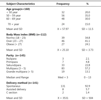
Table 1. Distribution of Subjects According to the Characteristic.
Subject Characteristics Frequency % Age group(n=160)
< 50 year 50 – 59 year 60 – 69 year
Body Mass index (BMI) (n=112)
Normo (18 – 23) Over (23 – 27) Obese (> 27)
Parity (n=145)
Nulipara Primipara Sekundipara Multipara (3 – 5) Grande multipara (> 5)
Delivery method (n=141)
Spontaneus Assisted delivery C section
131 8 2
92.9 5.7 1.4 Mean and SD
Mean and SD
Median and Range
Mean and SD
X = 57.97
X = 25.10
Med = 3
X = 3531
SD = 11.5
SD = 3.73
0 – 13
SD = 504
70 + year 24 15.0
32 56 48
39 46 27
3 7 33 83 19
Table2. Distribution of Subjects According to Pelvic Organ Prolapse Conditions
Uterine prolapse
0 Degree 1st Degree
2nd Degree
3rd Degree
4th Degree
Cystocele
0 Degree 1st Degree
2nd Degree
3rd Degree
4th Degree
Rectocele
0 Degree 1st Degree
2nd Degree
3rd Degree
4th Degree
1 3 41 88 27
5 12 108
24 11
0.6 1.9 25.6 55.0 16.9
3.1 7.5 67.5 15.0 6.9 24
45 52 39
15.0 28.1 32.5 24.4
0 0.0
Prolapse condition Frequency % Subject Characteristics Frequency % Menopause status (n=121)
Not yet menopause 1 – 5 year
6 – 10 year 11 + year
Median and range Med = 9 0 – 25
29 22 25 45
24,0 18,2 20,7 37,2
From table 1, it was observed that not all characteristic variables of the subjects were recorded in the medical record under study. Only the age variable of the subject was fully recorded. Most subjects aged between 50 to 69 years covered 65.6% with a mean age of 57,97 years and standard deviation was 11,5 year. Nutritional status was only present in 112 subgroups with the overweight group until obese reached 65.2% with an average body mass index of 25.1 +/- 3.73. Data on parity rate reached 145 subjects with multi parity reaching 70.3% and median averages of 3 deliveries with a maximum of 13 births.
A total of 141 subjects had a record of the last delivery method and 92.9% were spontaneous labor with an average birth weight of 3351 +/- 504 grams. The history of menopause was recorded in 121 medical records with a majority of more than 5 years, which reached 57.9% with a median of 9 years and the longest reaching 25 years.
Table 2 shows that the degree of uterine prolapse majority in the central compartment at third degrees with 32.5% and at fi rst degree as a asymptomatic reaches 15%. Based on the cystocele in the anterior compartment, it was also the third degree with 55.0% and there were normal subjects of 0.6% and the fi rst degree was 1.9%. Rectocel in the posterior compartment gives normal conditions in 3,1% subjects and fi rst degree at 7,5% while the majority was at second degrees with 67,5%.
Degrees by merge
0 Degree 1st Degree
2nd Degree
3rd Degree
4th Degree
Incontinent
no yes
Avulsion m. levator ani
Negative Unilateral Bilateral
145 8 7
90.6 5.0 4.4 154
6 96.23.
0 0 32 85 43
0.0 0.0 20.0 53.1 26.9
Prolapse condition Frequency %
Correlation between Clinical Examination and Hiatal Area
Correlation between uterine prolapse component and hiatal area measured using Spearman methods because the distribution of hiatal area were not homogen. R value of spearman for correlation between Gh size and hiatal area can be categories as weak correlation with R=0.43. Regression formula may be used to predict hiatal area based on Gh value according to Gh number, however with the weak R value, there were high error deviation number. And there were no correlation between Pb value with hiatal area with R=0.23, so we cannot make the regression formula from this relationship.
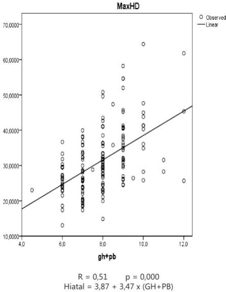
Figure 1 showed moderate correlation between combination of Gh+Pb with hiatal area that may reach R=0.51. Regression formula may be used to predict total hiatal area based on the combination of Gh+Pb may be used considering the accuracy of the formula that should also be considered.
Area under the curve of this condition was
69.8% with confi dence interval of 95% between
59.4% - 80.1%. The cut off point was observed to differentiate between 2nd degree and 3rd &
4th degree was 7.25 cm with sensitivity 64.8%,
specifi city 68.8%, positive predictive value 89.2% and negative predictive value 32.8%.
R = 0,51 p = 0,000 Hiatal = 3,87 + 3,47 x (GH+PB)
Figure 1. Correlation between addition of Gh and Pb with hiatal area (n=160)
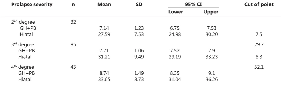
2nd degree
GH+PB
Hiatal 27.597.14 1.237.53 24.986.75 30.207.53
7.71
31.21 1.069.49 29.197.52 33.237.9
8.74
33.65 1.498.73 31.048.35 36.269.1
32
7.5
85 29.7
43 32.1
8.3 3rd degree
GH+PB Hiatal 4th degree
GH+PB Hiatal
Prolapse severity n Cut of point
Table 3. Cut off Point between Gh+Pb and Hiatal Area between Combined Prolapse
Mean SD 95% CI
Upper Lower
For hiatal area cut off point based on prolapse severity between 2nd degree and 3rd& 4th degree,
the area under the curve was 64.7% with
confi dence interval 95% between 53.7% until
75.6%. It was found that the cut off point to differentiate 2nd degree and 3rd or 4th degree were
28.15 cm2 with sensitivity 63.3%, specifi city 59.4%,
positive predictive value 86.2% and negative predictive value 28.8%.
Table 3 demonstrated that the cut off point between 2ndand 3rd degree of prolapse and 3rd and
4thdegree of prolapse accordingly based on data
distribution and overriding data calculated with
95% confi dence interval. For Gh+Pb combined
value we can fi nd cut off point of 2nd and 3rd
degree were 7.5 cm. For 3rd and 4th degree were
8.3 cm. For hiatal area, cut off point between2nd and 3rd degree were 29.7cm2. Between 3rd and 4th
degree were 32.1 cm2
DISCUSSION
POP prevalence were higher in older population until it reach the fi fth decade of female life, the prevalence relatively stable from the fi fth decade onwards. Wu et al mentioned that 45 years old female may not showed any POP signs / symptoms until she reached 50 or 60 years old.26 Tegersted et
al, in 2005 did research in Sweden and found that the prevalence of symptomatic POP increased
until the age of 60. In women aged 30-39, 40-49, and 50-59 found that the prevalence of POP were 4.1, 6.2, 11.8%. After that, the prevalence of POP remains constant. Between women age 60-69 and 70-79 years, the prevalence of POP were 12,2% and 11%. Using women aged 30-39 years old as a reference, Odd Ratio POP increased three folds after the age of 50 years old and remain the same in older generations.27 The mean age in this
research were 57.97 years old (range 26-80 years old)
Vaginal delivery caused pudenda nerve damage, levator ani muscle damage, and damage of the fascia surrounding the pelvic organ. Traumatic structural damage on the supporting facia and muscle during delivery was the main
contributors of urinary incontinence and POP.28
Dietz et al showed that spontaneous delivery increased the risk of POP more than three times the baseline level (OR 3.19; 95%, CI 1.07-9.49),
Figure 3. ROC curve with total hiatal area between 2nd
Assisted delivery increased the risk of POP more than 5 times of the baseline (OR 5.52; 95% CI
1.79-17.3) compared to C-Section.29 Parity may
have signifi cant correlation with dimension of
hiatal area during Valsalva maneuver and this effect may have infl uence on the fi rst delivery.30
In this research, the mean number of parity are 3 dominated with vaginal delivery 92.9%, assisted delivery 5.75% and C-section 1.4%.
Santoso BI showed that dysfunction of the
pelvic fl oor caused by biggest baby’s weight,
Receiver Operating Characteristic (ROC) curve found optimal cut off point of infant weight more than 3325 grams that may cause levator ani muscle trauma.31 Different with Boyles, found
vaginal delivery with baby weight more than 8 pounds (> 3600 grams) that may have signifi cant relationship with urinary incontinence post
partum.32 In this research, found that baby’s
body weight during vaginal delivery was 3,531, 31gr (range between 2500- 5200 grams)
Obesity was one of the risk factor of emerging signs and symptoms of prolapse, even though it’s relationship with objective measurement’s not apparent. In SWEPOP research (Swedish Pregnancy, Obesity and Pelvic Floor), symptomatic prolapse organ may increase 3% with each increase of BMI (Body Mass Index) with OR 1.03;
95% CI 1.01-1.05.33 There are some evidence
that obesity is a strong risk factor for incidence and progressivity of urinary incontinence
and incontinence alvi.34 Chen et al stated that
obese women was 4 times more likely to get urinary incontinence and 2 times more likely to get incontinence alvi compared to non-obese women.35 From this research, BMI was 2.51 (range
18-39.1) was considered as overweight.
Many researchers made conclusion that menopause was one of the risk factor for POP. A research on 5489 women found that 454 of those with POP signs/symptoms showed an increasing prevalence of POP based on their age, the prevalence will not increase further after 60 years
old.36 Tegerdst et al found that the prevalence
and risk factors of POP increased signifi cantly after 60 years old and the prevalence was stable at those older than 60 years old.26 Mothes et al in
their research showed that in those that already had had menopause for more than 10 years that menopause is an independent risk factors for
prolapse (p<0.001).27 In this research, we found
that the average length of menopause are 8.67 years and those that already had menopause >10 years in 45 samples (32.7%).
In combined diagnosis of POP we found that
2nd degree was found in 32 samples (20%), 3rd
degree was found in 85 samples (53.1%), 4th
degree was found in 43 samples (26.9%), there were no sample with non-prolapse and 1st degree,
according to criteria, symptomatic prolapse were those that higher than 2nd degree.28,29
Avulsion (macrotrauma) and overdistention traumatic (microtrauma) were the most common
etiology of POP.11 Majida et al (2012) done
research to compare the morphology and function of pelvic fl oor in 157 POP women with and without the defect of pubovisceral muscle. They found that he prevalence of major pelvic
fl oor muscle defect’s around 34%, similar with
other research that showed prevalence between 21-37%. This fi nding was different with previous opinion by Shek (2009) that stated 15-30% of female that experienced vaginal delivery had levator ani trauma (avulsion).11In this research, we
found that the incidence of avulsion in levator ani muscle’s around 9.4%.
Urinary incontinence that’s mainly stress incontinence have strong relationship with vaginal delivery.30 In female Swedish population
aged 20 years old during their fi rst labor, found that vaginal delivery had signifi cant relationship with degree of urinary incontinence severity (OR 1.68, 95% CI 1.40-2.03) and urinary incontinence problem (OR 1.85, 95% CI 1.42-2.39).30 In cohort
research in women between 5 years until 10 years after fi rst labor. History of one or more vaginal delivery has signifi cant relationship with odds of stress incontinence (OR 2.9, 95%, CI 1.5-5.5) but not with overactive bladder (OR 1.7, 95% CI 0.8-3.5). The effect of vaginal delivery in urinary incontinence mainly happened in postpartum periods. Different compared to the population in this research that’s dominated with vaginal delivery in 92.9% with incidence of urinary incontinence was 3.8%. This supported Dietz that compared pelvic organ mobility in Caucasians with Asian we found that there’s a difference in mobility in anterior compartment and posterior compartment that is smaller, and the central
The correlation between Gh length and hiatal
area showed the correlation coeffi cient was
0.43. This show weak but positive relationship between these 2 variables. Lowder et al in 2016 did a research that showed Gh length is a strong predictor of prolapse of apical structure with Gh length > 3.75 cm (ROC >0.8).32 Khunda et al also
showed positive correlation between Gh length and hiatal area with correlation coeffi cient of 0.52.17
Correlation between Pb length and hiatal area had correlation coeffi cient 0,24, this showed no relationship between these 2 variables. This
results is the same with previous fi ndings by
Dunivan et al in 2015 that showed Pb length does not have any relationship with prolapse degree but Gh length have relationship with prolapse degree until it reached 3rd degree.33
Correlation between the total of Gh and Pb length with hiatal area had correlation coeffi cient of 0.51 showed that there are positive correlation
with moderate relationship.25 This research
results was similar to those written by Khunda et al in the year of 2012 in which the total of Gh and Pb may have strong relationship with hiatal area
(r=0.722). 17We found formula to get the total
hiatal area from clinical examination of summing Gh and Pb length was 3.87 + 3.47 x (Gh+ Pb), this formula may be used in area without 3D / 4D USG. The sensitivity results of this examination was 64.8% and indicates that the total of Gh and
Pb length greater than 7.25 cm2could show POP
3rd and 4th degree in 64.8% of cases. Specifi city
results showed that 68.8%, means that total of Gh and Pb <7.25 cm2 may reveal 2nd degree of POP
in 68.8% of case. The result of positive predictive value of 89.2% is higher compared than negative predictive value (32.8%). This result showed that clinical examination result of Gh and Pb total >=7.25cm2 may predict 3rd and 4th degree POP with
high accuracy, clinical examination results of Gh and Pb less than 7.25 cm2 may have low predicting
power of 2nd degree of POP, in other words the
prevalence of grade 2 POP based on clinical examination may be in reality are undiagnosed
3rd and 4th degree of POP. This may be caused
by multifactorial cause of POP, and hiatal levator area (refl exion from pelvic fl oor muscles)’s not only the main causing factors of POP. Anatomy of pelvic fl oor supporting muscle’s divided into
passive and active structure. Passive structure encompassed pelvic bones and supporting tissue such as ligament and endopelvic fascia, active supporting tissue encompasses pelvic
fl oor muscle and the nerves that facilitates tonic contraction and contraction both voluntary and involuntary. Intermittent contraction. Active and passive component of pelvic fl oor function works as an integrated system that works with each
other.34 Other factors that may have important
repercussions were the damage of supporting tissue (both the collagen and elastin). POP’s closely related with a decrease in total collagen and the decrease of collagen solvability, an increase in intermediate intermolecular cross-links and advanced glycation cross links in prolapse tissue. Changes in collagen in prolapse tissue are four times more prevalent, this was clearly shown by matrix metalloproteinase and increased of collagenolytic activity that may in turn cause prolapse of tissue lose collagens. Fibroblast may decrease the collagen production in prolapse tissue; and an increase in the activity of MMP-1, 2 and 9 and a decrease of TIMP-1 activity may cause an increase in collagen turnover. Thus may cause the production of immature new collagen. Most of the researchers found that an increase type III collagen and a decrease of type 1 collagen, thus it may cause a decrease in type I/ type III collagen.35
Dietz conducted measurement of hiatal area during Valsalva on 544 women and classifi ed the results to this following fi ndings: mild 25-29.9
cm2, moderate 30-34.9 cm2, marked in 35-39.99
cm2, and severe ≥ 40cm29 In a study conducted
at Cipto Mangunkusumo Hospital, Jakarta, Indonesia, Punarbawa found that relationship between maximal area of levator hiatal area with degree of uterine prolapse, the optimal cut off point that have the highest sensivity and specifi city was 28.5 cm2.15 Santoso showed that
optimal cut off point to differentiate it with 1st-2nd
degree and 3rd-4th degree of cystocele was 29 cm2.
Optimal cut off point of hiatal levator ani muscle that differentiate it with 1st-2nd degree and 3rd
-4th degree of rectocele was 30 cm2.16 It is clearly
shown that hiatal area that may differentiate 2nd degree and 3rd-4th degree was smaller in this
research.
as “ballooning” were divided into mild, moderate, marked and severe according to total Gh+Pb divided with mean hiatal area which lies between 7.0-7.99cm/ 27.3cm2, 8.00-8.9 cm/ 27.3cm2,
9.0-9.99cm/ 35.1cm2 and>10cm/ 41.9 cm2. This result
was different from our fi ndings which showed
marked results in 7.5cm/ 29.7cm2 and severe 8.3/
32.1cm2. We can see in this research that severe
degree has more smaller result compare with western research, in accordance with Cheung
et al that compared pelvic fl oor biometry and
hip mobility in nullipara Caucasian and Asian women in Hongkong. It was found that Asian has
signifi cantly more dense puborectalis muscle,
smaller genital hiatus and less mobile pelvic
organ compared to Caucasian.30 Many research
also showed that in different ethnicity, the total hiatal area’s also different, and less pelvic organ mobility if compared to Caucasian. Many research also showed that In different ethnicity, total hiatal area may be different from each other.36,37
The limitations of this study were primarily cross sectional using secondary data, where sample selection cannot be randomized nor blinding because prolapse degree can be estimated when the patient performed a Valsalva
maneuver during pelvic fl oor transperineal
ultrasound examination
CONCLUSION
Clinical examination by summing Gh and Pb length have moderate correlation level (R=0.51) with hiatal area examination using 3D or 4D USG examination that can be used in many health care facilities with limited facility.
RECOMMENDATION
More research needed to fi nd hiatal area with
low severity as a cut off point in asymptomatic and symptomatic patient, to complete data of pelvic organ prolapse in South-East Asian Race especially Indonesian race.
REFERENCES
Ho MH, Bhatia NN. Pelvic organ prolapse in 1.
postmenopausal women. In: Lobo RA, editor. Treatment of the postmenopausal woman: basic and clinical aspects. Academic Press; 2007: 739–64.
Hendrix SL, Clark A, Nygaard I, Aragaki A, Barnabei V, 2.
McTiernan A. Pelvic organ prolapse in the Women’s Health Initiative: gravity and gravidity. Am J Obstet Gynecol. 2002;186(6):1160-6.
Slieker-ten Hove MCP, Pool-Goudzwaard AL, Eijkemans 3.
MJ, Steegers-Theunissen RP, Burger CW, Vierhout ME. The prevalence of pelvic organ prolapse symptoms and signs and their relation with bladder and bowel disorders in a general female population. Int Urogynecol J. 2009;20(9):1037-45.
Fritel X, Varnoux N, Zins M, Breart G, Ringa V. 4.
Symptomatic pelvic organ prolapse at midlife, quality of life, and risk factors. Obstet Gynecol. 2009 ;113(3):609-16.
Clark AL, Gregory T, Smith VJ, Edwards R. Epidemiologic 5.
evaluation of reoperation for surgically treated pelvic organ prolapse and urinary incontinence. Am J Obstet Gynecol. 2003;189(5):1261–7.
Olsen AL, Smith VJ, Bergstrom JO, Colling JC, Clark 6.
AL. Epidemiology of surgically managed pelvic organ prolapse and urinary incontinence. Obstet Gynecol. 1997;89(4):501-6.
Subak LL, Waetjen LE, Van Den Eeden S, Thom DH, 7.
Vittinghoff E, Brown JS. Cost of pelvic organ prolapse surgery in the United States. Obstet Gynecol. 2001;98(4):646-51.
Chow D, Rodríguez LV. Epidemiology and 8.
prevalence of pelvic organ prolapse. Curr Opin Urol. 2013;23(4):293-8.
Dietz HP, Shek C, De Leon J, Steensma AB. Ballooning of 9.
the levator hiatus. Ultrasound Obstet Gynecol Off J Int Soc Ultrasound Obstet Gynecol. 2008;31(6):676-80. Dietz HP, Franco AVM, Shek KL, Kirby A. Avulsion 10.
injury and levator hiatal ballooning: two independent risk factors for prolapse? An observational study. Acta Obstet Gynecol Scand. 2012 ;91(2):211-4.
Shek KL, Dietz HP. The effect of childbirth on hiatal 11.
dimensions. Obstet Gynecol. 2009;113(6):1272–8. DeLANCEY JO. Anatomy and biomechanics of genital 12.
prolapse. Clin Obstet Gynecol. 1993;36(4):897-909. Dietz H, Shek C, Clarke B. Biometry of the pubovisceral 13.
muscle and levator hiatus by three dimensional pelvic fl oor ultrasound. Ultrasound Obstet Gynecol. 2005;25(6):580-5.
Rodrigues AA, Bassaly R, McCullough M, Terwilliger HL, 14.
Hart S, Downes K, et al. Levator ani subtended volume: a novel parameter to evaluate levator ani muscle laxity in pelvic organ prolapse. Am J Obstet Gynecol. 2012;206(3):244-e1.
Punarbawa GM, Moegni F. Hubungan Area Hiatal 15.
Levator Dengan Derajat Prolaps Organ Panggul Yang Dinilai Dengan POP-Q. In Jakarta: FKUI-RSCM; 2013. 16. Santoso H, Moegni F. Hubungan derajat sistokel 16.
dan rektokel dengan area hiatal levator ani. In Jakarta: FKUI-RSCM; 2016.
Khunda A, Shek KL, Dietz HP. Can ballooning of the 17.
levator hiatus be determined clinically? Am J Obstet Gynecol. 2012 ;206(3):246.e1-4.
Gerges B, Kamisan Atan I, Shek KL, Dietz HP. How to 18.
Barry C, Dietz HP. The use of ultrasound in the 19.
evaluation of pelvic organ prolapse. Rev Gynecol Pract. 2005;5(3):182–95.
Dietz HP. Pelvic fl oor ultrasound in prolapse: what’s in 20.
it for the surgeon? Int Urogynecol J. 2011 ;22(10):1221-32.
Shek K, Dietz H. Pelvic fl oor ultrasonography: an update. 21.
Minerva Gynecol. 2013;65(1):1–20.
Stasi G, Ruoti EM. A critical evaluation in the delivery 22.
of the ultrasound practice: the point of view of the radiologist. Int J Med. 2015;9(1):5-10.
Digesu GA, Chaliha C, Salvatore S, Hutchings A, Khullar V. 23.
The relationship of vaginal prolapse severity tosymptoms and quality of life. BJOG. 2005;112(7):971-6.
Dunivan G, Ninivaggio C, Lyons K, Jeppson P, Komesu 24.
Y, Rogers R. Does Genital Hiatus and Perineal Body Measurements Change with Increasing Prolapse Stage? J Minim Invasive Gynecol. 2015;22(3):S52-3.
Mukaka M. A guide to appropriate use of Correlation 25.
coeffi cient in medical research. Malawi Med J J Med Assoc Malawi. 2012;24(3):69-71.
Tegerstedt G, Maehle-Schmidt M, Nyrén O, 26.
Hammarström M. Prevalence of symptomatic pelvic organ prolapse in a Swedish population. Int Urogynecol J. 2005;16(6):497-503.
Mothes A, Radosa M, Altendorf-Hofmann A, Runnebaum 27.
I. Risk index for pelvic organ prolapse based on established individual risk factors. Arch Gynecol Obstet. 2016;293(3):617-24.
Swift SE, Tate SB, Nicholas J. Correlation of symptoms 28.
with degree of pelvic organ support in a general population of women: what is pelvic organ prolapse? Am J Obstet Gynecol. 2003;189(2):372-7.
Dietz H, Mann K. What is clinically relevant prolapse? 29.
An attempt at defi ning cutoffs for the clinical assessment of pelvic organ descent. Int Urogynecol J. 2014;25(4):451-5.
Gyhagen M, Bullarbo M, Nielsen T, Milsom I. A 30.
comparison of the long term consequences of vaginal delivery versus caesarean section on the prevalence, severity and bothersomeness of urinary incontinence subtypes: a national cohort study in primiparous women. BJOG . 2013;120(12):1548-55.
Dietz H. Do Asian women have less pelvic organ mobility 31.
than Caucasians? Int Urogynecol J. 2003;14(4):250-3. Lowder JL, Oliphant SS, Shepherd JP, Ghetti C, Sutkin 32.
G. Genital hiatus size is associated with and predictive of apical vaginal support loss. Am J Obstet Gynecol. 2016;214(6):718-e1.
Dunivan G, Ninivaggio C, Lyons K, Jeppson P, Komesu 33.
Y, Rogers R. Does Genital Hiatus and Perineal Body Measurements Change with Increasing Prolapse Stage? J Minim Invasive Gynecol. 2015;22(3):S52-3.
Vergeldt TF, Weemhoff M, IntHout J, Kluivers KB. Risk 34.
factors for pelvic organ prolapse and its recurrence: a systematic review. Int Urogynecol J. 2015;26(11):1559– 73.
Lim VF, Khoo JK, Wong V, Moore KH. Recent studies 35.
of genetic dysfunction in pelvic organ prolapse: the role of collagen defects. Aust N Z J Obstet Gynecol. 2014;54(3):198-205.
Abdool Z, Dietz HP, Lindeque GB. Are there ethnic 36.
differences in the levator hiatus and pelvic organ descent? A Prospective observational study. Ultrasound Obstet Gynecol. 2016;
Shek K, Krause H, Wong V, Goh J, Dietz H. Is pelvic organ 37.