Brain Tumor Detection using Modified Fuzzy
Segmentation
Swati Deendyal Sona Malhotra
Department of Computer Science and Engineering Department of Computer Science and Engineering University Institute of Engineering and Technology
Kurukshetra University , Kurukshetra (India)
University Institute of Engineering and Technology Kurukshetra University , Kurukshetra (India)
Abstract
Designing a good measure for Medical segmentation is a known hard problem while the criteria of a good segmentation are often application-dependent and hard to explicitly define, for many applications the difference between a favorable segmentation and an inferior one is noticeable. Segmentation of medical images involves various image related problems. Medical Images contain noise that can alter the intensity of a pixel such that its classification becomes uncertain However It is possible and necessary to design efficient algorithms to find tumorous regions with segmentation algorithm this work is an effort in same direction utilizing Fuzzy Segmentation Algorithm.
Keywords: Image Segmentation, MRI Scans, Tumor Segmentation, Fuzzy Methods
_______________________________________________________________________________________________________
I.
INTRODUCTION
Image segmentation is an important processing step in many image, video and computer vision applications. Extensive research has been done in creating many different approaches and algorithms for image segmentation, but it is still difficult to assess whether one algorithm produces more accurate segmentations than another, whether it be for a particular image or set of images, or more generally, for a whole class of images. To date, the most common method for evaluating the effectiveness of a segmentation method is subjective evaluation, in which a human visually compares the image segmentation results for separate segmentation algorithms, which is a tedious process and inherently limits the depth of evaluation to a relatively small number of segmentation comparisons over a predetermined set of images. Another common evaluation alternative is supervised evaluation, in which a segmented image is compared against a manually segmented or pre-processed reference image [1].
It is often used to partition an image into separate regions, which ideally correspond to different real-world objects. It is a critical step towards content analysis and image understanding. Many segmentation methods have been developed, but there is still no satisfactory performance measure, which makes it hard to compare different segmentation methods, or even different parameterizations of a single method. However, the ability to compare two segmentations in an application-independent way is important: to autonomously select among two possible segmentations within a segmentation algorithm or a broader application; to place a new or existing segmentation algorithm on a solid experimental and scientific ground; and to monitor segmentation results on the fly, so that segmentation performance can be guaranteed and consistency can be maintained. Detection of the tumor is the main objective of the system. Detection plays a critical role in biomedical imaging.
Magnetic resonance imaging (MRI) of the brain is often used to monitor tumor response to treatment process. The segmentation of the brain tumor from the magnetic resonance images is important in medical diagnosis because it provides information associated to anatomical structures as well as potential abnormal tissues necessary to treatment planning and patient follow-up. It can also be helpful for general modeling of pathological brains and the construction of pathological brain atlases. Many techniques for finding MRI segmentation have been developed over the years based on several techniques. As designing a good measure for segmentation quality is a known hard problem some researchers even feel it is impossible. Each person has his/her distinct standard for a good segmentation and different applications may function better using different segmentations. While the criteria of a good segmentation are often application-dependent and hard to explicitly define, for many applications the difference between a favorable segmentation and an inferior one is noticeable.
II.
IMAGE SEGMENTATION PROBLEM
To reduce human effort and achieve better results, it is worthwhile to know in advance which images are difficult to segment and may require further user interaction or alternate processing. For this purpose, we introduce a new research problem: how to estimate the image segmentation difficulty level without actually performing image segmentation [2].
continuous domain such as on X-ray film, or in discrete space as in MRI. In 2-D discrete images, the location of each measurement is called a pixel and in 3-D images, it is called a voxel. For simplicity, we will often use the term “pixel” to refer to both the 2-D and 3-D cases [7].
Classically, image segmentation is defined as the partitioning of an image into non overlapping, constituent regions which are homogeneous with respect to some characteristic such as intensity or texture. If the domain of the image is given by I, then the segmentation problem is to determine the sets
Whose union is the entire image I. Thus, the sets that make up segmentation must satisfy
Where
is connected. Ideally, a segmentation method finds those sets that correspond to distinct anatomical structures or regions of interest in the image.
When the constraint that regions be connected is removed, then determining the sets Sk is called pixel classification and the sets themselves are called classes. Pixel classification rather than classical segmentation is often a desirable goal in medical images, particularly when disconnected regions belonging to the same tissue class need to be identified. Determination of the total number of classes K in pixel classification can be a difficult problem .Often, the value of K is assumed to be known based on prior knowledge of the anatomy being considered [7].
Several common approaches that have appeared in the recent literature on medical image segmentation. We define each method, provide an overview of how the method is implemented, and discuss its advantages and disadvantages. Although each technique is described separately, multiple techniques are often used in conjunction with one another for solving different segmentation problems [7].
We divide segmentation methods into eight categories: thresholding approaches,
region growing approaches, classifiers,
clustering approaches, Markov random field models, artificial neural networks, deformable models, and Atlas guided approaches.
III.
PROPOSED FUZZY BASED ALGORITHMThe most important part of any segmentation method is extension of feature space. Extension of feature space is based on simple idea, that neighboring pixels have approximately same values of lightness and Chroma. But in real images such as MRI Images, noise is corrupting the image data or image usually consists of textured segments. The images produced by MRI scans are usually gray images with intensity in the range gray scale. The MRI image of the brain consists of the cortex that lines the external surface of the brain and the gray nuclei deep inside of the brain including the thalami and basal ganglia. As Cancer is the leading cause of death for everyone as the cause of the disease remains unknown, early detection and diagnosis is the key to cancer control, and it can increase the success of treatment, save lives and reduce costs. Medical imaging is one of the most frequently used diagnostic tools to detect and classify defects.
Fuzzy Clustering involves the task of dividing data points into homogeneous classes or clusters so that items in the same class are as similar as possible and items in different classes are as dissimilar as possible The algorithm start with data scanning , in this items are identified and their domain is find out . for each domain similarity of each item is find out with another item, as similarity is determined for each and every item in a domain. Similar items are then identified in a domain by applying similarity algorithm. Candidates are generated after finding similar items in a domain. Exponential Weight of each candidate is calculated. Evaluation of candidates are done on the basis of weight calculated, such that weight is more or less then the given weights. After evaluation cluster center is identified and all the nearby points that having the distance similarity are grouped together and clusters are formed .Then this clustered output is generated on the basis of similarity. Cluster output that is generated is based on fuzzy similarity which make itemset in on cluster are more similar to one another and all candidates in one cluster are different from the candidates present in another cluster.
Image is transformed into feature space, stop condition ε, fuzziness parameter m . Let X={ , , ... , } into a collection of c fuzzy clusters with respect to some given criterion. Given a finite set of data, the algorithm returns a list of c cluster centers V, such that
V =
U = Uij, i =1, ..., c, j =1,..., n
Where Uij is a numerical value in [0, 1] that tells the degree to which the element xJ belongs to the ithcluster. 1) Step 1: Repeat for each pixel aij of image X.
2) Step 2: Select the number of clusters
,
Exponential weight µ (1< µ < ∞),
Initial partition matrix U0, and the termination criterion . Also, set the iteration index l to 0. 3) Step 3: Calculate the fuzzy cluster centers
using U1
4) Step 4: Calculate the new partition matrix
5) Step 5: Find out, whether in the closest surroundings of pixel aij exists segment, which points belong to same cluster center C and partition matrix Ui based on Energy of Pixels.
6) Step 6: Merge all Fuzzy segments which belong to one cluster center. 7) Step 7: Calculate the new partition matrix, for next iteration of segmentation
| |
| | 8) Step 8: if then, set i = i + 1, and recalculate Fuzzy cluster centers.
9) Step 9: if Terminate
IV.
RESULTS AND ANALYSIS
In our work we have taken many MRI Scans images containing tumor region in them at various locations, each tumor has its own unique properties such as size, shape, volume, energy and brain region.
Fig. 1: Sample Brain Tumor taken from MRI Scanner
In our work we have taken many MRI Scans images containing tumor region in them at various locations, each tumor has its own unique properties such as size, shape, volume, energy and brain region.
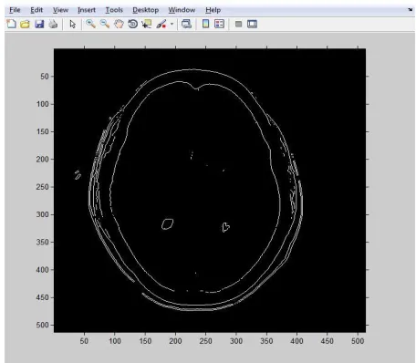
Fig. 3: Image Thresholding using Previous Method
The above figure shows the lack of proper segmentation in previous methods, due to the fact that tumor is a soft tissue and cannot by segmented by hard partitioning methods.
Fig. 4: Expert Region Selection in the proposed method
The Expert region is required to set the initial Seed region of the image as it allows the algorithm to search only in specified region of the image rather than the complete MRI Scan this makes algorithm faster.
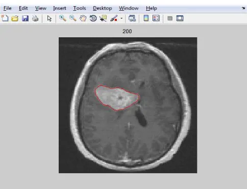
In above figure the Tumor region is clear identified by the first step of the algorithm, in figure below the region energy le vel is again adjusted for selecting the minimal fuzzy region for tumour segmentation
Fig. 6: Filtered Intensity Image for Brain showing Tumor Regions
Fig. 7: Initial Region Selected By the Algorithm for analysis
Fig. 8: Final segmented Region using Fuzzy based Approach
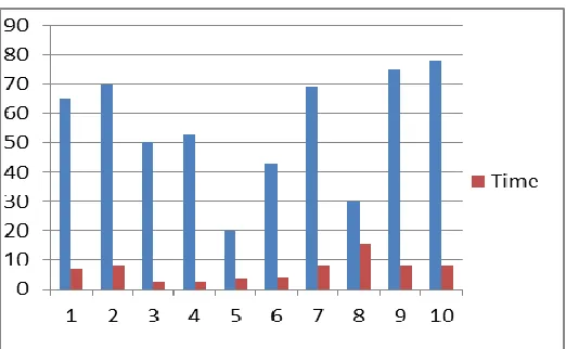
Fig. 9: Number of iterations vs Time required for each image
V.
CONCLUSION
To eliminate the dependence of the operator and improve the accuracy of diagnosis system aided diagnosis computer are a valuable and beneficial means for the detection of cancer and classification. Segmentation techniques based on gray level techniques such as threshold and techniques based on region are the simplest and find limited application techniques. However, their performance can be improved by incorporating them with the methods of hybrid clustering. techniques based on textural characteristics using atlas or look-up table can have excellent results on the segmentation of medical images , however, they need expertise in the construction of the atlas Limiting the technical atlas based is that , in some circumstances , it becomes difficult to choose correctly and label data has difficulty in segmenting complex structure with variable shape, size and properties in such situations it is best to use unsupervised methods such as fuzzy algorithms. In this work we proposed a novel fuzzy based MRI Image Segmentation algorithm, Fuzzy Segmentation involves the task of dividing data points into homogeneous classes or clusters so that items in the same class are as similar as possible and items in different classes are as dissimilar as possible. The results show that Fuzzy based Segmentation can successfully segment a tumor provided the parameters are set properly in the environment
REFERENCES
[1] Zhang, Hui, Jason E. Fritts, and Sally A. Goldman. "Image segmentation evaluation: A survey of unsupervised methods." computer vision and image understanding 110, no. 2 (2008): 260-280.
[2] Liu, Dingding, Yingen Xiong, Kari Pulli, and Linda Shapiro."Estimating image segmentation difficulty." In Machine Learning and Data Mining in Pattern Recognition, pp. 484-495. Springer Berlin Heidelberg, 2011.
[3] Pal, Nikhil R., and Sankar K. Pal. "A review on image segmentation techniques." Pattern recognition 26, no. 9 (1993): 1277-1294.
[4] Felzenszwalb, Pedro F., and Daniel P. Huttenlocher. "Efficient graph-based image segmentation." International Journal of Computer Vision 59, no. 2 (2004): 167-181.
[5] Senthilkumaran, N., and R. Rajesh. "Edge detection techniques for image segmentation–a survey of soft computing approaches." International Journal of Recent Trends in Engineering 1, no. 2 (2009).
[6] Fu, King-Sun, and J. K. Mui. "A survey on image segmentation." Pattern recognition 13, no. 1 (1981): 3-16.
[7] Pham, Dzung L., Chenyang Xu, and Jerry L. Prince. "Current methods in medical image segmentation 1." Annual review of biomedical engineering 2, no. 1 (2000): 315-337.
[8] Zhou, Guangyao, Stuart Geman, and Joachim M. Buhmann. "Sparse feature selection by information theory." In Information Theory (ISIT), 2014 IEEE International Symposium on, pp. 926-930. IEEE, 2014.
[9] Szirányi, Tamás, and Maha Shadaydeh. "Segmentation of Remote Sensing Images Using Similarity-Measure-Based Fusion-MRF Model." IEEE Geosci. Remote Sensing Lett. 11, no. 9 (2014): 1544-1548.
[10] Jui, Shang-Ling, Chao Lin, Haibing Guan, Ajith Abraham, Aboul Ella Hassanien, and Kai Xiao. "Fuzzy c-means with wavelet filtration for MR image segmentation." In Nature and Biologically Inspired Computing (NaBIC), 2014 Sixth World Congress on, pp. 12-16. IEEE, 2014.
[11] Ghosh, Susmita, Moumita Roy, and Ashish Ghosh. "Semi-supervised change detection using modified self-organizing feature map neural network." Applied Soft Computing 15 (2014): 1-20.
[12] Chuang, Yi-Fang, Dana Eldreth, Kirk I. Erickson, Vijay Varma, Gregory Harris, Linda P. Fried, George W. Rebok, Elizabeth K. Tanner, and Michelle C. Carlson. "Cardiovascular risks and brain function: a functional magnetic resonance imaging study of executive function in older adults." Neurobiology of aging 35, no. 6 (2014): 1396-1403.
[13] Carincotte, Cyril, Stéphane Derrode, and Salah Bourennane. "Multivariate fuzzy hidden Markov chains model applied to unsupervised multiscale SAR image segmentation." In Fuzzy Systems, 2005. FUZZ'05. The 14th IEEE International Conference on, pp. 288-293. IEEE, 2005.
[14] Chamorro-Martinez, Jesús, Daniel Sánchez, Belén Prados-Suárez, and Elena Galán-Perales. "Fuzzy Homogeneity Measures for Path-based Colour Image Segmentation." In Fuzzy Systems, 2005. FUZZ'05. The 14th IEEE International Conference on, pp. 218-223. IEEE, 2005.
[15] Dong, Guo, and Ming Xie. "Color clustering and learning for image segmentation based on neural networks." Neural Networks, IEEE Transactions on 16, no. 4 (2005): 925-936.