Bisphosphonates: Benefits, Basics, Potential Dental
Side Effects, and Management
1Vinayak Thorat, 2Rahul Shetty RevIew ARtICle
1,2Assistant Professor
1Department of Periodontology and Implantology, Bharati Vidyapeeth Dental College and Hospital, Navi Mumbai Maharashtra, India
2Department of Oral Medicine and Radiology, Bharati Vidyapeeth Dental College and Hospital, Navi Mumbai, Maharashtra, India Corresponding Author: Vinayak Thorat, Assistant Professor Department of Periodontology and Implantology, Bharati Vidyapeeth Dental College and Hospital, Navi Mumbai Maharashtra, India Phone: +919970748874, e-mail: dr.vinayakthorat@gmail.com ABSTRACT
Bisphosphonates are compounds used in the treatment of many skeletal disorders, such as bone metastases, osteoporosis, Paget’s disease, hypercalcemia of malignancy, and bone pain. Bisphosphonates have been found to be invaluable in control-ling pain and preventing fractures. But the long-term use of therapeutic doses of bisphosphonates may show side effects and dangers of using it. A new complication of bisphosphonate therapy administration, i.e., osteonecrosis of jaws also known as bisphosphonates-induced osteonecrosis (BIONJ) of the jaws, seems to be developing. A detailed knowledge of
con-flicting information regarding the impact of BIONJ in dentistry,
the development of osteonecrosis of the jaws, potential risk/ precipitating factors, and potential preventive measures for this oral complication need to be understood.
Keywords: Bisphosphonates, Bisphosphonates-induced osteo-necrosis, Bone diseases
How to cite this article: Thorat V, Shetty R. Bisphosphonates:
Benefits, Basics, Potential Dental Side Effects, and Manage -ment. Int J Prev Clin Dent Res 2017;4(1):64-68.
Source of support: Nil
Conflict of interest: None
INTRODUCTION
Bone disease can weaken the bones and cause the bones to become thinner (osteoporosis).1 They may even lead to lytic lesions causing holes in the bone. The weakened bones are vulnerable to fractures and are more likely to break under pressure (pathologic fracture). In bone dis-eases like myeloma, signals are sent to certain bone cells called osteoclasts, causing them to break down bone. In this process, the bones also release calcium, which may lead to a condition called hypercalcemia. Both myeloma bone disease and hypercalcemia can be treated with a group of drugs called bisphosphonates.2,3
Bisphosphonates are small inorganic molecules that bind to a substance called hydroxyapatite on the surface of damaged bones. Bisphosphonates prevent bone damage by inhibiting and destroying activity of osteoclast bone cells.
BENEFITS OF BISPHOSPHONATES
• Bisphosphonates are helpful in preventing damage of bone, hence, reducing bone pain and need for painkillers.
• They restore the calcium level by preventing hyper -calcemia.
• The chances of pathologic fractures reduce consider -ably, which helps the patients lead their lives confi-dently.
• Overall, it reduces the need of chemotherapy by improving the chances of healing and recovering the strength of the bone.4
HISTORICAL BACKGROUND
Bisphosphonates were first used in 1966. They were first administered to living animals, and an increase in bone mass was noted. In the late 1970s, low bone mass was shown to be associated with fracture, and by 1984, the two concepts had been linked. Eleven years later, the oral bisphosphonate alendronate was approved by the Food and Drug Administration (FDA) for the treatment of osteoporosis.5 After the approval, other oral agents, such as residronate and ibandronate, were approved. The first injectable form of bisphosphonate, pamidronate, was approved for the treatment of bone metastases in 1991. A more potent zolendronic acid received approval 10 years later.6 Didronel (etidronate) was first used over 20 years ago to treat patients with osteoporosis, but they devel-oped osteomalacia and clinical studies were stopped. A third injectable agent, ibandronate, was approved for use in osteoporosis patients in 2006.
The FDA has also notified the dental/medical com-munity about the side effects of using bisphosphonates that may lead to osteonecrosis of the jaws – primarily not only of the mandible, but also cases of the maxilla – occurring in association with bisphosphonates.
MECHANISM OF ACTION OF BISPHOSPHONATES
Bisphosphonates: Benefits, Basics, Potential Dental Side Effects, and Management IJPCDR
circulation is quite short, ranging from 30 minutes to 2 hours.7 However, once incorporated into bone tissue, they can persist for up to 10 years, depending on the skeletal turnover time.8
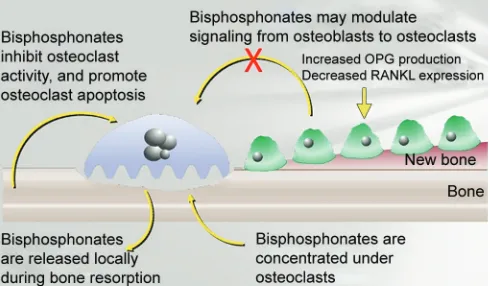
Bisphosphonates bind strongly to the bone surface at sites, particularly having increased bone turnover. Bisphosphonates act on bone mainly by two mechanisms simultaneously. They can both decrease osteoclast activity and decrease osteoclast numbers. The first is exemplified by internalization by osteoclasts, causing disruption of osteoclast-mediated bone resorption. The second is by inhibiting osteoclast recruitment and accelerating programmed cell death (apoptosis) of osteoclasts, thus reducing osteoclast numbers. Both mechanisms lead to reduction of bone resorption and to a decrease in bone turnover.9
Bisphosphonates may modulate the signaling from osteoblasts to osteoclasts (Fig. 1). It increases the osteo-protegerin (OPG) production, which inhibits both dif -ferentiation and function of osteoclasts. There is also reduced receptor activator of nuclear factor kappa-B ligand (RANKL) expression. The OPG protects bone from excessive resorption by binding to RANKL and prevent-ing it from bindprevent-ing to RANK.10
In summary, bisphosphonates are nonmetabolized analogs of pyrophosphate that are capable of localizing in bone and inhibiting osteoclastic function.11
ADVERSE EFFECTS OF BISPHOSPHONATES
Generally, the side effects seen are hypocalcemia, skeletal bone and joint pain, constipation or diarrhea, tiredness, etc. Oral bisphosphonates can cause upset stomach causing inflammation and erosions of the esophagus. Intravenous (IV) infusion can give rise to fever and flu-like symptoms after first infusion, but the greatest risk is that of development of bisphosphonate-related osteonecrosis of the jaw (BRONJ).12 It has been suggested that the bisphosphonates produce ischemic changes in the maxilla and mandible. Osteonecrosis of
jaw associated with bisphosphonate therapy in cancer patients was reported in 2003.13 The fact that these com-plications were not recognized during the trial phase of these drugs suggests that the effect of the drugs, when they become incorporated in the bones, might be cumulative in nature. The incidence of BRONJ is highest in patients with underlying malignancies, who receive high doses of IV bisphosphonates (e.g., zoledronic acid, 4 mg IV every 3–4 weeks) to decrease the risk of skel-etal complications of malignancy. Between 1 and 10% of these patients may go on to develop osteonecrosis of jaw. A study of multiple myeloma patients treated with zoledronic acid showed a progressively increas-ing incidence of osteonecrosis of the jaws to 10% by 36 months. With pamidronate, the incidence was lower to 4% by 36 months.14 The unique environment of the oral cavity could explain why the maxilla and mandible are solely involved. It can be hypothesized that patients who have received long-term bisphosphonate therapy may have a compromised blood supply to their maxilla and mandible. When dental extractions are performed on this group of patients, the open bony wound with a compromised healing ability cannot cope with the presence of oral microflora. The extraction wound then becomes infected and progresses into osteomyelitis due to the poor healing ability of the tissues.15 It then develops into osteonecrosis. It should be noted that all other bones in the skeleton are well enclosed in the soft tissue and, thus, protected from a resident microflora.
RISK FACTORS FOR BIONJ
A relevant number of secondary local and general risk factors have been identified. The addition of secondary factors can potentially shift a patient from a low to a high risk of developing of bisphosphonates-induced osteone-crosis (BIONJ).16
The local risk factors include:
• All invasive dental procedures (16- to 44-fold higher risk)
• Extractions of the lower molars (more than two-thirds of BIONJ cases are mandibular)
• Trauma related to unstable dentures (5-fold higher risk) • Thin mucosal coverage of the bones of the jaws
The general risk factors involved in BIONJ are: • Demographics (age > 60 years): Since blood circulation
and the ability to recover from trauma are decreased in older patients, advanced age is considered to place these patients at increased risk.
• Female gender: Women are at higher risk because they are more likely to receive nitrogen-containing bisphosphonates for the treatment of osteoporosis. • Other drugs: Concomitant therapy with corticoste
-roids, chemotherapeutics, or thalidomide increases the
suppression involving osteoclasts, osteoblasts, and osteocytes, thus increasing the risk of osteonecrosis, although this issue is debated in the literature. Con -comitant disease – immunocompromise and uncon-trolled diabetes are systemic conditions that can affect bone turnover.
• Lifestyle related: Heavy tobacco consumption and poor oral hygiene are associated with the delayed healing of surgical wounds and extraction sockets. • Genetic factors: Single-nucleotide polymorphisms
in the CYP2C8 gene influence the development of BIONJ in patients with multiple myeloma treated with bisphosphonates.
CLINICAL STAGING OF PATIENTS WITH BIONJ
Patient may be suffering from BIONJ if they fulfill the fol -lowing three criteria current or previous treatment with a bisphosphonate. Patients with BRONJ present with local -ized pain, neuropathy, halitosis, exposed bone, erythema, gingivitis, mobility of teeth with suppuration, and pus discharge. In order to standardize the criteria for BRONJ, the American Association of Oral and Maxillofacial Surgeons (AAOMS) in 2007 came up with the following three criteria:17,18
1. Current or previous treatment with a bisphospho -nate.
2. Exposed bone in the maxillofacial region that has persisted for more than 8 weeks.
3. No history of radiation therapy to the jaws.
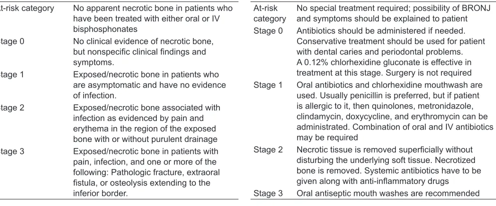
Ruggiero et al19 suggested the following clinical staging of BRONJ at AAOMS in 2009 (Table 1). Treat -ment strategy of BRONJ is as per the recommendation of AAOMS in 2009 (Table 2).20
Clinical symptoms and signs of BIONJ include pain, swelling, paresthesias, suppuration, along with soft tissue ulceration and intra- or extraoral sinus tracts. Radiographic abnormalities range from none to varying radiolucencies or radiopacities. The exact mechanism underlying this reaction is unknown; however, it has been postulated that bisphosphonates inhibit new vessel formation in the bone, which is associated with absent or delayed hard (alveolar) and soft tissue healing, usually after dental extractions.21 The oral lesions seen in BIONJ appear similar to those of radiation-induced osteonecrosis (Fig. 2).22 Clinically, there is oral ulceration with exposed underlying necrotic (“dead”) bone (Fig. 1). This oral condition causes chronic pain and severe, irreversible dysfunction and disfigurement of the jaw. Other symp -toms include soft tissue swelling, infection, and mobility of teeth. Patients may remain asymptomatic for many weeks or months, and BIONJ may be recognized only by the presence of exposed “painful” bone in the mouth. These lesions most likely become symptomatic when the necrotic sites become secondarily infected or if there is trauma to the soft tissue.
ROLE OF DENTISTS IN PREVENTION AND TREATMENT OF BIONJ
The role of the dentist can be summarized as follows: • Be aware of the risk of dentoalveolar surgical proce
-dures in the development of BIONJ.
• Recognize the clinical and radiographic features of osteonecrosis.
• Identify the local and systemic risk factors in patients on bisphosphonate therapy that place them into the low- or high-risk group for BIONJ.
Table 1: Clinical staging of BRONJ as suggested by Ruggiero et al19 at AAOMS in 2009
At-risk category No apparent necrotic bone in patients who have been treated with either oral or IV bisphosphonates
Stage 0 No clinical evidence of necrotic bone, but nonspecific clinical findings and symptoms.
Stage 1 Exposed/necrotic bone in patients who are asymptomatic and have no evidence of infection.
Stage 2 Exposed/necrotic bone associated with infection as evidenced by pain and erythema in the region of the exposed bone with or without purulent drainage Stage 3 Exposed/necrotic bone in patients with
pain, infection, and one or more of the following: Pathologic fracture, extraoral fistula, or osteolysis extending to the inferior border.
Table 2: Treatment strategy of BRONJ as per the recommendation of AAOMS in 2009 At-risk
category No special treatment required; possibility of BRONJ and symptoms should be explained to patient Stage 0 Antibiotics should be administered if needed.
Conservative treatment should be used for patient with dental caries and periodontal problems. A 0.12% chlorhexidine gluconate is effective in treatment at this stage. Surgery is not required Stage 1 Oral antibiotics and chlorhexidine mouthwash are
used. Usually penicillin is preferred, but if patient is allergic to it, then quinolones, metronidazole, clindamycin, doxycycline, and erythromycin can be administrated. Combination of oral and IV antibiotics may be required
Stage 2 Necrotic tissue is removed superficially without disturbing the underlying soft tissue. Necrotized bone is removed. Systemic antibiotics have to be given along with anti-inflammatory drugs
Bisphosphonates: Benefits, Basics, Potential Dental Side Effects, and Management IJPCDR
• Adopt preventive strategies in patients on bisphos -phonate therapy, and especially in those who require dentoalveolar surgical procedures.
• Consider the predictive value of biochemical markers of metabolic bone activity in determining the risk of BIONJ.
• Prior to treatment with bisphosphonates, any unsal -vaged teeth should be removed; all invasive dental procedures should be complete and optimal oral health should be achieved.
• Patient with established BRONJ should be treated by skillful dental specialists, and the purpose of treatment should be to release pain and infection of soft tissue and bone, and minimize osteonecrosis of the jaw.
• The conservative treatment should be the first choice because there is a possibility that periodontal surgery makes the surgical site to renecrotize, thus it should be delayed as long as possible.
Education
Patients should be educated regarding the warning signs and symptoms of osteoradionecrosis like edema, pain, or exposed bone, and the importance of maintaining good oral health throughout the treatment period.
Conservative
This is indicated in patients who have evidence of exposed bone, but no evidence of infection. It may not necessarily eliminate all the lesions, but it may provide patients with a long-term relief. This approach involves a combination of antiseptic mouthwashes and analgesics and the use of teriparatide.23
However, note that the teriparatide treatment should not be used in cancer patients, or patients with a history of skeletal radiation or active bone metasta-ses. Splints may be used to protect sites of exposed necrotic bone.
Nonsurgical Management
Indicated for patients with exposed bone with symptoms of infection. This treatment modality may also be utilized for patients with other comorbidities, which precludes invasive surgical methods. This approach requires antimi-crobial mouthwashes, systemic antibiotics, and antifungal medication and analgesics.
Surgical Management
Surgical intervention is indicated in patients with symp -tomatic exposed bone with fistula formation and one or more of the following: Exposed and necrotic bone extend-ing beyond the alveolar bone resultextend-ing in pathological fracture; extraoral fistula; oral antral communication, or osteolysis extending from the inferior border of the man-dible or the sinus floor. Surgical management involves necrotic bone resection, removal of loose sequestra of necrotic bone, and reconstructive surgery. The objective of surgical management is to eliminate areas of exposed bone to prevent the risk of further inflammation and infection. The amount of surgical debridement required remains controversial.
CONCLUSION
Bisphosphonate-associated osteonecrosis of the jaw is an important condition seen most commonly in oncol-ogy patients receiving high-dose IV bisphosphonates. A sound knowledge of the side effects and risk factors of using bisphosphonates can benefit the patients to lead a confident life. The dental practitioners can avoid the possible side effects like osteonecrosis of the jaw by taking into consideration the preventive and conserva-tive methods.
REFERENCES
1. Tonino RP, Meunier PJ, Emkey R, Rodriguez-Portales JA, Menkes CJ, Wasnich RD, Bone HG, Santora AC, Wu M, Desai R, et al. Skeletal benefits of alendronate: 7-year treatment of postmenopausal osteoporotic women. J Clin Endocrinol Metab 2000 Sep;85(9):3109-3115.
2. Ruggiero SL, Mehrota B, Rosenberg TJ, Engroff SL. Osteone -crosis of the jaws associated with the use of bisphosphonates:
a review of 63 cases. J Oral Maxillofac Surg 2004 May;62(5):
527-534.
3. Cheng A, Mavrokokki A, Carter G, Stein B, Fazzalari NL, Wilson DF, Goss AN. The dental implications of bisphospho
-nates and bone disease. Aust Dent J 2005 Dec;50(4 Suppl 2): S4-S13.
4. Khosla S, Bilezikian JP, Dempster DW, Lewiecki EM, Miller PD, Neer RM, Recker RR, Shane E, Shoback D, Potts JT. Benefits and risks of bisphosphonate therapy for osteoporosis. J Clin Endocrinol Metab 2012 Jul;97(7):2272-2282.
5. Fleisch H. Bisphosphonates – history and experimental basis.
Bone 1987 Feb;8(Suppl 1):S23-S28.
bone disease. Drugs 1991 Dec;42(6):919-944.
7. Martin TJ, Grill V. Bisphosphonates – mechanisms of action. Aust Prescr 2000 Jun;23(6):130-132.
8. Sparidans RW, Twiss IM, Talbot S. Bisphosphonates in bone diseases. Pharm World Sci 1998 Oct;20(5):206-213.
9. Fleisch H. Development of bisphosphonates. Breast Cancer
Res 2002;4(1)30-34.
10. Udagawa N, Takahashi N, Yasuda H, Mizuno A, Itoh K, Ueno Y, Shinki T, Gillespie MT, Martin TJ, Higashio K, et al. Osteoprotegerin produced by osteoblasts is an important
regulator in osteoclast development and function.
Endocrino-logy 2000 Sep;141(9):3478-3484.
11. Wang J, Goodger NM, Pogrel MA. Osteonecrosis of the jaws associated with cancer chemotherapy. J Oral Maxillofac Surg 2003 Sep;61(9):1104-1107.
12. Khosla S, Burr D, Cauley J, Dempster DW, Ebeling PR, Felsenberg D, Gagel RF, Gilsanz V, Guise T, Koka S, et al.
Bisphosphonate-associated osteonecrosis of the jaw: report
of a task force of the American Society for Bone and Mineral Research. J Bone Miner Res 2007 Oct;22(10):1479-1491. 13. Marx RE. Pamidronate (Aredia) and zoledronate (Zometa)
induced avascular necrosis of the jaws: a growing epidemic.
J Oral Maxillofac Surg 2003 Sep;61(9):1115-1117.
14. Durie BGM, Katz M, Crowley J. Osteonecrosis of the jaw and bisphosphonates. N Engl J Med 2005 Jul;353(1):99-102. 15. Melo MD, Obeid G. Osteonecrosis of the maxilla in a patient
with a history of bisphosphonate therapy. J Can Dent Assoc
2005 Feb;71(2):111-113.
diagnosis, staging and management. Oral Surg Oral Med Oral Pathol Oral Radiol Endod 2006 Oct;102(4):433-441. 17. Kurien J, Sunil EA, Mukunda A, Philip SR. An update on
bisphosphonate-related osteonecrosis of the Jaw. Indian J Clin Pract 2013 Aug;24(3):267-270.
18. Kim YG, Lee YD, Kwon YD, Suh JH, Jeen SM. Study on bisphosphonates-related osteonecrosis of the jaw (BRONJ): case report and literature review. J Korean Assoc Oral Maxil
-lofac Surg 2010 Aug;36:291-302.
19. Ruggiero SL, Dodson TB, Assael LA, Landesberg R, Marx RE, Mehrotra B; American Association of Oral and Maxillofacial Surgeons. American Association of Oral and Maxillofacial Surgeons position paper on bisphosphonate-related osteone
-crosis of the jaws – 2009 update. J Oral Maxillofac Surg 2009 May;67(5 Suppl):2-12.
20. Rizzolo D, Sedrak M. Managing the adverse effects of bisphosphonate therapy on the jaw. JAAPA 2009 Nov;22(11):
48-52.
21. Migliorati CA, Schubert MM, Peterson DE, Seneda LM.
Bisphosphonate-associated osteonecrosis of mandibular and maxillary bone: an emerging oral complication of supportive
cancer therapy. Cancer 2005 Jul;104(1):83-93.
22. Melo MD, Obeid G. Osteonecrosis of the jaws in patients with a history of receiving bisphosphonate therapy. Strategies for prevention and early recognition. J Am Dent Assoc 2005
Dec;136(12):1675-1681.
23. Bodenner D, Redman C, Riggs A. Teriparatide in the manage