Regenerative Endodontics
AbstractNon-vital infected teeth with mature or immature root apex have long been treated with root canal therapy. Current treatment modalities save millions of teeth are each year but fail to establish healthy pulp tissue in the teeth. But, what if we can once again make non-vital tooth vital? Root canal treatment offer high levels of success for many conditions, an ideal therapy will be regenerative approach, which deals with removal of diseased pulp tissues and replacing it with healthy pulp tissue to revitalize teeth. Regenerative endodontics is accomplished by performing regenerative endodontic procedures that maintain or restore the vitality of the tooth but also disinfect and remove the diseased tissue. Regeneration can be achieved through revascularization of root canal, stem cell therapy, pulp implant, scaffold implant and gene therapy. This article provides a review of regenerative endodontics, an emerging field focused on replacing diseased pulp with functional pulp tissue.
Key Words
Pulp regeneration; stem cells; scaffold; growth factors
INTRODUCTION
Regenerative endodontic therapy provides an
alternative treatment approach that is based on the principles of regenerative medicine and tissue engineering. Regenerative endodontic therapy has
been defined as “biologically based procedures
designed to replace damaged structures, including dentin and root structures, as well as cells of the
pulp-dentin complex.[1] In the immature tooth with
pulpal necrosis, this relates to complete restoration of pulpal function and subsequent completion of root development. The management of immature permanent teeth with pulpal necrosis is challenging as the root canal system is often difficult to debride and the thin dentinal walls are at an increased risk of a subsequent cervical fracture.[2] Regenerative
endodontics applies the principles of regenerative medicine, utilizing a combination of specific stem cells, three dimensional scaffolds and growth factors to regenerate pulp-dentin complex and revitalize teeth.[3]
Regenerating Endodontics and its Biologic Concern
Calcium hydroxide treatment is still used to induce apexification of the immature tooth with necrosed pulp before placing an obturation material
(gutta-percha) in the root canal system.[4] But
disadvantages include the time required for
formation of the calcified barrier, porosity of the barrier in comparison to MTA barrier, multiple appointments needed for reapplication of calcium hydroxide till an apical barrier is formed and the effect of long-term (months or more) use calcium hydroxide on the mechanical properties of tooth
dentin.[5] However, even with apexification
treatments further root development is not possible and immature teeth remain vulnerable to cervical root fractures. In contrast, regenerative endodontic therapy has the potential for increased root development with regeneration of pulpo-dentinal complex, and thereby, leading to a better long-term prognosis.[5]
Research Areas
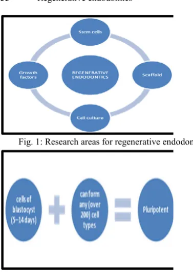
Regenerative endodontics comprises of research in adult stem cells, growth factors, organ-tissue culture, and tissue engineering materials (Fig. 1).
Amit Mahajan1, Bela Mahajan2,
Manish Gupta3, Sumit Agarwal4,
Nishant Singh5, Chetan Singh6
1Senior Lecturer, Department of Conservative
Dentistry and Endodontics, Rama Dental College and Hospital, Kanpur, India
2Senior Lecturer, Department of Oral
Pathology and Microbiology, Rama Dental College and Hospital, Kanpur, India
3Senior Lecturer, Department of Oral and
Maxillofacial Surgery, Rama Dental College and Hospital, Kanpur, India
4Reader, Department of Oral and Maxillofacial
Surgery, Rama Dental College and Hospital, Kanpur, India
5Reader, Department of Oral and Maxillofacial
Surgery, Rama Dental College and Hospital, Kanpur, India
6MDS, Department of Oral and Maxillofacial
Surgery, Consultant Oral and Maxillofacial Surgeon, India
I J Pre Clin Dent Res 2015;2(1):54-58 January-March
All rights reserved
The science of regenerative endodontics has a long history dating back to 1952 when Dr. BW Hermann reported on the application of calcium hydroxide in a case report of vital pulp amputation.[6] Presently,
two concepts exist in regenerative endodontics to treat non-vital infected teeth - one is the active pursuit of pulp-dentine regeneration to implant or regrow pulp (tissue engineering technology), and the other in which new living tissue is expected to form from the tissue present in the teeth itself,
allowing continued root development
(revascularization). Tissue engineering can be defined as 'an interdisciplinary field that applies the principles of engineering and life sciences toward the development of biological substitutes that restore, maintain, or improve tissue function.[7] The
three key components for tissue engineering[8] are:
Stem cells - to respond to growth factors.
Scaffold of extracellular matrix.
Growth factors.
Stem Cells
A stem cell is defined as a cell that has the ability to continuously divide and produce progeny cells that develop/ differentiate into various other types of
cells or tissues.[9] Stem cells are commonly
categorized as either embryonic or adult. Stem cells can also be classified by their source and their plsticity. According to the source of origin, stem cell are categorized as:
a) Autologous stem cells - are obtained from the same individual to whom they will be implanted.[8] Stem cells could be taken from the
bone marrow, peripheral blood,[10] the
periodontal ligament,[11] oral mucosa, or skin.
b) Allogenic stem cells - originate from a donor of the same species.[8] Examples of donor allogenic
cells include blood cells used for a blood transfusion,[12] bone marrow cells used for a
bone marrow transplant.[13]
c) Xenogenic cells - are those isolated from individuals of another species.[8] Pig tooth pulp
cells have been transplanted into mice, and these have formed tooth crown structures. This suggests use of donated animal pulp stem cells to create tooth tissues in humans.[14]
Stem cells are also commonly subdivided into totipotent (Fig. 2), pluripotent (Fig. 3), and multipotent (Fig. 4) categories according to their plasticity. The plasticity of the stem cell defines its ability to produce cells of different tissues.[14]
Various postnatal dental areDental pulp stem cells
(DPSC) which are derived from third molar, stem cells from human-exfoliated deciduous teeth (SHED) which are present within the pulp tissue of deciduous teeth, periodontal ligament stem cells (PDLSC), stem Cells from apical papilla (SCAP), stem cells from teeth extracted for orthodontic purposes.[8]
Growth Factors
Growth factors are proteins that bind to receptors on the cell and act as signals to induce cellular proliferation and/or differentiation.[1] Examples of
key growth factors in pulp and dentin formation
include bone morphogenetic proteins (BMPs),[15]
Fig. 1: Research areas for regenerative endodontics Fig. 2: Totopotent stem cells
transforming growth factor-beta[16] and fibroblastic
growth factor.[17,5] Transforming growth factor-beta
growth factors are secreted by odontoblasts and deposited within the dentin matrix, where they remain protected in an active form through interaction with other components of the dentin matrix[18] whereas BMPs stimulates differentiation
of adult pulp stem cells into an odontoblastoid morphology in culture.[19]
Scaffolds
Pulp stem cells should be organized into a three-dimensional structure that can support cell organization and vascularization for tissue engineering. This can be accomplished using a porous polymer scaffold seeded with pulp stem cells.[20] A scaffold should have:
a) Growth factors to aid stem cell proliferation and differentiation, leading to improved and faster tissue development.[21]
b) Nutrients promoting cell survival and growth.[22]
c) Antibiotics to prevent any bacterial in-growth in the canal systems.
In pulp-exposed teeth, dentin chips have been found to stimulate reparative dentin bridge formation. Dentin chips may provide a matrix for pulp stem cell attachment and also be a reservoir of growth factors. Many types of biodegradable or permanent scaffolds made of natural (collagen, hyaluronic acid, chitosan and chitin) or synthetic (polylactic acid, polyglycolic acid, tricalcium phosphate, hydroxyapatite) materials are available.[5] Recently,
peptide hydrogel nanofibers and various fibrin gels have been investigated as potential scaffolds for dental pulp tissue engineering.[5] A scaffold should
meet following requirements:
a) Biodegradability: scaffolds need to be absorbed by the surrounding tissues without the necessity of surgical removal.[23]
b) High porosity and an adequate pore size: to facilitate cell seeding and diffusion throughout the whole structure of both cells and nutrients.[24]
c) The rate at which degradation occurs has to match with the rate of tissue formation; this means that the scaffold provides structural integrity till the time cells are fabricating their own natural matrix structure around themselves, and it will eventually break down, leaving the newly formed tissue that will take over the mechanical load.[25]
The simplest approach to pulp tissue regeneration would be to regrow pulp over remaining pulp tissue
but attempts to regenerate pulp tissue under conditions of inflammation or partial necrosis have
proved unsuccessful,[26] and it is generally
recognized that the long-term prognosis of direct pulp capping infected tissue is poor and not recommended.[27] In the presence of infection, the
pulp stem cells that survive appear to be incapable of mineralization and deposition of a tertiary dentin bridge. Therefore, the majority of the available evidence suggests that necrotic and infected tooth pulp does not heal and it will be necessary to disinfect the root canal systems and remove infected hard and soft tissues before using regenerative endodontic treatments.[14]
Importance of Disinfection of Root Canal
Current regenerative endodontic protocols rely on irrigants to disinfect the canal and bleeding from the periapical area to bring cells and growth factors into the root canal and the blood clot and dentin walls to provide scaffolds for the generation of new tissue.[28] Disinfection is one of the prime objectives
of root canal preparation. The irrigant plays an important role as the irrigant acts as a lubricant during instrumentation and removes debris and microorganisms out of the canal. Sodium hypochlorite has been extensively used for several decades for this purpose.[29] Its excellent properties
of tissue dissolution and antimicrobial activity make it the irrigant of choice for the treatment of teeth with pulp necrosis, even though it has several undesirable characteristics, such as tissue toxicity at high concentrations.[30,31] The presence of a smear
layer on root canal walls is also a concern as it may inhibit the adherence of implanted pulp stem cells, leading to the failure of regenerative endodontic treatment. It is important to remove the smear layer from the root canal walls appear to be necessary to help promote the success of regenerative endodontics. The smear layer comprise of denatured cutting debris produced on instrumented cavity surfaces, and is composed of dentin, odontoblastic processes, nonspecific inorganic contaminants, and
microorganisms.[32] Its removal provides better
sealing of the endodontic filling material to dentin, and avoids the leakage of microorganisms into oral tissues.[33] Chemical chelating agents are used to
remove the smear layer from root canal walls, most
commonly a 17% solution of
ethylenediaminetetraacetic acid (EDTA) that is applied as a final flush.[34]
Measuring Clinical Success
Clinical success of regenerative endodontic therapy will depend on vascular blood flow, mineralizing odontoblastoid cells, intact afferent innervations and lack of signs or symptoms.[8] The clinical outcome
of regenerative endodontic procedure can be measured by invasive (histopath) technique or non invasive methods. It is not ethical to remove functioning tissues to conduct a histological analysis. Therefore, it will not be possible to
histologically investigate mineralizing
odontoblastoid cell functioning or nerve innervation. Clinicians will have to rely on the noninvasive tests that are in use, such as laser Doppler blood flowmetry in teeth; pulp testing involving heat and cold; and lack of signs or symptoms. Magnetic resonance imaging (MRI) has shown the potential to distinguish between vital and nonvital tooth pulps.[35] The ideal clinical outcome
will be a nonsymptomatic tooth that never needs retreatment.[14]
CONCLUSION
Regenerative endodontic methods have the potential for regenerating both pulp and dentin tissues and therefore may offer an alternative method to save teeth that are compromised. The success of regenerative endodontic therapy is dependent on the ability of researchers to create a technique that will allow clinicians to create a functional pulp tissue within cleaned and shaped root canal systems. The source of pulp tissue may be from revascularization, which involves enlarging the tooth apex to allow bleeding into root canals and generation of vital tissue that appears capable of forming hard tissue under certain conditions; stem cell therapy, involving the delivery of stem cells into root canals; scaffold, or pulp implantation, involving the surgical implantation of synthetic pulp tissue grown in the laboratory. One of the most challenging aspects of developing a regenerative endodontic therapy is to understand how the various component procedures can be optimized and integrated to produce the outcome of a regenerated pulp-dentin complex.
REFERENCES
1. Murray PE, Garcia-Godoy F, Hargreaves KM.
Regenerative endodontics: a review of current status and a call for action. J Endod 2007;33:377-90.
2. Cvek M. Prognosis of luxated non-vital
maxillary incisors treated with calcium hydroxide and filled with gutta-percha. A
retrospective clinical study. Endod Dent Traumatol 1992;8:45-55.
3. Maobin Yang. Regenerative endodontics: a
new treatment modality for pulp regeneration. JSM Dent 2007;1(2):10-11
4. Frank AL. Therapy for the divergent pulpless
tooth by continued apical formation. J Am Dent Assoc 1966;72:87-93.
5. Endodontics: colleagues for excellence.
Regenerative endodontics. Spring 2013. www.aae.org/colleagues
6. Herman BW. On the reaction of the dental
pulp to vital amputation and calxyl capping. Dtsch Zahnarztl Z 1952;7:1446-7.
7. Langer R, Vacanti JP. Tissue engineering.
Science 1993;260:920-6.
8. Bansal R, Bansal R. Regenerative
endodontics: A state of the art. Indian J Dent Res 2011;22:122-31.
9. Rao MS. Stem sense: a proposal for the
classification of stem cells. Stem Cells Dev 2004;13:452-5.
10. Jansen J, Thompson JM, Dugan MJ.
Peripheral blood progenitor cell
transplantation. Ther Apher 2002;6:5-14. 11. Seo BM, Miura M, Sonoyama W, Coppe C,
Stanyon R, Shi S. Recovery of stem cells from cryopreserved periodontal ligament. J Dent Res 2005;84:907-12.
12. Amin M, Fergusson D, Aziz A, Wilson K, Coyle D, Hebert P. The cost of allogeneic red blood cells: a systematic review. Transfus Med 2003;13:275-85.
13. Murphy WJ, Blazar BR. New strategies for preventing graft-versus-host disease. Curr Opin Immunol 1999;11:509-15.
14. Murray PE, Garcia-Godoy F, Hargreaves KM. Regenerative endodontics: a review of current status and a call for action. JOE 2007;33(4). 15. Nakashima M. Bone morphogenetic proteins
in dentin regeneration for potential use in endodontic therapy. Cytokine Growth Factor Rev 2005;16:369-76.
16. Chan CP, Lan WH, Chang MC. Effects of TGF-betas on the growth, collagen synthesis and collagen lattice contraction of human dental pulp fibroblasts in vitro. Arch Oral Biol 2005;50:469-79.
from gelatin hydrogels. J Endod 2009;35:858-65.
18. Smith AJ, Matthews JB, Hall RC. Transforming growth factor-beta1 (TGF-beta1) in dentine matrix. Ligand activation and receptor expression. Eur J Oral Sci 1998;106(Suppl 1):179-84.
19. Saito T, Ogawa M, Hata Y, Bessho K. Acceleration effect of human recombinant
bone morphogenetic protein-2 on
differentiation of human pulp cells into odontoblasts. J Endod 2004;30:205-8.
20. Nakashima M. Tissue engineering in endodontics. Aust Endod J 2005;31:111-3. 21. Oringer RJ Biological mediators for
periodontal and bone regeneration. Compend Contin Educ Den. 2002;23:501-4, 506-10. 22. Karande TS, Ong JL, Agrawal CM. Diffusion
in musculoskeletal tissue engineering
scaffolds: design issues related to porosity, permeability, architecture, and nutrient mixing. Ann Biomed Engl 2004;32:1728-43. 23. Schopper C, Ziya-Ghazvini F, Goriwoda W.
HA/TCP compounding of a porous Ca P biomaterial improves bone formation and scaffold degradation: a long-term histological study. J Biomed Mater Res B Appl Biomater 2005;74:458-67.
24. Sachlos E, Czernuszka JT. Making tissue engineering scaffolds work. Review: the application of solid freeform fabrication technology to the production of tissue engineering scaffolds. Eur Cell Mater 2003;30:29 –39.
25. Freed LE, Vunjak-Novakovic G, Biron RJ. Biodegradable polymer scaffolds for tissue engineering. Biotechnology 1994;12:689-93. 26. Rutherford RB, Gu K. Treatment of inflamed
ferret dental pulps with recombinant bone morphogenetic protein-7.1. Eur J Oral Sci 2000;108:202-6.
27. Barthel CR, Rosenkranz B, Leuenberg A, Roulet JF. Pulp capping of carious exposures: treatment outcome after 5 and 10 years: a retrospective study. J Endod 2000;26:525-8. 28. Galler KM, D’Souza RN. Tissue engineering
approaches for regenerative dentistry. Regen Med 2011;6:111-24.
29. Kaufman AY, Keila S. Hypersensitivity to sodium hypochlorite. J Endod 1989;15:224-6. 30. Oncag O, Hosgor M, Hilmioglu S, Zekioglu
O, Eronat C, Burhanoglu D. Comparison of
antibacterial and toxic effects of various root canal irrigants. Int Endod J 2003;36:423-32. 31. Takeda FH, Harashima T, Kimura Y,
Matsumoto K. A comparative study of the removal of smear layer by three endodontic irrigants and two types of laser. Int Endod J 1999;32:32-9.
32. Czonstkowsky M, Wilson EG, Holstein FA. The smear layer in endodontics. Dent Clin North Am 1990;34:13-25.
33. Sen BH, Wesselink PR, Turkun M. The smear layer: a phenomenon in root canal therapy. Int Endod J 1995;28:141-8.
34. Menezes AC, Zanet CG, Valera MC. Smear layer removal capacity of disinfectant solutions used with and without EDTA for the irrigation of canals: a SEM study. Pesqui Odontol Bras 2003;17:349-55.
35. Kress B, Buhl Y, Anders L, Stippich C, Palm F, Bahren W, Sartor K. Quantitative analysis of MRI signal intensity as a tool for evaluating tooth pulp vitality. Dentomaxillofac Radiol 2004;33:241-4.