Tanaffos (2009) 8(1), 56-61
©2009 NRITLD, National Research Institute of Tuberculosis and Lung Disease, Iran
Surgical Treatment of Pulmonary Hydatid Cyst in 72
Children
Ali Reza Mirshemirani 1, Sajad Razavi 2, Sina Sadeghian 1
1 Department of Pediatric Surgery, 2 Department of Pediatric Anesthesiology, Mofid Chidlren's Hospital, Shahid Beheshti University, M.C, TEHRAN-IRAN.
ABSTRACT
Background: Hydatid disease is a parasitic infestation which is endemic in many sheep and cattle raising areas (such as
Iran) and is still an important health hazard in the world. The aim of this study was to evaluate the outcome of surgical
treatment in patients with hydatid disease.
Materials and Methods: This retrospective study evaluated 72 consecutive patients who presented with pulmonary hydatid
cyst to Mofid Children’s Hospital from 1992 to 2007. Patients’ medical records were reviewed and their gender, age, clinical
features, cyst localization, diagnostic tools, operative techniques, pathologic report, morbidity and mortality, recurrence,
hospital stay and outcome of treatment were evaluated.
Results: The patient group consisted of 40(55.56%) boys and 32(44.44%) girls in the age range of 2 to 14 yrs. In general, 72
patients had a total of 87 cysts. Fifty-five patients (76.38%) had single cysts. Fifty-five lung cysts (63.21%) were in the right
side, and 31(35.64%) were in the right lower lobe. Cough was the most common symptom and chest radiography gave a
correct diagnosis in 68(94.44%) patients. Conservative surgical treatment was carried out in 70 children (97.22%). There
were no mortality or recurrence in our cases.
Conclusion: Due to the high accuracy of chest X-ray in diagnosis of lung hydatid cyst, it is the preferred method of diagnosis
in endemic regions. Parenchyma-saving surgical procedures such as cystotomy and capitonnage as well as cyst delivering
by lung expansion are the preferred methods of treatment for pulmonary hydatid disease in childhood. These methods are
safe, reliable and successful. (Tanaffos 2009; 8 (1): 56-61)
Key words: Hydatid cyst, Lung, Conservative surgical treatment, Children
INTRODUCTION
Hydatid disease is a public health hazard in Iran (1,2). It is common in rural areas where
cattle and dogs are kept. The primary hosts for the
Correspondence to: Mirshemirani AR
Address: Department of Pediatric Surgery, Mofid Children's hospital, Shahid
Beheshti University, M.C. Tehran-Iran.
Email address: almirshemirani@yahoo.com
Received: 1 June 2008
Accepted: 30 November 2008
Echinococcus granulosus tapeworms were dogs and canines. They produce eggs in the intestine and pass
them in the stool. Eggs are ingested by intermediate hosts such as sheep and cows. Humans become accidental hosts by eating tapeworm eggs (3).
liver due to less elasticity of the lungs. This may explain the high incidence of disease in these organs in children (4,5).
Diagnosis of the hydatid disease of the lung is made by history, physical exam findings and radiological evaluation, but serological investigations
have a limited value.
Symptoms are related with size, localization, pressure on surrounding tissues and rupture of the
cyst. The most common symptom is cough.
The cyst is identified on X-ray as a round or oval
homogenous opacity that can be differentiated from pulmonary parenchyma (6).
In the present retrospective study, we reviewed
the gender, age, clinical features, cyst localization, diagnostic tools, operative techniques, complications, morbidity and mortality, hospital stay, recurrence,
and outcome of treatment in children with pulmonary hydatid cyst.
MATERIALS AND METHODS
This study was conducted on hospitalized patients operated for pulmonary hydatid cysts at Mofid
Children’s Hospital in Tehran, Iran during 1992 to 2007.
A total of 72 patients were evaluated in this study.
Data were collected from the medical records of patients in the archives and analyzed in terms of age, gender, clinical features, cyst localization, diagnostic
tools, operative techniques, complications, morbidity and mortality, hospital stay, recurrence and outcome of treatment. ELISA test was performed for all
patients and IFA test for the suspected ones. Pathological report was the main factor in selecting the patients. We performed chest radiography for all
and lung CT-scan for suspected ones, which was effective for diagnosis. For abdominal evaluation, we performed ultra-sonography for all, and abdominal
CT-scan for 6 patients. Cystotomy and capitonnage were mostly used for those with unruptured hydatid
cysts but in 15 cases, cysts were delivered after incising the pericyst layer and lung expansion by the anesthesiologist. For ruptured cysts, we performed
debridement, 20% hypertonic saline irrigation, and drainage.
All patients were followed up in the outpatient
clinic and all the relevant information was recorded in their files for at least two to four years.
RESULTS
Out of 72 children, 40(55.56%) were boys and the
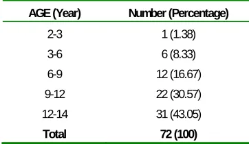
remaining 32(44.44%) were girls. Age ranged from 2 to 14 years old and the youngest one was 2 years-old. (Table 1)
Table 1. Age distribution in 72 patients.
AGE (Year) Number (Percentage)
2-3 1 (1.38)
3-6 6 (8.33)
6-9 12 (16.67)
9-12 22 (30.57)
12-14 31 (43.05)
Total 72 (100)
Incidence rate of hydatid disease increased by age. There were 18(25%) patients in the age range of 3 to 9 years old and 53(73.62%) patients in the age group of 9 to 14 years old.
Seventy-two patients had a total of 87 cysts out of which 55(63.21%) cysts were in the right lung and 32(36.79%) were in the left lung. Also, cysts were most commonly situated in the right lower lobe (35.64%) (Table 2).
Table 2. Distribution of 87 lung cysts
Lung Side
Upper Lobe
Middle Lobe
Lower
Lobe Total/Percentage
Right 14 10 31 55 (63.21)
Left 13 __ 19 32 (36.79)
The most commonly involved segment was posterior basal segment of the lower lobe (35%).
Cyst localization was single in 76.38%, unilateral
multiple in 20.84% and bilateral multiple in 2.78% of patients. Twelve (16.64%) patients had coexisting liver cysts. Sixty-three cysts (72%) were intact and
uncomplicated.
Of the total of 87 cysts, 62(71.26%) were larger than 4cm in diameter.
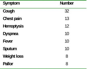
Cough was the most common symptom (Table 3) and 20 patients had no symptom.
Table 3. Symptoms in 72 patients with lung hydatid cysts.
Symptom Number
Cough 32
Chest pain 13
Hemoptysis 12
Dyspnea 10
Fever 10
Sputum 10
Weight loss 8
Pallor 8
Chest radiography gave a correct diagnosis in
68(94.44%) patients (Figure1). Ultrasound and computed tomography scanning were used in those who were suspected to have coexisting liver cysts
(Figure 2)
Conservative surgical treatment was carried out in
65 of 72 children (90%). Cystotomy and capitonnage, as well as cyst delivering by lung expansion were the preferred methods.
Other surgical approaches were segmentectomy in 5 and lobectomy in 2 patients.
Of 10 patients with concomitant liver and
pulmonary hydatid cysts, 9 were approached by right thoracopherenotomy and one by median sternotomy and right pherenotomy.
Atelectasis and wound infection were the most
frequent post-operative complications. The morbidity rate was 12.5%.
The mean length of hospital stay was 9 days
(range 5-28 days) and no recurrence or mortality were observed during the follow-up period. Echinococcus granulosus was detected in 98% of our
patients and only 2% showed Echinococcus multilocularis.
Figure 1. Chest radiography of a 7 year-old boy with multiple bilateral
cysts.
Figure 2. CT scan of a patient with a coexisting liver cyst
DISCUSSION
Of our 72 cases, 40(55.56%) were boys and
that in children, males were more likely to be infected with pulmonary hydatid cyst (7,8), but in Bulent's report incidence of hydatid cyst was equal in
males and females (9,10).
Patients’ age ranged from 2 to 14 years old and the youngest one was 2 years old. We believed that 2
years of age is nearly the youngest age of this pathology, but Prashant has reported hydatid disease in a six month-old infant (11).
Incidence rate of hydatid cyst increased by age in our patients; 53(74%) cases were more than 9 years
old. Children’s hydatid cyst is more common in lungs than in liver, our study as well as Montazeri’s study confirmed this finding (7). But Sehitogullari
(6) believed that liver cyst was more common in childhood and Talaiezadeh (8) found nearly equal incidence of hydatid cyst in lung and liver in children
(41% and 43%).
In our study, 72 patients had a total of 87 cysts, 55(63.21%) in the right lung and 32(36.79%) in the
left lung. In Ozvaran (12) series, 62.5% of the cysts were in the right lung and 37.5% were in the left lung.
Cysts were most commonly situated in the right lower lobe (35.64%) in our study, but in Sarifimood report (13) this rate was 27.4% and others reported a
rate of 51.9% (6).
In our study, cyst localizations were single in 76.38%, unilateral multiple in 20.84% and bilateral
multiple in 2.78% of patients and the most commonly involved segment was posterior basal segment of the lower lobe (35%). Isolated pulmonary
cysts are more common in children (14).
Twelve (16.67%) patients had coexisting liver cysts and 63(72%) cysts were intact and
uncomplicated. Of the total of 87 cysts, 62(71.26%) were larger than 4cm in diameter. But Kanat reported huge cysts (10cm) in his case series. Cysts also tend
to become bigger in children than in adults (14). Cough was the most common symptom in our
patients, similar to others studies (7,15,16).
Cysts may grow faster in the lung than in the liver due to less elasticity of the lung. This may explain
the high incidence of disease in children’s lungs (7). In Talaiezadeh (8) series chest pain was the most common symptom.
Chest radiography indicated a correct diagnosis in 94.44% of our patients, nearly similar to the rate
96.4% given by Koeseoglu (17). But the accuracy of chest X-ray was 84% in Montazeri series (7). Conservative surgical treatment was carried out in 65
of 72 children (90%). Cystotomy and capitonnage, as well as cyst delivering by lung expansion were the preferred methods of surgery similar to Kanat (14)
study.
Other surgical approaches were segmentectomy in 5 and lobectomy in 2 patients. Of 10 patients with
concomitant liver and pulmonary hydatid cysts in our series, 9 were approached by right thoracopherenotomy and one by median sternotomy
and right pherenotomy, similar to other series (17,18,19).
One stage surgical management of lung and liver
hydatid cysts by right thoracotomy + pherenotmy has been performed by Biswas et al (20). Recently thoracoscopy was used for diagnosis and treatment of
hydatid cyst (11,21).
Atelectasis and wound infection were the most frequent post-operative complications in our study
which was similar to others’ (14,17,19,22).
In our study, the morbidity rate was 12.5%; this rate was 14.4% in Balcy's study (22). There were no
recurrence or mortality in our study, similar to other series (6,17). The mean length of hospital stay was 9 days (range 5-28 days), similar to Balcy's report (22).
In our study, 12 patients had severe air-leakage which prolonged the hospital stay, and we had to maintain the chest-tube for a longer period of time. In
treatment for cases with air-leakage.
Echinococcus granulosus was detected in 98% of our patients and only 2% had Echinococcus
multilocularis.
CONCLUSIONS
Due to the high accuracy of chest X-ray in diagnosis of hydatid cyst of the lung, we recommend it as the method of choice for its diagnosis in
endemic regions.
Surgery is the treatment of choice and
parenchyma-saving surgical procedures such as cystotomy and capitonnage, as well as cyst delivering by pulmonary expansion are the preferred methods of
treatment for pulmonary hydatid disease in childhood. These simple procedures are safe, reliable and successful. Meticulous surgery, suturing the
traumatized bronchioles with proline sutures and maintaining the chest-tube for a longer period of time may stop air-leakage and no further surgical
intervention is usually required.
Pulmonary hydatid cyst tends to be bigger in children than adults.
Acknowledgment
The authors would like to thank Mrs. M. Saeedi
and A. Karimi who cooperated in typing and editing the text and helped us during the analysis.
REFERENCES
1. Saidi F. Surgery of hydatid disease. Philadelphia (PA): WB
Saunders; 1976. 8.
2. Harandi MF, Hobbs RP, Adams PJ, Mobedi I, Morgan-Ryan
UM, Thompson RC. Molecular and morphological
characterization of Echinococcus granulosus of human and
animal origin in Iran. Parasitology 2002; 125 (Pt 4):
367-73.
3. King CH. Cestodes (tapeworms). In: Mandell GL, Bennet JE,
Dolin R. Principles and practice of infectious disease 6th ed.
New York: Churchill Livingston, 2005; 3290-2.
4. Beggs I. The radiology of hydatid disease. AJR Am J
Roentgenol 1985; 145 (3): 639- 48.
5. Karaoglanoglu N, Kurkcuoglu IC, Gorguner M, Eroglu A,
Turkyilmaz A. Giant hydatid lung cysts. Eur J Cardiothorac
Surg 2001; 19 (6): 914- 7.
6. Sehitogullari A. Our results in surgical treatment of hydaid
cyst of the lungs. European Journal of General Medicine
2007; 4(1): 5-8.
7. Montazeri V, Sokouti M, Rashidi HR. Comparison of
pulmonary hydatid disease between children and adult.
Tanaffos 2007; 6(1): 13-18.
8. Talaiezadeh AH, Maraghi Sh. Hydatid disease in children: A
different pattern than adults. Pakistan Journal of Medical
Science 2006; 22(3): 329-32.
9. Bulent K, Gultekin G, Serdar H, Mustafa Necmi I, Koray D,
Unal S. Analysis of Pulmonary Hydatidosis According to
Their Segmentary Locations. Clinical Pulmonary Medicine
2008; 15(1): 8-12
10.Cangir AK, Sahin E, Enön S, Kavukçu S, Akay H, Okten I,
et al. Surgical treatment of pulmonary hydatid cysts in
children. J Pediatr Surg 2001; 36 (6): 917- 20.
11.Prashant J, Beejal S, Hemanshi Sh, SV Parelkar, SS
Borwankar. Thoracoscopic excision of mediastinal cysts in
children. Journal of Minimal Access Surgery 2007; 3(4):
123-6
12.Ozvaran M, Unver E, Uskul B, etal. An evaluation of
diagnosis and treatment of pulmonary hydatid cyst. Turkish
respiratory journal 2000; 1(2): 11-13.
13.Sharifimood B, Fazaeli A, Izadi SH, et al. Fifteen years
experience, with pulmonary hydatidosis in Zahedan, Iran.
Iranian Journal of Parasitology 2007; 2(4): 7-11.
14.Kanat F, Turk E, Aribas OK. Comparison of pulmonary
hydatid cysts in children and adults. ANZ J Surg 2004; 74
(10): 885- 9.
15.Rattan KN, Sharma A, Sharma Anita. Hydatid disease in
children. Indian Journal of Chest Diseases and Allied
Sciences 1998; 40 (1): 73-7.
16.Anadol D, Göçmen A, Kiper N, Ozçelik U. Hydatid disease
in childhood: a retrospective analysis of 376 cases. Pediatr
17.Koeseoglu B, Bakan V, Onem O, Bilici S, Demirtas I.
Conservative Surgical Treatment of Pulmonary Hydatid
Disease in Children: An Analysis of 35 Cases. Surg Today
2002; 32(9): 779-83.
18.Muharrem C, Canon S, Murat K, et al. Surgical treatment of
pulmonary hydatid disease in children: report of 122 cases. J
Pediatr Surg 2000; 35(12): 1710-3.
19.Ulkü R, Onen A, Onat S. Surgical treatment of pulmonary
hydatid cysts in children: report of 66 cases. Eur J Pediatr
Surg 2004; 14 (4): 255- 9.
20.Biswas B, Ghosh D, Bhattacharjee R, Patra A, Basuthakur S.
One stage surgical management of hydatid cyst of lung &
liver-by right thoracotomy & phrenotomy. Indian Journal of
Thoracic and Cardiovascular Surgery 2004; 20(1): 47.
21.Mallick MS, Al-Qahtani A, Al-Saadi MM, Al-Boukai AA.
Thoracoscopic treatment of pulmonary hydatid cyst in a
child. J Pediatr Surg 2005; 40 (12): e35- 7.
22.Balcý AE, Ülkü R, Eren &Thorn;, Eren MN, Cebeci E,
Erdem K. Surgical Treatment of Pulmonary Hydatid Cysts
in Children. Thorac Cardiovasc Surg 2002 Thema: Tuesday,
February 19, 2002 Special Session – Thoracic and Vascular