135
KLECZKOWSKI, J. & KLECZKOWSKI, A. (1953). J . gen. Mimobiol. 8, 135-144.The Behaviour of Rhizobium Bacteriophages during and
after Exposure to Ultraviolet Radiation
BY J. KLECZKOWSKI AND A. KLECZKOWSKI
Rothamsted Experimental Station, Harpenden, Hertfordshire
SUMMARY : After inactivation by ultraviolet radiation, particles of two Rhizobium
bacteriophages interfered temporarily with the multiplication of active particles of the homologous phage, in liquid cultures of their respective host bacteria. Inactivated particles did not affect the number of plaques produced by active particles in bac- terial cultures on agar.
No evidence was found that particles that were inactive singly became active when two or more of them infected the same bacterial cell.
The rate of inactivation approximated closely to that of a first-order reaction. Exposing infected bacteria to visible Iight increased the residual activities of irradiated phage preparations by amounts equivalent to decreasing the doses of ultraviolet irradiation by a constant factor. Exposing either the irradiated phage preparations or the bacterial cultures separately to visible light had no effect.
Those ultraviolet irradiated phage particles which remained active were so altered that they became relatively unstable.
Much work has been done on the effects of ultraviolet radiation on bacterio- phages of Bacterium coli, but relatively little on others. The course of inactiva- tion, i.e. of becoming unable to multiply and cause lysis, was found to approximate to that of a first-order reaction, though slight deviations have been recorded (Latarjet & Wahl, 1945; Latarjet & Morenne, 1951). Some inactivated phages have proved able to interfere with multiplication of host bacteria and of active phages (Luria & Delbruck, 1942). Residual activity of some irradiated phage preparations could be increased by increasing their concentration at which they were brought into contact with susceptible bac- teria (' multiplicity reactivation ' Luria & Dulbecco, 1949) or by exposure of infected host bacteria to visible light (' photo-reactivation
'
Dulbecco, 1950).The work described in the present paper was started to see how far these phenomena applied to bacteriophages of strains of Rhixobium and was done simultaneously with a comparable work on plant viruses (Bawden &
Kleczkowski, 1953). During the work a new phenomenon was encountered; when irradiated phage preparations were stored their residual activity de- creased at a much higher rate than did the activity of unirradiated phage preparation stored in the same conditions. A few experiments were made to study some details of this phenomenon.
MATERIAL AND METHODS
producing plaques, have been described previously (Kleczkowska, 1945 ; Kleczkowski & Kleczkowski, 1952). Phage cultures were lysed liquid bac- terial cultures passed through a Chamberland L3 filter and stored at 2'.
The source of ultraviolet (u.v.) radiation was a low-pressure mercury dis- charge lamp made by the Thermal Syndicate Ltd. and fitted with a cylindrical chromium-plated reflector. According to the makers' specifications the inten- sity of radiation, 99
%
of which was of wave-length 2536A, was lZlpW./sq.cm. at a distance of 100 cm.Undiluted phage cultures were irradiated as layers 0.15 em. deep in a Petri dish. They were exposed to the lamp a t a distance of 20cm. and rocked during the whole time of exposure. The rocking is assumed to have ensured that all phage particles were equally exposed to the radiation.
The intensity of radiation and its absorption by irradiated materials were not measured. The measurements would be useless because the amount of absorption by phage particles, as distinct from that caused by other con- stituents of crude bacterial lysates, would still remain unknown.
RESULTS
Interference by inactivated phage with multiplication of active phage and of host bacteria
This problem is considered first because if there were any interference it might need to be allowed for whenever residual activity of an irradiated phage preparation is tested.
The presence of inactivated phage does not interfere with formation of plaques by active phage. This is shown by the fact that adding untreated phage to an irradiated phage preparation, which is then incubated suitably diluted in a culture of host bacteria and plated, has no effect on the number of plaques. Nor is the plaque number affected by adding an irradiated phage preparation to an equal volume of a culture of host bacteria in which untreated phage preparation is then diluted and plated. Thus no active phage particles are destroyed or otherwise made permanently unable to multiply. On the other hand, irradiated phage preparations can stop completely, though tem- porarily, the multiplication of active phage in liquid cultures of host bacteria and can make the host bacteria unable to multiply and form colonies on agar medium.
Ultraviolet irradiated bacteriophages
137
After 24 hr. incubation, the concentration of active phage in the culture containing even the most concentrated irradiated phage was almost as high as that of the control. It is obvious, therefore, that the inhibitory effect of the irradiated phage preparation lasted less than 24 hr.
Table 1. Effect of an ultraviolet-irradiated preparation of phage 31'7 on multiplication of phage 317
Materials. Untreated preparation of 317 phage : 5 x lo8 plaques/ml. U.V. irradiated preparation of 317 phage: irradiation time was 20 min.; activity fell from 5 x lo8 to 50 plaques/ml. U.V. irradiated preparation of Cl, phage: irradiation time was 20 min. ;
activity fell from 18 x lo8 to 80 plaques/ml. 24 hr. culture of 317 bacteria: 15 x lo6
cells/ml. (haemocytometer count). U.V. irradiated medium : irradiation time was 20 min
.
The mixtures: 24 hr. culture of 317 bacteria+an equal volume of:
Irradiated 317 phage (undiluted) Irradiated 317 phage (dil. 1/41) Irradiated 317 phage (dil. 1/16?> Irradiated C1, (undiluted) Untreated Cl, (undiluted) Irradiated medium Untreated medium
Phage concentration in the mixtures
(in terms of numbers of plaques/ml.)
r A
\
Immediately After 3 hr. After 24 hr.
530 460 39 x 108"
600 1,500 -
550 19,000 -
560 7,500 52 x lo8
500 15,000 54 x 108
570 8,000 50 x lo8
550 16,500 53 x 108
One volume of the untreated preparation of 317 phage diluted 1/10, was added to nine volumes of each mixture. Samples of the mixtures were taken for assay of concentration of phage 317 by the plaque count method immediately, after 3 hr. and after 24 hr. incubation a t 25'.
*
The mean diameter of the plaques was about half that of the control.p Diluted in untreated medium.
Although after 24 hr. incubation the culture containing irradiated phage produced almost as many plaques as did the control phage culture, the diameter of the plaques (mean=1.9 mm.) was smaller than that of the control (mean = 3.4 mm.). No explanation of this phenomenon can be offered. It is not caused by a phage mutation, because phage isolated from the small plaques produced plaques of normal size.
Table 2 shows that U.V. inactivated phage Cl, also temporarily prevented
the multiplication of active phage Cl, in a liquid culture of Cl, bacteria. Com- parison of Tables 1 and 2 shows that the multiplication of active phage was prevented only by irradiated preparations of the homologous phage. Irradiated preparations of the other inhibited only to the same extent as did the irradiated medium.
of active phage, it does so only in a bacterial strain which is its host and then it interferes with the multiplication of any phage in it.
Luria & Delbriick (1942) found that the U.V. inactivated phage that inter- fered with phage multiplication also rendered the bacteria unable to multiply, whereas the other inactivated phage had no such effect. Moreover, the ability of the irradiated phage to interfere with phage multiplication could be destroyed by excess of U.V. irradiation, when their ability to make their host
Table 2. Effect of an ultraviolet irradiated Preparation of C1, phage on multiplication of Cl, phage
Materials. Untreated preparation of CI, phage : 18 x 108 plaques/ml. U.V. irradiated preparation of C1, phage: irradiation time was 20 min.; activity fell from 18 x lo* to 80 plaques/ml. U.V. irradiated preparation of 317 phage: irradiation time was 20 m h ; activity fell from 5 x 10⁸ to 50 plaques/ml. 24 hr. culture of Cl, bacteria: 16 x lo8 cells/ml. (haemocytometer count). U.V. irradiated medium: irradiation time was 20 min.
Phage concentration in the mixtures (in terms of numbers of plaques/ml.)
The mixtures: % hr. culture of r A \
After 5 hr.
C1, bacteria
+
an equal volume of: ImmediatelyIrradiated phage C1, 950 810
Irradiated phage 317 890 2500
Untreated phage 317 930 6700
Irradiated medium 860 3000
Untreated medium 870 7000
One volume of the untreated preparation of C1, phage diluted 1/10, was added to nine volumes of each mixture. Samples of the mixtures were taken for assay of concentration of phage C1, by the plaque count method immediately and after 5 hr. incubation at 25".
bacteria unable to multiply also disappeared. It is probable, therefore, that the interference with both phage and host multiplication are expressions of the same disturbance of host metabolism caused by the U.V. inactivated phage. If so, U.V. inactivated Rhixobium phages would be expected to be able to interfere with multiplication of their host bacteria, and Table 3 shows that addition of an irradiated preparation of phage 317 to an equal volume of a 24 hr. culture of 317 bacteria did decrease considerably the number of colonies formed by the bacteria on agar medium. The decrease was probably caused by U.V. inactivated phage and not by the remaining active phage or by some other constituent of the irradiated phage preparation. The number of plaques formed by the irradiated preparation was 50/ml. Thus, if the propor- tion of active phage particles that formed plaques when the preparation was
Ultraviolet irradiated bacteriophages
to do so. The fact that irradiated medium or irradiated (as well as unirradiated) preparations of a heterologous phage (CI,) caused no drop in the number of colonies, shows that the constituent of the irradiated preparation of the homo- logous phage (317) that caused the drop was probably inactivated phage 317.
Table 3. Eflect of an ultraviolet irradiated preparation of phage 317 on multiplication of its host bacteria
Materials. All the materials used in this experiment were the same as those used in the experiments shown in Tables 1 and 2.
Mean numbers of colonies/plate formed by the mixtures used
at a dilution of The mixtures: 24 hr. culture of
317 bacteria
+
an equal volume of: A \11104 11105
Untreated phage 317 42 4.5
Irradiated phage C1, 370 39
Irradiated phage 317 264 25
Untreated phage C15 380 42
Untreated medium 355 36
Irradiated medium 363 39
The mixtures were incubated for 15 min. at room temperature then diluted l / l O p and 1/106 in untreated medium, and 1 ml. of each dilution was plated by mixing with 9 ml. of melted agar medium cooled to 42' and pouring into a Petri dish which was then incubated for 7 days at 25". There were four replications of each treatment.
However, irradiated medium or irradiated preparations of the heterologous
phage can slow bacterial multiplication in liquid cultures if mixed in equal volumes, This agrees with other observations that U.V. irradiation makes media less suitable for bacterial growth, an effect usually attributed to the action of peroxides formed by U.V. irradiation (Coblenz & Fulton, 1924; Bedford, 1927; Proks, 1933; Wyss, Haas, Clark & Stone, 1950). The slight inhibitory effect on phage multiplication given by irradiated medium and irradiated preparations of heterologous phages (Tables 1 and 2) probably resulted from their effect on growth of the host bacteria.
The common feature of the effect of u.v. inactivated phages, on the one hand, and of such inhibitors as ribonuclease or some polysaccharides (Kleczkowski &
Kleczkowski, 1952), on the other, is that they interfere with phage multi- plication in liquid cultures of host bacteria while having no effect on the numbers of plaques formed on agar. This effect on the former and not the latter, showing an apparent contradiction, has been discussed elsewhere (Kleczkowski & Kleczkowski, 1952).
Tests for ' multiplicity reactivation'
The ' multiplicity reactivation ' observed with a few coli bacteriophages (Luria
was observed only in a proportion of a few coli phages tested by Luria &
Dulbecco (1949), and it could not be observed with a Staphylococcus phage (Price, 1950), or with three different plant viruses (Bawden & Kleczkowski, 1953).
T h e rate of inactivation and the effect of visible light
Table 4 shows that the rate of inactivation of the 317 phage by u.v. radiation approximates closely to that of a first-order reaction because the value of
k,
obtained from the equation p =e-kt (where p is the proportion of remaining activity and t the time of exposure), was almost constant when experimental conditions were constant.
Table 4. T h e rate of inaxtivation of phage 317 Time of
exposure to
U.V. radiation
(min.) 0 0.283 1 2 4
Dilution a t which Total nos. of the preparation plaques formed on
was plated 14 plates”
1/108 519
1/108 278
1/107 498
11106 713
11104 956
Proportion of remaining
1 -00
0.54 2-15
0.096 2.34
0.014 2-13
0*00018 2.16
activity k t -
*
There were seven separate experiments with the same times of exposure. In each experiment two platings were made with each preparation giving 14 as the total number of platings.
t
The value of k is obtained from the equation p =e-kt, where p is the proportion of remaining activity and t is the time of exposure (in minutes).Table 5. T h e eflect of visible light o n the activity of ultra.violet irradiated 317 phage
Plates not exposed to visible light
r h 3
Time of Dilution a t Total nos.
irradiation which the of plaques Proportion of of phage preparation formed on remaining
(min.) was plated 4 plates activity k*
2.25 1/106 131 0.0079 2.15
4.5 l / l O S 130 0.000079 2.10
0 it107 165 1 so0 -
*
Obtained as in Table 4.Plates exposed for 3 hr. to daylight immediately after plating
I A 7
Total nos.
of plaques Proportion of formed on remaining
4 plates activity k*
158 1 -00 -
336 0.021 1 -72
765 0.00048 1.70
Table 5 shows that a smaller value of k is obtained if the agar plates inoculated with mixtures of host bacteria and irradiated phage are exposed for some time to visible light than if they are kept all the time in darkness. In each condition, however, the value of lc is constant. The decrease in the value of lc arises from the fact that U.V. irradiated phage shows increased
Ultraviolet irradiated bacteriophages
141
ciple ' of Kelner (1949), according to which exposure to visible light is equiva- lent to reducing the dose of U.V. radiation by a constant factor. The principle was formulated for U.V. irradiated bacteria and found by Dulbecco (1950) to apply to U.V. irradiated coli phages and by Bawden & Kleczkowski (1953) to some U.V. irradiated plant viruses.
Visible light increases the residual activity of U.V. irradiated phage prepara- tions only if the infected host bacteria are exposed to it. Exposureof the irradiated phage preparations, or of the host bacteria, or of both, separately, i.e. before they are mixed, has no effect. Similarly, the residual activity of U.V. irradiated preparations of some plant viruses is increased by exposing the inoculated host plant to visible light, whereas exposure of irradiated virus preparations in vitro, or of the host plant before inoculation, has no such effect (Bawden & Kleczkowski, 1953).
Visible light can counteract lethal and some other effects of U.V. radiation
on unicellular (see Kelner, 1951) and on some multicellular (Bawden & Kleczkowski, 1952) organisms by acting on them directly, whereas it can counteract comparable effects of U.V. radiation on bacteriophages and plant viruses only when they have combined with their cellular host organisms. Cellular structure is thus apparently essential for the occurrence of this phenomenon.
The fact that illumination of infected host bacteria cannot increase residual activity of U.V. irradiated phage above a certain limit, so that a proportion
of phage particles will remain inactive irrespective of the amount of illumina- tion, led Dulbecco (1950) to conclude that there are two different kinds of injury inflicted by U.V. radiation on phage particles : ' photoreactivable
'
and' nonphotoreactivable
'.
The conclusion that there are at least two and possibly more different kinds of injury indeed seems inescapable. The fact that the value ofk
is constant when the conditions of illumination are constant means that the process of inflicting each kind of injury is a separate first-order reaction. Conclusions that can be drawn from this are given elsewhere (Bawden & Kleczkowski, 1953).Relative instability of active phage in irradiated preparations
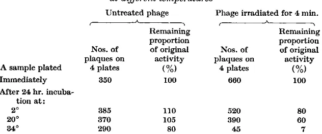
Tables 6 and 7 show that when U.V. irradiated and unirradiated phage preparations are incubated in identical conditions, the former lose their activity at a proportionally higher rate than the latter. The more a preparation is irradiated the higher is the relative rate of the decrease of its remaining activity (Table 6). The difference between irradiated and control preparations is noticeable when the incubation temperature is near O", but it becomes more obvious as the temperature increases, i.e. when both irradiated and control preparations become less stable (Table 7).
controls. The instability is, therefore, a property of the irradiated phage particle and it is a result of either a change in the structure of the particle caused by U.V. radiation, or a selective effect of the radiation. The latter possibility seems unlikely, because it necessitates the assumption that the
Table 6. Eflect of incubation at 34' of preparations of phage 317
ultraviolet irradiated for various lengths of time Numbers of plaques formed on
4 plates by samples plated
After 3 hr. h
r \
Time of Dilution a t which Immediately incubation a t
irradiation the preparation after 34' following Drop in (min.) was plated irradiation irradiation activity to
0 1.75 3.5 5-0
11107 305 295 97 %
l/lOB 84 60 71 %
11104 225 74 33 %
1/102 980 257 26 %
Table 7. Effect of incubating an ultraviolet irradiated preparation of 317 phage at different temperatures
Untreated phage Phage irradiated for 4 min.
Remaining Remaining
proportion proportion
Nos. of of original Nos. of of original
-
I h \plaques on activity plaques on activity
A sample plated 4 plates (%) 4 plates (%I
Immediately 350 100 660 100
After 24 hr. incuba- tion at:
2 O 385
20° 370
34O 290
110 520
105 390
80 45
80 60 7 Samples of unirradiated phage preparation were plated at a dilution of 1/10' and those of the irradiated preparation a t 1/108.
more stable particles are more easily inactivated by U.V. radiation than the less stable ones. Moreover, it would have to be assumed that phage particles differ in susceptibility to U.V. radiation, and this means that the value of k should decrease as the residual activity of irradiated phage preparations decreases to the values reached in the experiments shown in Tables 6 and 7, whereas in fact the value of k remained approximately constant (Tables 4 and 5). It seems, therefore, more likely that U.V. radiation can so alter a phage particle that, although still active, it is rendered unstable.
Ultraviolet irradiated bacteriophages
143
may have resulted from failure to adjust experimental conditions to the much greater stability of the viruses used.Alper (1952) has just reported that inactivation of a phage by X-rays still goes on after the irradiation of its preparation has ended. Two different causes of this could be distinguished. In the first place X-rays alter phage particles making them susceptible to the inactivating effect of hydrogen peroxide, and, secondly, X-rays produce hydrogen peroxide in the medium. Diluting irradiated phage in unirradiated medium can, therefore, prevent its further quick inactivation.
Instability of residual activity of U.V. irradiated preparations of the Rhizo-
bium bacteriophages is not likely to be caused by a similar combination of two causes, for diluting the preparations l / l O O O in unirradiated medium did not make them more stable. On the contrary, they even became less stable, suggesting the presence of some protective material in the crude bacterial lysate.
When residual activity of U.V. irradiated phage preparations falls as a result of ageing, the ability of the preparations to respond to exposure of infected host bacteria to visible light seems to remain unchanged. The ratio of the number of plaques formed with exposure to visible light to that formed with- out exposure does not seem to alter appreciably. It is concluded that irradiated phage particles that still are fully active and those active only if the infected host is exposed to visible light, both lose their activities at the same relative rate. The ability of an U.V. irradiated phage particle to be active only if the infected host is exposed to visible light may be a transition stage between fully active and fully inactive states, but there is no evidence for this.
REFERENCES
ALPER, T. (1952). A new after-effect of X-rays on dilute aqueous suspensions of bacteriophage. Nature, Lond. 169, 964.
BAWDEN, F. C. & KLECZKOWSKI, A. (1952). Ultraviolet injury to higher plants counteracted by visible light. Nature, Lond. 169, 90.
BAWDEN, F. C. & KLECZKOWSKI, A. (1953). The behaviour of some plant viruses after exposure to ultraviolet radiation. J . gen. Microbiol. 8 , 145.
BEDFORD, T. H. B. (1927). The nature of action of ultra-violet light on micro- organisms. Brit. J . exp. Path. 8, 437.
COLBENZ, W. W. & FULTON, H. R. (1924). A radiometric investigation of the germi- cidal action of ultra-violet radiation. Sci. Pap. U.S. Bur. Stand. 19, 641. DULBECCO, R. (1950). Experiments on photoreactivation of bacteriophages in-
activated with ultraviolet radiation. J . B a t . 59, 329.
HENLE, W. (1950). Interference phenomenon between animal viruses : a review. J. Immunol. 64, 203.
KELNER, A. (1949). Photoreactivation of ultraviolet treated Escherichia coli, with special reference to the dose-reduction principle and to ultraviolet-induced mutations. J . Bact. 58, 511.
KELNER, A. (1951). Action spectra for photoreactivation of ultraviolet-irradiated Escherichia coli and Streptomyces griseus. J . gen. Physiol. 34, 835.
KLECZKOWSKI, J. & KLECZKOWSKI, A. (1952). Effect of specific polysaccharides
produced by the host bacteria and of ribonuclease on the multiplication of
Rhixobium phages. J. gen. Microbiol. 7 , 340.
LATARJET, R. & MORENNE, P. (1951). Inactivation d’un bacteriophage par un
rayonnement ultra-violet de tr&s faible intensit& Ann. Inst. Pasteur, 80, 220.
LATARJET, R. & WARL, R. (1945). Precisions sur l’inactivation des bacteriophages
par les rayons ultraviolets. Ann. Inst. Pasteur, 71, 336.
LURIA, S. E. & DELBRUCK, M. (1942). Interference between inactivated bacterial
virus and active virus of the same strain and of a different strain. Arch. Bio-
chem. 1, 207.
LURIA, S. E. & DULBECCO, R. (1949). Genetic recombination leading to production
of active bacteriophage from ultraviolet inactivated bacteriophage particles.
Genetics, 34, 95.
LURIA, S. E., WILLIAMS, R. C. & BACKUS, R. C. (1951). Electron microscopic counts
of bacteriophage particles. J. Bact. 61, 179.
PRICE, W. H. (1950). Phage formation in Staphylococcus muscae cultures. IX. Effect of multiple infection on virus synthesis in the absence and presence of specific substrates. J. gen. Physiol. 34, 251.
PROKS, J. (1933). Recherches sur I’irradiation des milieux nutritifs bacteriologiques.
Lait, 13, 331.
WYSS, O., HAAS, F., CLARK, J. B. & STONE, W. S. (1950). Some effects of ultraviolet irradiation on micro-organisms, J. cell. comp. Physiol. 35, Suppl. no, 1, 133.