R E S E A R C H
Open Access
Selective killing of spinal cord neural stem
cells impairs locomotor recovery in a
mouse model of spinal cord injury
Melania Cusimano
1, Elena Brambilla
1, Alessia Capotondo
1, Donatella De Feo
1, Antonio Tomasso
1, Giancarlo Comi
2,
Patrizia D
’
Adamo
3, Luca Muzio
1*†and Gianvito Martino
1*†Abstract
Background:Spinal cord injury (SCI) is a devastating condition mainly deriving from a traumatic damage of the spinal cord (SC). Immune cells and endogenous SC-neural stem cells (SC-NSCs) play a critical role in wound healing processes, although both are ineffective to completely restore tissue functioning. The role of SC-NSCs in SCI and, in particular, whether such cells can interplay with the immune response are poorly investigated issues, although mechanisms governing such interactions might open new avenues to develop novel therapeutic approaches. Methods:We used two transgenic mouse lines to trace as well as to kill SC-NSCs in mice receiving SCI. We used Nestin CreERT2 mice to trace SC-NSCs descendants in the spinal cord of mice subjected to SCI. While mice carrying the suicide gene thymidine kinase (TK) along with the GFP reporter, under the control of theNestinpromoter regions (NestinTKmice) were used to label and selectively kill SC-NSCs.
Results:We found that SC-NSCs are capable to self-activate after SCI. In addition, a significant worsening of clinical and pathological features of SCI was observed in the NestinTKmice, upon selective ablation of SC-NSCs before the injury induction. Finally, mice lacking in SC-NSCs and receiving SCI displayed reduced levels of different neurotrophic factors in the SC and significantly higher number of M1-like myeloid cells.
Conclusion:Our data show that SC-NSCs undergo cell proliferation in response to traumatic spinal cord injury. Mice lacking SC-NSCs display overt microglia activation and exaggerate expression of pro-inflammatory cytokines. The absence of SC-NSCs impaired functional recovery as well as neuronal and oligodendrocyte cell survival. Collectively our data indicate that SC-NSCs can interact with microglia/macrophages modulating their activation/responses and that such interaction is importantly involved in mechanisms leading tissue recovery.
Keywords:Spinal cord injury, Endogenous neural stem cells, Neurotrophic factors, Inflammation
Background
Spinal cord injury (SCI) is a chronic disease that generally affects young people, showing incidences that range from 10 to 900 per million worldwide [1]. The management of SCI requires impressive health care resources, and the clinical amelioration achieved so far can only modestly in-crease the quality of life in patients. Recently, neural stem cell (NSC)-based treatments have received a great deal of
scientific interest. Accordingly, transplanted NSCs are neuroprotective because they secrete neurotrophic factors and offer bystander effects that sustain tissue recovery in several models of neurodegenerative disorders [2,3]. This multifaceted therapeutic activity promotes the re-programming of the hostile microenvironment to a more instructive one, which eventually induces tissue repair [4]. Besides transplanted NSCs, also endogenous spinal cord NSCs (SC-NSCs) are critically involved in the central nervous system (CNS) recovery after SCI. Indeed, SC-NSCs self-activate after a traumatic injury and secrete neurotrophic factors that promote neuronal survival [5]. SC-NSCs are embedded in the ependymal cell layer, which
* Correspondence:muzio.luca@hsr.it;gianvito.martino@hsr.it
†Equal contributors
1Neuroimmunology Unit, Division of Neuroscience, Institute of Experimental
Neurology (INSPE), San Raffaele Scientific Institute, Via Olgettina 58, 20132 Milan, Italy
Full list of author information is available at the end of the article
is lining the central canal of the SC, arranged in specific germinal niches. Such niches show a different cyto-architecture, if compared with niches of the brain sub-ventricular zone (SVZ), [6,7]. A key distinction is related to the position of proliferating cells within SC-confined niches. Indeed, dividing SC-NSCs are directly located in the ependymal layer rather than in the sub-ependymal one. Moreover, proliferating SC-NSCs typically self-renew themselves, rather than producing transit-amplifying pro-genitors. Their descendants remain within the ependymal layer and do not share any lineage relationship with sub-ependymal cells [8,9]. In response to tissue damage, such as after SCI, SC-NSCs increase rates of cell proliferation, giving rise to astrocytes that contribute to the formation of the astroglial scar [5]. However, a fraction of SC-NSCs also express Olig-2 and acquire the cell morphology of immature oligodendrocytes that eventually migrate to the injury site. Later on, such cells differentiate in myelinating oligodendrocytes that finally enwrap axons [10,11].
Similarly to other neuroinflammatory disorders, im-mune cells dominate the early post traumatic events oc-curring upon SCI and the prolonged secondary injury phase, which may last up to weeks or months. Indeed, the pathophysiology of SCI involves an initial injury which is followed by a sequela of secondary damages that includes many multifaceted pathological mecha-nisms. Among them, the inflammatory cascade arises from innate (neutrophils, macrophages, and microglia) and adaptive (B and T) immune responses. Such cells re-lease inflammatory cytokines and chemokines, nitric oxide oxidants, proteases, and complement proteins [12]. About myeloid cells, microglia are rapidly activated in response to the SCI, while monocyte-derived macro-phages enter the CNS few days after the trauma and their number generally peaks 7–10 days after SCI [13]. It is likely that either microglia or macrophages undergo activation acquiring a complex functional phenotype which involves the expression of pro-inflammatory as well as anti-inflammatory gene sets. The acquisition of such phenotypes is highly flexible and influenced by complex cascades of agonistic and antagonistic cues [14]. Infiltrating monocyte-derived macrophages are centrally involved in tissue repair processes, as shown in SCI mice receiving infusion of a supplemental amount of monocytes [15]. Similarly, the depletion of monocytes, but not of microglia, impairs recovery of mice upon SCI [16]. Infiltrating myeloid cells express CD11b, CD115, F4/80, Gr1, and activation markers such as CD11c and MHC-II. The selective ablation of CD11c in infiltrating myeloid cells affects recovery of SCI mice [16]. However, also activated microglia express CD11c at high levels [17] and considering that activated microglia and infil-trating macrophages could differentially modulate tissue repair, is not surprising that SCI mice transplanted with
NSCs display a functional recovery and a remarkable re-duction of CD11c+ cells [4]. In this study, we aimed at determining contribution of SC-NSCs to the functional recovery of mice receiving SCI. We selectively ablated SC-NSCs using transgenic mice that express the suicide thymidine kinase 1 (TK) gene under the control of
Nestin regulatory regions. Our results show that SC-NSCs depletion causes a substantial reduction of growth factors in the injured tissue, increased demyelination and impaired locomotion recovery.
Methods Study approval
Mice were maintained under pathogen-free conditions at San Raffaele Hospital mouse facility (Milan, Italy). All ef-forts were made to minimize animal suffering and to re-duce the number of mice used, in accordance with the European Communities Council Directive of 24 November 1986 (86/609/EEC). All procedures involving animals were executed according to the guidelines of the Institutional Animal Care and Use Committee (protocol number: 622) of San Raffaele Scientific Institute, Milan.
Spinal cord neural stem cells culture
Immunofluorescence on SC-NSC cultures was done according our published methods [22]. Briefly cells were fixed with 4% paraformaldehyde 10′ at room temperature, then rinsed three times with PBS, and then incubated for 60 min with a blocking solution [PBS, 10% normal goat serum (NGS, Sigma), 0.1% albumin bovine serum (BSA, Sigma)] to avoid a-specific binding of anti-bodies. For intracellular staining, we added 0.1% Triton X-100 in blocking solution. Cells were incubated with the appropriate primary antibody for 2 h. Cells were then washed in PBS and then incubated for 45 min with fluorescent secondary antibodies. The nuclei were stained with 4, 6-diamine-2-fenilindole (1 μg/ml, DAPI, Roche). Cells were then washed and mounted with Fluorescent mounting medium (Dako). The following antibodies were used: Rb-α GFAP (Dako), mouse-α O4 (clone 81, Millipore), and mouse-α NeuN (clone A60, Millipore). Imaging was done using Leica SP5 confocal microscope equipped with LasX software.
Transgenic mice and ganciclovir regimen
Selective ablation of SC-NSCs was done in NestinTKmice that were originated from NestinfloxGFPflox-TK mice [19] in which GFP excision was done using CMV-Cre mouse line [23]. NestinTKmice express thymidine kinase 1 (TK) under the control of the second intron enhancer ofNestin
[24]. Mice received 100 mg/kg/day of ganciclovir (GCV) dissolved in distilled water by subcutaneously implanted osmotic minipumps (ALZET® Model 2002, DURECT Cor-poration) [19]. After 14 days, pumps were replaced with new ones, thus allowing 28 days of continuous treatment. Controls were raised using WT liters with or without GCV regimen. To evaluate the ablation of SC-NSCs, 5-Bromo-2′-deoxyuridine (BrdU, 1 mg/ml dissolved in 1% sucrose) was administered in the drinking water continu-ously for a month. Depending on the experimental setting, cell proliferation in SCI mice was also evaluated by BrdU intraperitoneal injections (100 mg/kg). Cell tracing was done using mice carrying the NestinCreERT2allele [25] that were crossed with the reporter mouse line Rosa 26YFP [26]. Double transgenic mice were supplemented with Tamoxifen (Sigma) for 5 days, washed out for further 5 days, and finally subjected to SCI.
Contusion spinal cord injury
Twelve-week male mice (22–25 g) were anesthetized with ketamine/xylazine and a laminectomy was performed in thoracic spinal cord at the level of the T12 vertebra. Contu-sions were generated applying 75 k-dynes with an Infinite Horizons Impactor (Precision systems and Instrumenta-tion), [16]. As postoperative care, we administered enroflox-acin (Baytril, Bayer; 2.5 mg/kg, subcutaneously) once daily for 2 weeks. Urine was expelled by manual abdominal pressure twice daily for 1 week and then once daily for the
duration of the experiment. The recovery of locomotor per-formance was evaluated using the Basso Mouse Scale (BMS), as previously described [16]. Briefly, mice were observed individually for 4 min each in an open field by three investigators blinded to surgery procedures and geno-type. Hind limb motor function was recorded and scored according to the BMS guidelines. For statistical analyses we calculated means of left and right hind limb scores for each animal. Catwalk XT Gait Analysis System (Noldus Informa-tion Technology, Asheville, NC, USA) was used to assess walk ability of mice. Mice were allowed to freely ambulate along an illuminated glass plate within a confined corridor (L 50 cm × W 8 cm) in a darkened room and footprints were recorded with a high-speed camera for following analyses with CatWalk XT 10.0 software (Noldus).
Tissue processing and histopathology
10 magnifications and lesion volumes were expressed in cubic millimeter. The lesion volume contours were traced at the boundaries between positive and GFAP-negative regions according published methods [27]. Demye-lination volumes were calculated as contours on Luxol-Fast blue-stained spinal cord segment-representative 10-μm-thick axial sections accordingly our published method [4]. Calculations were made imaging four to five mice per group with × 2.5 or × 10 magnifications and lesion volumes were expressed in cubic millimeter.
Quantification of cell number
The number of GFP+/BrdU+cells was calculated in corres-pondence of the closest spared central canal to lesion epi-center for up to 2 mm both rostrally and caudally (n= 3 mice per group). Olig2+ and NeuN+ cells were counted from the closest spared cross-section to lesion epicenter for up to 4 mm, both rostrally and caudally. The number of Olig2+cells was calculated in correspondence of the dorsal funiculus in a trapezoidal area (0.08 mm2) per section (n= 3 mice per group). The number of NeuN+cells was calcu-lated in correspondence of four different regions (lamina X, ventral horn, dorsal horn and lateral gray matter) for a total area of 0.1 mm2per section (n= 3 mice per group).
RNA isolation, reverse transcription, and quantitative real-time polymerase chain reaction
Segments encompassing the thoracic spinal cord T11-T13 region were rapidly sampled and lysed for total RNA ex-traction using RNeasy® Mini kit (Qiagen) kit according to the manufacturer’s recommendations including DNase di-gestion. cDNA synthesis was performed by using Thermo-ScriptTM RT-PCR System (Invitrogen) and Random Hexamer (Invitrogen). Real-time PCR was done using the following TaqMan® probes (Applied Biosystems):
Gene Cod AB
GAPDH Mm99999915_g1
IGF1 Mm00439561_m1
LIF Mm00434761_m1
CNTF Mm00446373_m1
Vegfa Mm00437304_m1
TNF Mm00443258_m1
IL1b Mm01336189_m1
iNos(NOS2) Mm00440502_m1
IL6 Mm00446190_m1
Ex vivo cytofluorimetric analyses of immune cells
Mice subjected to spinal cord injury were killed by an overdose of anesthetic and their spinal cords, processed for flow cytometry after saline transcardial perfusion.
The lesioned cord segments extending between T11 and T13 were homogenized and reduced to single-cell sus-pensions by Neural Tissue Dissociation Kit (P) (Miltenyi Biotec) according to the manufacturer’s instructions. Cell suspension was filtered through a 70-μm strainer, centrifuged over a 30% discontinuous Percoll Gradient (GE Healthcare) and myelin debris removed from the surface (modified from Cardona et al., [28]). Cells were labeled with Zombie Aqua™ die (Biolegend), then incu-bated with 5% FBS, 1%BSA, and 5 mg/ml rat anti-mouse Fc III/II receptor (CD16/CD32) blocking antibodies (BD), and then stained using the following monoclonal antibodies: CD45-Pb (Biolegend), CD11b-PeCy7 (BD), Ly6C-APCCy7 (BD), Ly6G-PerCP Cy5.5 (BD), and CD11c-APC (ebioscence). Cells were analyzed by LSR Fortessa (Beckton Dickinson) and data analyses by FlowJo (Treestar) software.
Statistics
Sample size for each experiment was estimated at the beginning of the study with the PS power and sample size calculator software (version 3.0.43) based on variance data obtained in previous experiments [4]. Data are expressed as the mean ± standard error of the mean (S.E.M.) or mean ± standard deviation (S.D.) of independent experiments. Normality was assessed in each experiment by applying either Kolmogorov–Smirnov test (with Dallas–Wilkinson– Lille for P value) or D’ Agostino & Pearson omnibus normality test. Comparisons were made using the unpaired
t test and one-way or two-way analysis of variance (ANOVA) tests, followed by Tukey’s multiple comparison test. Nonparametric data have been compared using Mann Whitney test or Kruskal-Wallis test followed by Dunn’s multiple comparison test. Statistical tests were carried out using PRISM5.01 (GraphPad Software, La Jolla, CA, USA). Value less than 0.05 was considered statistically significant.
Results
Central canal-derived cells from Nestin floxGFPflox-TK mice generate neurospheres
Nestin promoter restricts the transgene expression to NSCs [30]. Furthermore, none of the GFP+ cells co-expressed the GFAP marker in the SC (Additional file1: Figure S1A), while, some GFP+ependymal cells were posi-tive for Id1, which is a dominant-negaposi-tive helix-loop-helix transcriptional regulator that identifies self-renewing NSCs of the SVZ (Additional file1: Figure S1B), [31].
We next raised NSCs cultures from the SC of Nestin flox
GFPflox-TK mice to further demonstrate that SC-restricted GFP+ cells possess features of bona fide NSCs. We included in our experiments a functional control rep-resented by SVZ-derived NSC cultures [32]. Dissociated SCs from adult Nestin floxGFPflox-TK mice gave raise to primary neurospheres that were characterized by the pres-ence of GFP+ cells in virtually all spheres we assayed (Fig. 1a). We next assayed self-renewal potentials of SC-NSCs comparing their ability to growth in vitro as normally observed in SVZ-derived NSC cultures. We found that both NSCs cultures displayed similar growth curves (Fig. 1b). In the SVZ, Nestin is transiently expressed by activated type-B1 cells, while quiescent B1 cells are negative and levels of this marker generally drop
when cells undergo to cell differentiation [33–35]. Because we initially examined cultures that were composed by a mixed cell population containing either GFP+ or GFP− cells, we next sorted GFP+ cells from Nestin floxGFPflox -TK-derived SC cultures to generate neurospheres. Dissoci-ated cells from SCs of Nestin floxGFPflox-TK mice were expanded in vitro for two passages, then GFP+cells were sorted by flow cytometry (Additional file2: Figure S2B). GFP+cells were plated at the concentration of 8000/cm2, left to grow in standard NSC medium and examined for the generation of spheres. Three days after plating several cells started to generate small spheres that reached the morphology and the size of classical SVZ-derived NSCs neurospheres after 7 days (Additional file2: Figure S2C, D). We next examined some markers that are commonly expressed by SVZ-derived NSCs cultures [18]. We observed that around 64% of the GFP+ cells derived from SCs of NestinfloxGFPflox-TK mice expressed SOX2. CD44 and VLA4 were expressed by the vast majority of these GFP+ cells, while CXCR4 expression was confined in a small percentage of them (Fig. 1c). Dissociated SC-NSC neurospheres were plated in differentiation medium and
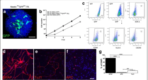
Fig. 1Ependymal cells lining the spinal cord central canal display neural stem cells properties in vitro.aRepresentative confocal image of a primary
neurosphere raised from NestinfloxGFPflox-TK mice. Nestin+(GFP) cells are embedded inside the sphere.bGrowth curves of NSCs derived from the SC
of WT (black dots) and of NestinfloxGFPflox-TK (white dots) mice. SVZ-derived neurospheres cultures were raised from WT mice and used as control
(white square),n= 3 for each group.cFlow cytometry analysis of GFP+cells labeled for SOX-2, CD44, VLA4, and CXCR4. Cells were obtained from in
P2-dissociated neurospheres of SC of NestinfloxGFPflox-TK mice. The GFP gate was established using cultures from WT SCs (n= 3).d–gRepresentative
confocal images showing SC-NSCs from NestinfloxGFPflox-TK mice plated on Petri dishes in the absence of growth factors and labeled for GFAP (d),
TuJ1 (e), and APC (f). Percentages are calculated on the total number of nuclei (DAPI) and quantifications are shown in panel (g). Data are collected
from one representative experiment that has been replicated two times. One-way ANOVA followed by Tukey multiple comparisons post-test has been
labeled for GFAP, TuJ1, and APC. We observed that SC-NSCs from NestinfloxGFPflox-TK mice can differentiate in astrocytes, neurons, and oligodendrocytes (Fig. 1d–g). Thus, in agreement with previously published results, we observed that GFP+ ependymal cell cultures from Nestin flox
GFPflox-TK mice have bona fide features of NSCs [10].
Traumatic spinal cord injury triggers endogenous SC-NSC proliferation
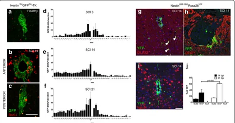
We next investigated cell proliferation of GFP+cells in the intact and above all in the injured SC [11]. Cell proliferation was assayed in mice receiving a severe contusion of the SC that was obtained using IH impactor at T12 thoracic level. After the injury, Nestin floxGFPflox-TK mice received daily injections of BrdU and proliferating cells were scored 3, 14, and 21 days after the contusion. As expected, unaffected Nestin floxGFPflox-TK mice displayed few GFP+BrdU+ cells in the SC (Fig. 2a), while their numbers were substantially increased upon SCI (Fig.2b, c). Most of the GFP+BrdU+cells was located anteriorly to the site of the injury, peaking in a region that was positioned 0.1–0.2 mm far for the lesion site (Fig.2d–f). These results confirm and extend previously published data, showing increased BrdU uptake in the SC of SCI mice [36]. However, our data showed that cell proliferation persisted
in regions flanking the site of the injury for more than 2 weeks (Fig. 2f), [11]. We next performed a fate map experiment tracing ependymal cells descendants using a transgenic mouse line in mice that carry the NestinCreERT2 allele [25]. NestinCreERT2mice were crossed with Rosa26YFP reporter mice [26], injected with Tamoxifen for 5 days, washed out for further 5 days, and finally subjected to SCI. Tracing of ependymal descendants was done 14 and 31 days after the induction of the injury. Sections were labeled for YFP and markers of oligodendrocytes precursors, astrocytes, and neurons (Fig. 2g–i). At both time points, most of the YFP+cells co-expressed GFAP, a subset of them was Olig2+, while NeuN+YFP+ cells were never detected (Fig. 2j). These results show that SC-NSC descendants can give rise to oligodendrocytes precursors and astrocytes in response to tissue damage.
Spontaneous functional recovery is impaired in GCV-NestinTKmice
We next genetically ablated SC-NSC to investigate their functional role in healing processes that spontaneously occur after SCI. We crossed Nestin floxGFPflox-TK mice with mice carrying the CMV-Cre allele that ubiquitously express the Cre recombinase [23]. We successfully gen-erated NestinTK mice in which the loxP- GFP cassette
Fig. 2SC-NSC activation and differentiation upon SCI.aRepresentative confocal images showing the central canal of healthy NestinfloxGFPflox-TK
mice labeled for GFP and BrdU.b,cSegments proximal (b) and distal (c) to the lesion epicenter from a NestinfloxGFPflox-TK mouse sampled
14 days after SCI.d–fQuantifications of GFP+BrdU+cells (means ± S.E.M) within the ependymal layer of the central canal in NestinfloxGFPflox-TK
mice sacrificed 3, 14, and 21 days after SCI (n= 3 mice/time point).g–jNestinCreERT2Rosa 26YFPmice were injected with TAM for 5 days and,
following a washout of further 5 days, subjected to injury. Confocal images of OLIG2/YFP (arrows indicate double positive cells), GFAP+YFP+, and
NeuN+YFP+are provide ing–i. Quantifications (means ± S.E.M.) are plotted onj, (n= 3 for each group). Two-way ANOVA, followed by Bonferroni’s
was excised, allowing mice to express a functional TK gene. We proved the efficiency of SC-NSC ablation, treating NestinTKmice along with their wild type (WT) littermates with GCV for 28 days. Mice received BrdU, and SC sections were scored for Vimentin (VIM) and BrdU to demonstrate the effective ablation of ependymal cells. We observed a substantial reduction of VIM+cells in ependymal layer of NestinTK mice. Accordingly, the number of ependymal BrdU+ cells was severely diminished in GCV-NestinTK mice (Additional file 3: Figure S3A–C).
We next subjected GCV-NestinTKand GCV-WT mice to SCI. We included in our experimental setting two additional groups of mice, which were composed by NestinTKand WT mice receiving saline, instead of GCV. Mice were scored for neurological functions until day 31 using the BMS score, which measures hind limb motor ability in an open field arena [37]. GCV-NestinTKmice, lacking the SC-NSCs, exhibited an impaired recovery, when compared to GCV-WT mice. Comparing BNS curves, we observed that differences in motor limb ability started to become statistically significant on day 10 and remained significant until the end of the experi-ment, at day 31 (Fig. 3a, b). We next induced SCI on further GCV-WT and GCV-NestinTK mice to evaluate their locomotion abilities using the Catwalk XT gait ana-lysis [38]. Mice were collected at day 18 upon SCI and scored with the BMS. GCV-NestinTK mice displayed a significant worsening of scores when compare to GCV-WT mice (Additional file 4: Figure S4A). Mice were next assayed with the Catwalk to confirm the general impair-ment occurring to the GCV-NestinTKgroup. Anterior paws
prints did not show any alteration in both experimental groups, while posterior paws prints were hardly detectable on the glass walkway. However, examining movie files, we observed that GCV-NestinTKmice showed a general wors-ening of locomotion that was featured by a lack of both hind leg prints due to an almost complete paralysis of the legs. On the other hand, GCV-WT mice showed a major rescue charged to the RH paw and partially to the LH paw (Additional files 5 and6 show two representative animals from both groups). Accordingly, step timing views of two representative mice from each group, confirmed the sub-stantial impairment of locomotion in the GCV-NestinTK mouse (Additional file4: Figure S4 B, C).
To find out the mechanisms underlying the functional worsening of GCV-NestinTK mice, we first evaluated pathological features of mice at day 31. Since BMS scores of both NestinTK and WT mice receiving SCI and the saline treatment substantially overlapped, we only per-formed histological assessments on GCV-NestinTK and GCV-WT mice. We stereologically calculated the volumes of the glial scar, scoring the distribution of GFAP-positive and GFAP-negative areas around the site of the injury. GCV-NestinTKand GCV-WT mice displayed similar scar volumes, indicating that the lesion volume was not affected by SC-NSC ablation (Fig. 4a–b). On the other hand, demyelination was significantly increased in regions located anteriorly to the site of the injury in GCV-NestinTK mice (Fig. 4e–g). Parallel to demyelination, we also observed a significant reduction of Olig2+progenitors in GCV-NestinTKmice (Fig.4h–j). Finally, we noticed that the depletion of SC-NSCs caused a significant reduction of neurons in regions flanking the site of the injury.
Fig. 3SC-NSCs are involved in the spontaneous process of functional recovery form spinal cord injury. Histogram ina, shows locomotor functions of
saline control mice (black dots,n= 5), saline NestinTKmice (gray dots,n= 5), GCV-WT mice (blued dots,n= 8), and GCV-NestinTKmice (red dots,n= 5).
Data are represented as Basso Mouse Scale (± S.E.M.) mean values of the locomotor score of each group.bBasso Mouse Scale (± S.E.M.) mean values
of the locomotor score of individual mice on day 31 (each dot represent a single animal). Data derived from one representative experiment that has
been replicated two times. Statistical analysis inahas been done comparing at each time point GCV-WT and GCV-NestinTKmice. Day 10,p= 0.044; day
14,p= 0.026; day 18,p= 0.007; day 21,p= 0.008; day 24,p= 0.0044; day 28,p= 0.0045; day 31,p= 0.0044. Results inbhave been assayed by repeated
measures ANOVA followed by Bonferroni’s multiple comparison test. **p= 0.0045 for GCV versus NestinTK+ GCV;p= 0.0015 for NestinTK+ GCV versus
Interestingly, such alteration included the ventral horn and affected motor neurons, (Fig.4k–m). Altogether, these data suggest that the functional worsening of GCV-NestinTKmice subjected to SCI parallels with a general re-duction of oligodendrocytes and neurons.
Loss of neurotrophic factors affects GCV-NestinTKmice
Besides cell replacement, endogenous SC-NSCs support wound healing through the secretion of a complex array of neurotrophic factors, which directly or indirectly enhances neuronal survival [5]. Among such factors,
SC-Fig. 4GCV-NestinTKmice show enhanced tissue damage after SCI.a–dStereological quantification of the lesion volume as GFAP−tissue injury in
GCV-NestinTKand GCV-WT mice.a,bRepresentative axial images of GFAP-labeled sections from GCV-WT (a) and GCV-NestinTK(b) mice. In the 3D rendering
c, the solid light blue is the central canal and the solid red is the volume negative for GFAP. Quantifications (means ± S.E.M.) are shown ind(n= 5–8 per
group).e–gStereological quantification of demyelination by Luxol fast blue (LFB) in GCV-NestinTKand GCV-WT mice, (e,fviolet solid,n= 4–5 per group).
Quantifications of the demyelination volumes (means ± S.E.M.) are shown ing, *p= 0.023.h–iOlig2+p= 0.0005.k–l, NeuN+cells (**p= 0.0058 anterior and
**p= 0.0052 posterior) by immunohistochemistry in GCV-NestinTKand GCV-7WT mice. Quantifications (means ± S.E.M.,n= 3 per group), performed and day
31, are provided inj,m. One-way ANOVA followed by Bonferroni’s multiple comparison test has been used to analyze data ofg,j, andm. Scale
NSC-derived astrocytes secreteIgf1andCntf, which play a crucial role in supporting neuronal survival after SCI [5]. We next investigated mechanisms regulating the lack of clinical recovery observed in GCV-NestinTKmice measuring the expression levels of such growth factors in a region encompassing T11–T13 spinal segments from SCs sampled at days 7 and 31. We also included in our analysis two genes that encode for Lif and Vegfa, which are two growth factors released by NSCs that upon transplantation mediate neuroprotective effects in animal models of multiple sclerosis and brain ischemia, respectively [39, 40]. Igf1 expression levels were signifi-cantly dampened in GCV-NestinTK mice when com-pared to levels measured in GCV-WT litters at both days 7 and at 31 (Fig.5).LifmRNA levels were reduced at day 7 in GCV-NestinTK mice, although such reduc-tion was a trend that did not reach the statistical signifi-cance (Fig. 5b). The inhibition of the SC-NSC cell proliferation observed in FoxJ1-rasless mice affects SCI outcomes and induces a general reduction of CNTF levels in perilesional regions [5]. We observed a modest reduction of Cntf mRNA levels in GCV-NestinTK mice sampled at day 31 (Fig. 5c). At this time point, also
Vegfa mRNA levels were affected in GCV-NestinTK mice, although such reduction was a trend, not reaching the statistical significance (Fig.5d). However, we also no-ticed that the simple administration of GCV to unin-jured mice reduced Vegfa levels, suggesting that such alteration was probably relied on the pharmacological treatment (Fig. 5d). Overall, these data corroborate the idea that SC-NSC ablation affects the general expression
of neurotrophic factors in the lesioned SC, which ham-pers endogenous mechanisms of tissue repair.
The ablation of SC-NSCs modulates the perilesional in-flammatory milieu
Besides activation of microglia and astrocytes, a local inflammatory response involving blood-borne cells dom-inates the site of the injury [36,41]. To find out the pos-sible interplay between SC-NSCs and the inflammatory response, we analyzed the identity and the phenotype of inflammatory cells located at the injury site. We dissoci-ated lesioned SCs from GCV-NestinTK and GCV-WT mice at day 7 and using flow cytometry we evaluated the distribution of different cell subpopulations that were taking part to the inflammatory response. The lack of SC-NSCs modulate neither percentages of bona fide lymphocytes (CD45+CD11b−), (Fig. 6a, b) nor percent-ages of CD45+CD11b+expressing cells (Fig. 6a, c). We next scored the cell morphology of activated myeloid cells analyzing sections labeled for Iba1+F4/80+ cells at day 18. Double positive cells were well ramified in both experimental groups when examined 3 mm far from the site of the injury (Additional file 7: Figure S5A, A’, C, and C’). At the site of the injury, Iba1+F4/80+ cells in-creased in their number, were amoeboid, and highly expressed F4/80. However, judging their cell morphology as well as their distribution around the lesion site, we did not find any relevant difference between GCV-WT and GCV-NestinTKmice (Additional file 7: Figure S5 B, B’, D, and D’). We next wanted to discriminate between activated microglia and infiltrating myeloid cells. We
Fig. 5Downregulation of neurotrophic factors in GCV-NestinTKmice. Real-time PCR forIgf1(a),Lif(b),CNTF(c), andVegfa(d) performed on total
RNA extracts derived from T11–T13 spinal cord segments. Explants were collected at days 7 and 31. Values indicate the mean fold changes ± S.D.
(n= 3–6 for each group) calculated on the expression levels measured in healthy mice (black bars). Comparisons were done using thetStudent
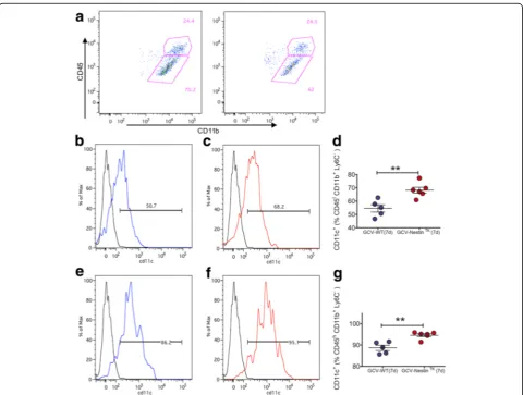
Fig. 6Modulation of the inflammatory response in GCV-NestinTKmice. Representative dot plots showing the gating strategy for the identification
of lymphoid (CD45+CD11b-)and myeloid (CD45+CD11b+) cells, gated on the CD45+cell populations isolated from injured spinal cord at day 7 (a).
Quantification of lymphoid (b) and myeloid cells (c).Representative dot plots showing the gating strategy for the identification of macrophage lineage
cells (CD45+CD11b−Ly6C+) and microglia cells (CD45+CD11b−Ly6C+) cells (d). Quantification of macrophage lineage cells (e) and microglial lineage cells
(f). For all quantifications red dots are GCV-NestinTKmice while blue dots are GCV-WT mice, each dot represents a single animal. Horizontal bars
exploited the flow cytometry and the expression of the marker Ly6C to discriminate infiltrating CD45+CD11b+ cells (Fig. 6d), [42]. We observed that there was a slight decrease of microglia, which paralleled with a modest increment of CD45+CD11b+Ly6C+ cell number in GCV-NestinTKmice, although these differences were a trend that did not reach the statistical significance (Fig.6d–f). We an-alyzed the activation profiles of microglia, scoring expres-sion levels of CD11c in CD45+CD11b+Ly6C−cells. On the basis of CD45 expression levels, we further subdivided this cell population in CD45 intermediate (CD45I) and CD45 high (CD45h) cells. Percentages of CD11c+ cells were sig-nificantly increased in CD11b+CD45Ias well as in CD11b +
CD45h cells of GCV-NestinTK mice (Fig. 7b–f). These results mirrored our published data, showing that SCI mice receiving an extra amount of NSCs by transplantation re-duced CD11c expression levels in myeloid cells [4]. Since
CD11c+ cells usually participate to the pro-inflammatory response secreting inflammatory cytokines [4], we next assayed mRNAs levels of some pro-inflammatory cues in GCV-NestinTKmice. At day 7, SCI mice lacking SC-NSCs significantly increased the expression of TNFα and IL-1β
mRNA levels when compared with GCV-WT mice, (Additional file8: Figure S6A–B). Similarly,iNOSandIL-6
mRNA levels were increased in GCV-NestinTK, although these differences were a trend not reaching the statistical significance (Additional file 8: Figure S6D). However, the elevation ofTNFα andIL-1βmRNA was transient and at day 31 and we did not find any significant difference between control mice and mice lacking SC-NSCs (Additional file8: Figure S6A–D). All in all, the ablation of SC-NSCs greatly impairs locomotion recovery in mice and increases the content of M1-like myeloid cells in the perile-sional area.
Fig. 7CD11c is increased in microglia of GCV-NestinTKmice. Representative dot plots showing the gating strategy for the identification of CD45high
and CD45Intalso expressing CD11b, gated on the CD45+Ly6C−cells (a). Quantifications of the expression level CD11c on CD45ICD11d+Ly6C−cells (b-c)
and CD11c on CD45hCD11d+Ly6C−cells (e-g). For all quantifications red dots are GCV-NestinTKmice while blue dots are GCV-WT mice, each dot
Discussion
The central canal of the adult SC contains SC-NSCs that undergo to functional activation in response to tissue damage [43]. The remarkably activation of SC-NSCs, well documented in mice receiving the SCI, involves cell prolif-eration and the secretion of diffusible molecules [11]. Blocking their proliferation capacity resulted in a reduc-tion of spontaneous tissue recovery [5]. This is partially due to the innate ability of SC-NSC descendants to secrete growth factors that promote tissue recovery. To assess the role of endogenous SC-NSCs in the spontaneous recovery occurring in the injured SC, we performed the genetic ab-lation of Nestin-expressing SC-NSCs. Our approach is based on the concept that besides Vimentin, SC-NSCs also express Nestin, and transgenic mice carrying a TK suicide gene under the control of Nestin regulatory region can be used to ablate SC-NSCs [10, 19]. We initially characterized our transgenic mice, carrying a floxable GFP cassette before the TK gene, to demonstrate that they can be definitely used to trace and to target SC-NSCs. We confirmed that GFP+ cells deriving from Nestinflox GFP-flox
-TK mice are able to generate neurospheres in vitro as well as that such cells can give rise to oligodendrocytes, astrocytes, and neurons upon growth factor withdrawal. A key aspect of the SC-NSCs response to tissue damage includes the cell division. In agreement with published data [5,36], we also demonstrated that GFP+BrdU+ cells increased in the injured spinal cord of Nestin floxGFPflox -TK mice. We observed that GFP+BrdU+cells were mainly located anteriorly to the site of the lesion, suggesting that some signals can differentially regulate the SC-NSC cell proliferation during SCI. Using transgenic mice expressing the CreERT2 allele under the control of the Nestin
promoter [25], we were able to fate mapping SC-NSC descendants in SCI mice. Starting from 2 weeks after the injury a considerable high number of YFP+cells differenti-ated in astrocytes that mainly surrounded the injury site, while few of them remained undifferentiated. In addition to astrocytes, some SC-NSCs differentiated in Olig2+cells, re-inforcing the idea that SC-NSCs sustain the myelination of the injured SC. Considering that NG2+progenitors increase rates of cell proliferation in response to SCI [36], we could speculate that some GFP+BrdU+ cells scored in the gray matter of SCI mice may belong to the oligodendrocytes precursors cell population.
Our knowledge about SC-NSCs in the SCI model is based on experimental approaches that inhibit their abil-ity to enter the cell cycle or to migrate toward perile-sional sites [5,44]. In our experimental setting, we killed a cohort of proliferating Nestin+ cells. Such cells are bona fide slowly dividing NSCs that may be relevantly involved in tissue repair. Such approach significantly dampened the number of proliferating ependymal cells but not completely abolish the entire population of
SC-NSCs. However, the net result of this manipulation was a substantial worsening of clinical outcomes in mice. Thus, despite the fact that proliferating Nestin+ cells are normally few in the healthy ependymal layer, their ablation resulted in a less responsiveness of the CNS to micro environmental changes occurring upon SCI and in turn to a worsening of the functional recovery.
Published results indicate that a reduction of SC-NSC cell proliferation affects the formation of a compact scar [5]. On the other hand, the inactivation ofβ1-integrin in SC-NSCs enhances astrocyte migration to the injury site and contributes to the scar formation [44]. Both manipulations are detrimental for mice and affect their locomotion, indi-cating that SC-NSCs regulate the formation of the scar and such aspect is relevant for repair processes [5, 44]. Unex-pectedly, the ablation of proliferating SC-NSCs occurring in GCV-NestinTKmice worsened clinical outcomes in mice without modulating the size of the scar. Such difference could be explained considering that we transiently depleted the proliferating pool ependymal Nestin+cells. In principle, other ependymal cells, as well as surviving Nestin+ cells, could participate to the injury response at later time points. Such cells could contribute to seal the injury, preventing an excessive enlargement of the damaged tissue, but they do not rescue the locomotion of mice.
We observed that demyelination was significantly increased in regions located anteriorly to the site of damage. Interestingly, this is the region of the SCI that is also featured by an intense cell proliferation of SC-NSCs, as we have shown in Nestin floxGFPflox-TK mice. Thus, we can speculate that the depletion of these cells deeply influences the remyelination process, reducing the number of Olig2+ cells and\or affecting the release of neurotrophic factors in the injured tissue.
Interestingly, parallel to the lack of SC-NSCs, we ob-served increasing percentages of microglia/macrophages that express the CD11c marker. This result is in line with our previously published data showing that SCI mice receiving NSCs transplantation display a substan-tial restoration of locomotion that parallel a striking re-duction of CD11c+CD206−cells in SC [4]. We explained these results considering that transplanted NSCs can skew microglia/macrophages to acquire neuroprotective phenotype in the injured SC [4]. Here, we observed that mice lacking SC-NSCs increased percentages of CD11c expressing cells as well as the expression levels of pro-inflammatory cytokines. Thus, as previously described for transplanted NSCs, also resident SC-NSCs can modulate the activation of microglia/macrophages that occurs in response to tissue damaging [18,45,46].
Conclusion
on NSCs transplanted mice [4], we can speculate that both cell types exert a significant immunomodulatory function in the injured CNS, once they are facing inflammation. Such observation is not trivial, since transplanted NSCs are expanded in vitro for weeks or even months before being transplanted. Nonetheless, in vitro manipulations do not obstacle NSCs to perform a modulation of the inflamed environment, once transplanted. Similarly, we could specu-late that endogenous SC-NSCs can exert a substantial“ by-stander”effect in the damaged tissue, limiting inflammatory cascades arising from parenchymal microglia and/or CNS-infiltrating immune cells. If we consider that the degree of inflammation correlates with the severity of SCI outcomes, the identification of signals released by SC-NSCs ought to give us new strategies in the perspective of a therapeutic approach based on NSC transplantation or on fostering the beneficial functions of the endogenous NSCs.
Additional files
Additional file 1:Figure S1.Characterization of the spinal cord
ependymal canal of a NestinfloxGFPflox-TK mouse. Representative
confocal images showing immunofluorescences for GFP/GFAP (A), and
GFP/Id1 (B) in the ependymal layer of a NestinfloxGFPflox-TK mice (n= 3).
Scale bar is 30μm. (TIFF 1498 kb)
Additional file 2:Figure S2.Sorted GFP+ cells from NestinfloxGFPflox-TK
mice give rise to neurospheres. PanelAandBshow the gating strategy
for sorting GFP+cells from SCs bulk cultures obtained from Nestin
floxGFPflox-TK mice. WT litters (A) were used to set up the gating strategy
that we used to sort GFP+cells (B). GFP+cells were plated at the density
of 8000 cells/cm2and daily examined for the presence of neurospheres.
Small spheres were observed after 3 days (C), while spheres with
diameters larger than 100μm were easily observed after 7 days (D). Scale
bar 50μm (n= 3 independent preparations). (TIFF 9717 kb)
Additional file 3:Figure S3.GCV treatment ablates proliferating
SC-eNSCs in NestinTKmice. PanelsAandBshow representative confocal
im-ages of VIM (red) and Brdu (green) in the ependymal layer of GCV-WT (A)
and GCV-NestinTK(B) mice (n= 3 for each group). Mice were sacrificed at
the end of the GCV treatment. Quantifications (means ± S.E.M.) are shown
in panelC. Two-way ANOVA followed by Bonferroni’s multiple
Compari-son test has been used to analyze data. **p= 0.011 andp= 0.045 in
thor-acic and lumbar segments, respectively. Scale bar 20μm. (TIFF 2754 kb)
Additional file 4:Figure S4.Catwalk assay on GCV-NestinTKmice.
Histo-gram in panelA, shows locomotor functions of GCV-WT mice (blued dots,
n= 6) and GCV-NestinTKmice (red dots,n= 6). Data are represented as
Basso Mouse Scale (± S.E.M.) mean values of the locomotor score of each group. Statistical analysis in panel A has been done comparing at each time
point GCV-WT and GCV-NestinTKmice. Day 3p= 0.015; day 5p= 0.00047;
day 7p= 0.00014; day 10p= 0.00031; day 14p= 0.00046; day 18p=
0.00019. Footprint and timing view of a GCV-WT (A) and a GCV-NestinTK(B)
mouse acquired by the Catwalk XT Gait Analysis System. RF (right front), LF (left front), LH (left hind), RH (right hind) paws. (TIFF 4588 kb)
Additional file 5:Movie S1Video of Catwalk gait analysis of a representative GCV- WT mouse. WT mice were treated with GCV, subjected to SCI and followed for locomotor recovery by BMS score when behavioral amelioration reached a plateau value mice, were subjected to Catwalk gait analysis. The video shows recording of a representative GCV-WT animal (BMS score = 3). Free ambulation along an illuminated glass plate in a dark-ened room has been recoded for 4.30 s. Video has been captured at day 18 after SCI. (AVI 139494 kb)
Additional file 6:Movie S2Video of Catwalk gait analysis of a
representative GCV-NestinTKmouse. NestinTKmice were treated with GCV,
sub-jected to SCI and followed for locomotor recovery by BMS score when
behavioral amelioration reached a plateau value, mice were subjected to
Cat-walk gait analysis. The video shows recording of a representative GCV-NestinTK
animal (BMS score = 1). Free ambulation along an illuminated glass plate in a darkened room has been recorded for 3.22 s. Video has been captured at day 18 after SCI. (AVI 104621 kb)
Additional file 7:Figure S5.macrophages/macrophages activation affects
both GCV-WT and GCV-NestinTKmice. (A) Histological analysis of a SC region
located 3 mm far from the injury site from a GCV-WT mice (18 days post injury). Microglia/macrophages are labeled for Iba1 and F4/80. Arrowhead
indicates cells that are shown at high magnification in panelA’. Panel B shows
the site of the injury in the SC of GCV-WT mouse. Arrowhead indicates cells
that are shown at high magnification in panelB′. A representative section of
the SC located 3 mm far from the site of the injury from a GCV-NestinTKmouse
(18 days post injury) is shown in panelC. Arrowhead indicates cells that are
shown at high magnification in panelC′. PanelDshows the site of the injury
while the arrowhead indicates cells that are shown at high magnification inD’
(n= 3 for each group). Scale bar 50μm (TIFF 4124 kb)
Additional file 8:Figure S6.Upregulation of inflammatory cues in
GCV-NestinTKmice. Real-time PCR analysis of pro-inflammatory genes (A–D)
in T11–T13 spinal cord tissues at different time points after the injury induction.
GCV-NestinTKmice (red bars) have a increased expression of pro-inflammatory
genes after injury compared with control mice (blue bars). Values indicate
mean fold changes ± S.E.M (n= 3–6 for each group). Comparisons were done
using thetStudent test:TNFα:*p= 0.021;IL-1β:*p= 0.046;Vegfa:p= 0.008. (TIFF 7997 kb)
Abbreviations
BMS:Basso Mouse Scale; BrdU: 5-Bromo-2′-deoxyuridine; CNS: Central
nervous system; GCV: Ganciclovir; NSC: Neural stem cell; SC: Spinal cord; SCI: Spinal cord injury; SC-NSCs: Spinal cord neural stem cells; SVZ: Sub-ventricular zone; TK: Thymidine kinase 1; VIM: Vimentin; WT: Wild type
Acknowledgements
We deeply acknowledge P. Brown for the helpful discussion and E. Butti for
sharing NestinTK mice. We would like to thank G. Di Campli from D’Adamo’s
lab for helping with the behavioral protocol.
Funding
This project was supported by EU-FP7 TargetBrain project, Neurokine-ITN and BMW, Italy.
Availability of data and materials
All data generated or analyzed during this study are included in this published article and its additional files.
Authors’contributions
MC conceived and performed the experiments. EB and AT performed the in vivo experiments. AC and DD did the flow cytometry. PDA did the behavioral test. LM, GC, MC, and GM conceived the design of experiments. LM and MC wrote the final manuscript. All authors read and approved the final manuscript.
Ethics approval
All experiments were conducted in accordance with the European Communities Council Directive of 24 November 1986 (86/609/EEC). All procedures involving animals were executed according to the guidelines of the Institutional Animal Care and Use Committee (protocol number. 622) of San Raffaele Scientific Institute, Milan.
Consent for publication Not applicable.
Competing interests
The authors declare that they have no competing interests.
Publisher’s Note
Author details
1Neuroimmunology Unit, Division of Neuroscience, Institute of Experimental
Neurology (INSPE), San Raffaele Scientific Institute, Via Olgettina 58, 20132 Milan, Italy.2Department of Neurology, Institute of Experimental Neurology (INSPE), Vita Salute San Raffaele University, 20132 Milan, Italy.3Molecular
Genetics of Intellectual Disabilities Unit, Division of Neuroscience at San Raffaele Scientific Institute, Milan, Italy.
Received: 14 September 2017 Accepted: 1 February 2018
References
1. Singh A, et al. Global prevalence and incidence of traumatic spinal cord
injury. Clin Epidemiol. 2014;6:309–31.
2. Kokaia Z, et al. Cross-talk between neural stem cells and immune cells: the
key to better brain repair? Nat Neurosci. 2012;15(8):1078–87.
3. Martino G, et al. Brain regeneration in physiology and pathology: the
immune signature driving therapeutic plasticity of neural stem cells. Physiol
Rev. 2011;91(4):1281–304.
4. Cusimano M, et al. Transplanted neural stem/precursor cells instruct
phagocytes and reduce secondary tissue damage in the injured spinal cord. Brain. 2012;135(Pt 2):447–60.
5. Sabelstrom H, et al. Resident neural stem cells restrict tissue damage and
neuronal loss after spinal cord injury in mice. Science. 2013;342(6158):637–40.
6. Barnabe-Heider F, Frisen J. Stem cells for spinal cord repair. Cell Stem Cell.
2008;3(1):16–24.
7. Alvarez-Buylla A, Herrera DG, Wichterle H. The subventricular zone: source of
neuronal precursors for brain repair. Prog Brain Res. 2000;127:1–11.
8. Alfaro-Cervello C, et al. Biciliated ependymal cell proliferation contributes to
spinal cord growth. J Comp Neurol. 2012;520(15):3528–52.
9. Hamilton LK, et al. Cellular organization of the central canal ependymal
zone, a niche of latent neural stem cells in the adult mammalian spinal
cord. Neuroscience. 2009;164(3):1044–56.
10. Meletis K, et al. Spinal cord injury reveals multilineage differentiation of
ependymal cells. PLoS Biol. 2008;6(7):e182.
11. Barnabe-Heider F, et al. Origin of new glial cells in intact and injured adult
spinal cord. Cell Stem Cell. 2010;7(4):470–82.
12. Popovich PG, Wei P, Stokes BT. Cellular inflammatory response after spinal cord
injury in Sprague-Dawley and Lewis rats. J Comp Neurol. 1997;377(3):443–64.
13. Mawhinney LA, et al. Differential detection and distribution of microglial
and hematogenous macrophage populations in the injured spinal cord of
lys-EGFP-ki transgenic mice. J Neuropathol Exp Neurol. 2012;71(3):180–97.
14. Kigerl KA, et al. Identification of two distinct macrophage subsets with
divergent effects causing either neurotoxicity or regeneration in the injured
mouse spinal cord. J Neurosci. 2009;29(43):13435–44.
15. Shechter R, et al. The glial scar-monocyte interplay: a pivotal resolution
phase in spinal cord repair. PLoS One. 2011;6(12):e27969.
16. Shechter R, et al. Infiltrating blood-derived macrophages are vital cells
playing an anti-inflammatory role in recovery from spinal cord injury in mice. PLoS Med. 2009;6(7):e1000113.
17. Immig K, et al. CD11c-positive cells from brain, spleen, lung, and liver
exhibit site-specific immune phenotypes and plastically adapt to new
environments. Glia. 2015;63(4):611–25.
18. Pluchino S, et al. Neurosphere-derived multipotent precursors promote
neuroprotection by an immunomodulatory mechanism. Nature. 2005; 436(7048):266–71.
19. Butti E, et al. Subventricular zone neural progenitors protect striatal neurons
from glutamatergic excitotoxicity. Brain. 2012;135:3320–35.
20. Pluchino S, et al. Immune regulatory neural stem/precursor cells protect
from central nervous system autoimmunity by restraining dendritic cell function. PLoS One. 2009;4(6):e5959.
21. Pluchino S, Martino G. The therapeutic use of stem cells for myelin repair in
autoimmune demyelinating disorders. J Neurol Sci. 2005;233:117.
22. Pluchino S, et al. Injection of adult neurospheres induces recovery in a
chronic model of multiple sclerosis. Nature. 2003;422(6933):688–94.
23. Su H, et al. A targeted X-linked CMV-Cre line. Genesis. 2002;32(2):187–8.
24. Kawaguchi A, et al. Nestin-EGFP transgenic mice: visualization of the self-renewal
and multipotency of CNS stem cells. Mol Cell Neurosci. 2001;17(2):259–73.
25. Imayoshi I, et al. Roles of continuous neurogenesis in the structural and
functional integrity of the adult forebrain. Nat Neurosci. 2008;11(10):1153–61.
26. Soriano P. Generalized lacZ expression with the ROSA26 Cre reporter strain.
Nat Genet. 1999;21(1):70–1.
27. Galvan MD, et al. Deficiency in complement C1q improves histological and
functional locomotor outcome after spinal cord injury. J Neurosci. 2008; 28(51):13876–88.
28. Cardona AE, et al. Isolation of murine microglial cells for RNA analysis or
flow cytometry. Nat Protoc. 2006;1(4):1947–51.
29. Li X, et al. Regenerative potential of ependymal cells for spinal cord injuries
over time. EBioMedicine. 2016;13:55–65.
30. Zimmerman L, et al. Independent regulatory elements in the nestin gene
direct transgene expression to neural stem cells or muscle precursors.
Neuron. 1994;12(1):11–24.
31. Nam HS, Benezra R. High levels of Id1 expression define B1 type adult
neural stem cells. Cell Stem Cell. 2009;5(5):515–26.
32. Weiss S, et al. Is there a neural stem cell in the mammalian forebrain?
Trends Neurosci. 1996;19(9):387–93.
33. Doetsch F, Garcia-Verdugo JM, Alvarez-Buylla A. Cellular composition and
three-dimensional organization of the subventricular germinal zone in the
adult mammalian brain. J Neurosci. 1997;17(13):5046–61.
34. Lim DA, Alvarez-Buylla A. Adult neural stem cells stake their ground. Trends
Neurosci. 2014;37(10):563–71.
35. Frederiksen K, McKay RD. Proliferation and differentiation of rat
neuroepithelial precursor cells in vivo. J Neurosci. 1988;8(4):1144–51.
36. Horky LL, et al. Fate of endogenous stem/progenitor cells following spinal
cord injury. J Comp Neurol. 2006;498(4):525–38.
37. Basso DM, et al. Basso mouse scale for locomotion detects differences in
recovery after spinal cord injury in five common mouse strains. J
Neurotrauma. 2006;23(5):635–59.
38. Hayakawa K, et al. Intrathecal injection of a therapeutic gene-containing
polyplex to treat spinal cord injury. J Control Release. 2015;197:1–9.
39. Laterza C, et al. iPSC-derived neural precursors exert a neuroprotective role
in immune-mediated demyelination via the secretion of LIF. Nat Commun. 2013;4:2597.
40. Bacigaluppi M, et al. Neural stem cell transplantation induces stroke
recovery by upregulating glutamate transporter GLT-1 in astrocytes. J
Neurosci. 2016;36(41):10529–44.
41. Popovich PG, Hickey WF. Bone marrow chimeric rats reveal the unique
distribution of resident and recruited macrophages in the contused rat
spinal cord. J Neuropathol Exp Neurol. 2001;60(7):676–85.
42. Greter M, Lelios I, Croxford AL. Microglia versus myeloid cell nomenclature
during brain inflammation. Front Immunol. 2015;6:249.
43. Johansson CB, et al. Identification of a neural stem cell in the adult
mammalian central nervous system. Cell. 1999;96(1):25–34.
44. North HA, et al. beta1-integrin alters ependymal stem cell BMP receptor
localization and attenuates astrogliosis after spinal cord injury. J Neurosci. 2015;35(9):3725–33.
45. Butovsky O, et al. Microglia activated by IL-4 or IFN-gamma differentially
induce neurogenesis and oligodendrogenesis from adult stem/progenitor cells. Mol Cell Neurosci. 2006;31(1):149–60.
46. Pluchino S, et al. Persistent inflammation alters the function of the
endogenous brain stem cell compartment. Brain. 2008;131(Pt 10):2564–78.
• We accept pre-submission inquiries
• Our selector tool helps you to find the most relevant journal
• We provide round the clock customer support
• Convenient online submission
• Thorough peer review
• Inclusion in PubMed and all major indexing services • Maximum visibility for your research
Submit your manuscript at www.biomedcentral.com/submit