R E S E A R C H
Open Access
Characterization of a functional insertion
sequence IS
Sau2
from
Staphylococcus
aureus
Liangliang Wang
1,2,3†, Wei Si
1†, Huping Xue
1and Xin Zhao
1,4*Abstract
Background:ISSau2has been suggested as a member of the IS150f subgroup in the IS3 family. It encodes a fusion transposase OrfAB produced by programmed−1 translational frameshifting with two overlapping reading framesorfA andorfB. To better characterize ISSau2, the binding and cleaving activities of the ISSau2transposase and its transposition frequency were studied.
Results:The purified ISSau2transposase OrfAB was a functional protein in vitro since it bound specifically to ISSau2 terminal inverted repeat sequences (IRs) and cleaved the transposon ends at the artificial mini-transposon pUC19-IRL-gfp-IRR. In addition, the transposition frequency of ISSau2in vivo was approximately 1.76 ± 0.13 × 10⁻³, based on a GFP hop-on assay. Furthermore, OrfB cleaved IRs with the similar catalytic activity of OrfAB, while OrfA had no catalytic activity. Finally, either OrfA or OrfB significantly reduced the transposition of ISSau2induced by OrfAB.
Conclusion:We have confirmed that ISSau2is a member of IS150/IS3family. The ISSau2transposase OrfAB could bind to and cleave the specific fragments containing the terminal inverted repeat sequences and induce the transposition, suggesting that ISSau2is at least partially functional. Meanwhile, both OrfA and OrfB inhibited the transposition by ISSau2. Our results will help understand biological roles of ISSau2in its hostS. aureus.
Keywords:ISSau2, Functional insertion sequence, IS150, Transposition frequency
Background
Insertion sequences are ubiquitous in prokaryote and eukaryotes. They exert a major effect on genome evolu-tion. Previously, we have suggested that an insertion se-quence ISSau2in Staphylococcus aureuswas probably a member of the IS150 subgroup in the IS3 family based on the sequence structure and searching results from the ISfinder database [1]. Members of the IS3 family have a general structure, consisting of a single transpo-sase gene flanked by terminal inverted repeats (IRs) and the transposase gene contains two open reading frames,
orfA and orfB. The transposase contains a DNA-binding helix-turn-helix (HTH) motif which specifically recognizes
the transposon inverted repeats [2] and a DDE domain which catalyzes transposition reactions [3]. The IS3family can be further divided into six subgroups (IS150, IS407, IS51, IS3, IS2 and IS911) based on the structure of inser-tion sequences and alignment of their OrfB sequences [4]. Whether ISSau2functions as a member of the IS150 sub-group remains to be determined.
While searching the ISfinder database, 11 subgroups/ groups of insertion sequences contain two open reading frames, orfA and orfB. Among them, 8 (IS1, IS150, IS407, IS51, IS3, IS2, IS427, and IS630) definitely use OrfAB as the transposase. At the same time, which pro-tein (OrfA, OrfB or OrfAB) functions as a transposase in the other 3 subgroups/groups (IS21, IS605, IS607) re-mains elusive [4, 5]. For the vast majority of insertion se-quences in the IS3 family, the OrfAB transposase is produced by programmed−1 translational frameshifting with the motif AnG as the frameshifting region [5, 6].
Translational frameshifting is essential for expression of OrfAB in the IS3 family [7]. A bioinformatics analysis
* Correspondence:[email protected] †Equal contributors
1
College of Animal Science and Technology, Northwest A&F University, No.3 Taicheng Road, Yangling 712100, Shaanxi Province, People’s Republic of China
4Department of Animal Science, McGill University, Quebec, Canada
Full list of author information is available at the end of the article
revealed that ISSau2contains bothorfA and orfB [8]. A frameshift could occur at the A6G sequence site to
pro-duce a single functional OrfAB. This needs to be experi-mentally confirmed.
The transposition of IS3/IS150 subgroup elements, catalyzed by the OrfAB transposase, occurs by a cut and paste mechanism. In the first step of transposition, the transposase OrfAB specifically binds to one IR and cleaves it to generate a“figure of eight” loop [9–11]. In the second step of transposition, the figure of eight is processed into a transposon circle and completes the transposition process to another position of the genome. However, only a few studies have proved the transpos-ition function of IS150 elements either in vitro or in vivo. To better understand the biological activity of ISSau2, the binding and cleaving activities of the ISSau2
transposases in vitro and the transposition of ISSau2in vivo deserve exploration.
Besides OrfAB, OrfA and OrfB are also produced dur-ing the transposition of the IS3family suggesting poten-tial roles of OrfA and OrfB for transposition in nature. OrfA contains a HTH motif and is considered to com-pete with OrfAB for the DNA binding site, while OrfB might compete for the catalytic site to reduce the trans-posase activity [4]. It has been reported that both OrfA and OrfB of the IS3 subgroup inhibited transposition, based on generation of circles or linear molecules using an artificial plasmid product in vitro [10]. In addition, OrfB of IS629, a member of the IS3family, was also able to reduce the transposition frequency [12]. However, there was no study on the function of OrfA or OrfB of the IS150 subgroup. It would be interesting to detect whether the OrfA or OrfB of ISSau2has the same inhib-ition activity.
In our previous work, the distribution and sequence di-versity of ISSau2 were determined [1]. The goals of this study were to determine binding and cleaving activities of
the transposase OrfAB of ISSau2and its transposition fre-quency and to investigate the inhibitory function of OrfA and OrfB in the transposition of ISSau2.
Results
ISSau2belongs to the IS150subgroup of the IS3family
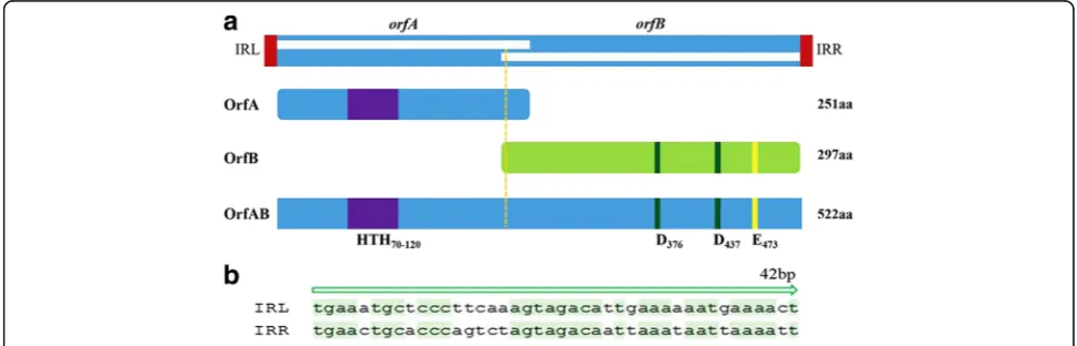
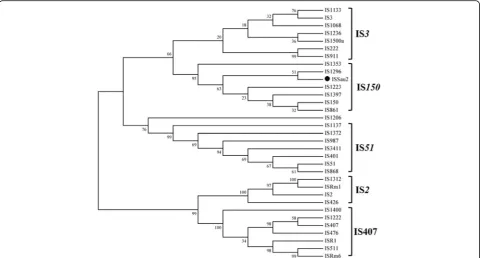
ISSau2 from bovine S. aureus isolates was flanked by imperfect inverted 42 bp nucleotide repeats (IRL and IRR) (Fig. 1). Examination of ISSau2 OrfAB revealed a HTH motif within the N-terminal region and an Aspartate-Aspartate-Glutamate (DDE) catalytic domain in the C-terminal of OrfAB. There were 60 amino acid residues between the two D residues and 35 amino acid residues between the second D and E residues. A lysine (K) residue was six amino acids downstream of the E residue. For members of the IS150 subgroup, lysine (K) residue should be six amino acids downstream of the E residue. To sub-categorize ISSau2, a phylogenetic tree was constructed based on the amino acid sequence alignment of OrfB (Fig. 2). The result from the phylo-genetic analysis confirmed that ISSau2 belongs to the IS150subgroup of the IS3family.
Translational frameshift of OrfAB occurred in ISSau2
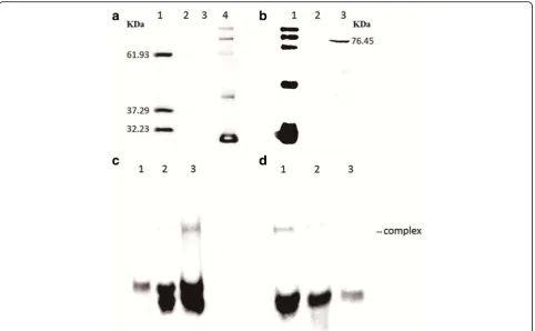
To verify the frameshifting phenomenon during expres-sion of ISSau2, the whole transposase sequence was inserted into pET28b to create ISSau2-pET28b, which was transformed intoE. coli BL21 (DE3) for expression. As shown in Fig. 3a, three proteins were expressed with similar concentrations and expected to be OrfAB, OrfB and OrfA from top to bottom. The results indicated that
OrfAB of ISSau2 was produced by −1 translational
frameshifting.
OrfAB bound to ISSau2IRs in vitro
In order to ascertain the binding between transposase OrfAB and the target DNA, an ISSau2mutant, containing
a single guanine insertion in the A6G sequence to generate
A6G2, was inserted into pET32a to create pET32-OrfAB
and expressed in E. coli BL21 (DE3). The transposase OrfAB was expressed according to the Western blot ana-lysis (Fig. 3b) and purified.
To test whether the transposase can bind to DNA, an electrophoretic mobility shift assay (EMSA) was carried out with the purified transposase OrfAB and the biotin-labeled 42 bp oligonucleotides (IRL and IRR) which included the putative transposase binding sites. Transpo-sase OrfAB bound to IRL42 in the presence of an excess of the poly (dI-dC) competitor and Mg2+(Fig. 3c) and also bound to ISSau2 IRR42 with a similar affinity
(Fig. 3d). Crude extracts from E. coli with pET32a
failed to bind to both left and right ISSau2inverted re-peat sequences as shown in lane 2 of Fig. 3c-d. There-fore, the binding was specifically carried out by the purified ISSau2transposase OrfAB.
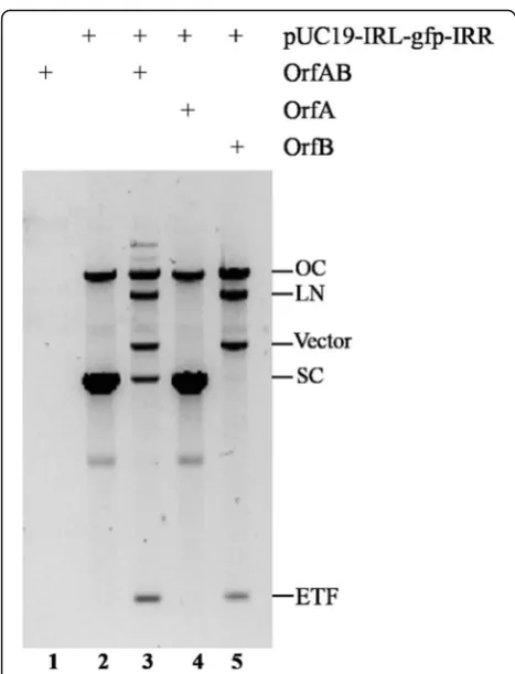
ISSau2transposase OrfAB cleaved transposon ends
To test whether the ISSau2 transposase had a catalytic activity, a plasmid (pUC19-IRL-gfp-IRR), an artificial mini-transposon with IRL and IRR flanking a gfp gene, was incubated in a buffer with purified full-length trans-posase OrfAB. The reaction produced linearized plasmid and two fragments of the correct size to be excised
mini-transposon (ETF) and the vector backbone (Fig. 4, lane 3). Restriction digestion confirmed that these prod-ucts were produced by double-strand cleavage at IRR and IRL. As shown in lane 3 and 5 of Fig. 4, both the linearized plasmid (LN) and nicked plasmid (OC) were also produced by transposase OrfAB in the cleavage re-action. These results indicated that transposase OrfAB cleaved both single and double strands in vitro.
To test which domain in the transposase OrfAB is re-quired for cleavage, purified OrfA and OrfB were used in the cleavage assay. OrfA did not have the catalytic activity to cleave transposon end (Fig. 4, lane 4), while OrfB and OrfAB had the similar activity for the cleavage (Fig. 4, lane 3 and lane 5). These results proved that OrfB was involved in the cleavage reaction and in ISSau2transposition reactions.
Measurement of transposition frequency of ISSau2inE.
coli
To measure the transposition frequency of ISSau2 in vivo, a modified GFP hop-on assay was used. A GFP hop-on assay plasmid pET28a-ISgfp was constructed. It contained a gfp gene which lacks transcriptional and translational signals and was located between IRL and IRR of ISSau2. Thegfpgene in the GFP-hopper was not expressed at its original location in the plasmid. In the
presence of pET32a-OrfAB, it could be expected that ex-pression of the ISSau2transposase gene led to transpos-ition of ISgfp, into an expressed gene in the bacterial genome in the correct orientation and reading frame, resulting in the fusion of the gene and gfpgene and ex-pression of a green fluorescent fusion protein.
To detect transpositional events of ISgfp by FACS, we in-troduced both pET28a-ISgfp and pET32a-OrfAB into E. coliDH5α. The strain harboring pET28a-ISgfp acted as a negative control. Per 106events through the FACS, 622.32 ± 18.84 events were detected in the negative control group (Fig. 5a). As shown in Fig. 5b, 2381.82 ± 105.68 events for 106total events were detected with the transposase induc-tion and the number was significantly higher than that in the negative control group, demonstrating that the appear-ance of the extra fluorescent signals depended on the OrfAB transposase. The transposition frequency of the ISgfp, representing the transposition frequency of ISSau2
in vivo, was approximately 1.76 ± 0.13 × 10⁻³.
Effect of OrfA and OrfB on the transposition of ISSau2
To investigate whether OrfA or OrfB affected the trans-position activity, pET32a-OrfA and pET32a-OrfB were introduced into the strains harboring the pET28a-ISgfp and pET32a-OrfAB. As shown in Fig. 5c and D, OrfA (1209.85 ± 3.66 events for 106 total events) and OrfB (1341.95 ± 4.32 events for 106 total events) significantly decreased the transposition frequency of ISgfp. These data suggested that OrfA and OrfB were the inhibitors for the transposition of ISSau2.
Discussion
Until now, only 138 insertion sequences have been iden-tified as members of IS150 subgroup in the IS3 family (data from ISfinder). There were only a few studies on IS150 insertion sequences. Early studies identified an unknown insertion sequence based on the homology analyses of nucleotide sequences or protein sequences [13, 14]. Later on, presumed members have also been
required to possess the common features of IS150: 1200– 1400 bp in length, containing twoorf, a HTH motif, a D (57–60) D (35) E motif, and an AnG frameshifting window
(OrfAB as a transposase) [4, 9]. In accordance with these, our results confirmed that ISSau2 belongs to the IS150
subgroup of IS3 family not only supported by the se-quences analysis (Fig. 1) but also by results from the mo-lecular experiments (Fig. 3a). First, ISSau2 contains two open reading frames encoded two proteins. Secondly, OrfA contains a HTH motif, while OrfB contains a D (60) D (35) E motif and the OrfAB transposase produced by programmed−1 translational frameshifting with the motif A6G (Fig. 1). Additionally, three proteins were expressed
with similar concentrations (Fig. 3a) and this is consistent with the idea that the frequency of frameshifting in IS150
is approximately 50% [15]. Last, the classification was fur-ther confirmed by the phylogenetic analysis using OrfB protein sequence (Fig. 2).
ISSau2was first found in HA-MRSA252 strain by the complete genome sequence analysis [16]. Our previous study found that ISSau2 was only present in S. aureus
[1]. Except our work, only anther paper analyzed the in-sertion sites of ISSau2 in CC30 S. aureus by bioinfor-matics analysis [8] without any other functional studies. Our results in this study confirmed that ISSau2 was at least partially functional with binding and cleavage activ-ities by the transposase OrfAB and the transposase was able to catalyze the transposition in vivo.
The ISSau2 transposase OrfAB bound to the inverted repeat sequences, where it carries out cleavage and strand transfer reactions. The conserved HTH motif in the N terminal of OrfAB is supposed to bind the IRs specifically. The low binding of purified OrfAB to the IRL or IRR (Fig. 3c and d) was presumably due to the fact that the full-length transposase binds poorly to the
Fig. 5Flow cytometric analysis of ISgfp transposition and measurement of the transposition frequency of ISSau2.aE. colistrain harboring pET28a-ISgfp as a negative control.bE. colistrain harboring pET28a-ISgfp and pET32a-OrfAB.cE. colistrain harboring pET28a-ISgfp, pET32a-OrfAB and pET32a-OrfA.
dE. colistrain harboring pET28a-ISgfp, pET32a-OrfAB and pET32a-OrfB. Experiments were performed in triplicates and representative results from one experiment are shown. The range of gated events is indicated as marker 1 (M1)
ends in vitro [17]. Additionally, we showed that OrfAB was able to cleave the transposon ends, otherwise, OrfB alone could also cleave the transposon in vitro (Fig. 4) with an explicable reason that it contained the DDE do-main which catalyzes transposition reactions [3]. Our
re-sults at least partly support the idea that the
transposition occurred by a cut and paste mechanism like other members in the IS3family [18].
The transposition frequency of ISSau2 was estimated using a modified“GFP hop-on assay”method, which has been used to determine the transposition of transposons in both Bacillus subtilis and E. coli [19, 20]. This method, making it possible to detect transposition at the single cell level, was more efficient to estimate the trans-position frequency than other methods [19, 20], such as papillation assays [21, 22], mutation accumulation ex-periment [23] and mating-out assay [24]. Thus, the GFP hop-on assay was adapted to measure the transposition frequency of ISSau2 in the presence of OrfA, OrfB or OrfAB. As shown in Fig. 5, OrfAB of ISSau2 induced transposition at the frequency of 1.76 ± 0.13 × 10⁻³. Nevertheless, the observed transposition frequency (1.76 ± 0.13 × 10⁻³) might be over-estimated than the real transposition in bacteria, due to the laboratory condi-tions which had few limiting factors and thus favored the transposition. In nature, the transposition rates of in-sertion sequences generally must be maintained at a low level, acting as a possible strategy to limit any negative effects on the host genome [25]. These data demon-strated that the transposition of ISSau2depended on the transposase OrfAB and indicated that ISSau2 was at least partially functional.
It is interesting that both OrfA and OrfB inhibited sig-nificantly the transposition of ISSau2 induced by OrfAB. In the IS3 family, OrfA inhibits reactions promoted by transposases by binding to transposon ends [26]. OrfB might form a complex with OrfA to inhibit the transpos-ition of ISSau2by blocking formation of an active trans-pososome consisting of transposase, two terminal IRs and target DNA or by preventing the transposase from catalyzing the strand transfer reaction [26].
In summary, we characterized a functional insertion sequence ISSau2inS. aureus. To the best of our know-ledge, this is the first study to investigate the transpos-ition of ISSau2 and to characterize the effects of OrfA and OrfB of ISSau2 on transposition. Our results pro-vide a solid basis for future studies of the molecular mechanisms involved in ISSau2 transposition and its biological function in its host S. aureus. Meanwhile, we now have an experimental system that allow us to characterize any novel insertion sequence in bacterial cells and even in other eukaryotic cells especially for the insertion sequences which contain twoorfgenes and use the fusion OrfAB as the transposase.
Conclusion
We have confirmed that ISSau2 is a member of IS150/ IS3family. The ISSau2 transposase OrfAB could bind to and cleave the specific fragments containing the terminal inverted repeat sequences and induce the transposition, suggesting that ISSau2 was functional. Meanwhile, both OrfA and OrfB were the inhibitors for transposition of ISSau2. Our results will help understand biological roles of ISSau2in its hostS. aureus.
Methods
Construction of prokaryotic expression vectors
A known ISSau2 transposase sequence, located insdrC
ofS. aureusE48 [27], was isolated by PCR with the spe-cific primers pairs ISSau2-F/ISSau2-R (Table 1) using the Pfu polymerase, and inserted between BamH I and
Xho I sites of pET28b to create pET28-ISSau2. Within
pET28-ISSau2, an N-terminal 6-His tag and a
C-terminal 6-His tag were linked to either side of the ISSau2 sequence. Meanwhile, a G residue was inserted downstream of A6G in the ISSau2sequence, forming an
A6G2 region [2], by overlap PCR using the two primer
pairs ISSau2-F/OrfAB-overlap-R and OrfAB-overlap-F/ ISSau2-R (Table 1). In theory, addition of the G would not change the OrfAB amino acid sequence [28]. The
mutated sequence was inserted between BamH I and
Xho I sites of pET32a, which contains an N-terminal
6-His tag, to create pET32-OrfAB. The orfA region
(753 bp) andorfBregion (891 bp) of ISSau2were ampli-fied by PCR using the primers ISSau2-F/OrfA-R and OrfB-F/ISSau2-R respectively (Table 1) and inserted into pET32a to construct pET32a-OrfA and pET32a-OrfB.
Expression and purification of ISSau2transposase
The constructed expression vectors were transformed into BL21 (DE3) competent cells, separately. The over-night culture of BL21 (DE3) was diluted 1:50 in fresh LB broth containing different antibiotics (pET28-ISSau2:
50 μg/mL kanamycin; pET32-OrfAB: 100 μg/mL
am-picillin) with vigorous shaking at 37 °C to an OD600 of
0.4–0.6. Expression of the transposase was induced by the addition of Isopropyl-β-Dthiogalactopyranoside (IPTG) to 0.2 mM. Bacterial cells were grown for 6 h at 28 °C and harvested by centrifugation.
All the following steps were carried out on ice. Cul-tured bacterial cells (BL21 with pET32-OrfAB) were sus-pended in a PBS buffer (137 mM NaCl, 2.7 mM KCl, 10 mM Na2HPO4, and 2 mM KH2PO4) and disrupted by
acid (Ni-NTa) column equilibrated with PBS. The col-umn was washed with 10 colcol-umn volumes of PBS followed by 6 column volumes of PBS plus 20 mM imid-azole, impure proteins were eluted in PBS plus 75 mM imidazole and the purified OrfAB was obtained in PBS plus 500 mM imidazole. The dialyzed protein was made up to 50% glycerol and stored at−80 °C.
Electrophoretic mobility shift assays (EMSA)
Two 42 bp biotin-labeled DNA fragments containing
IRL and IRR were generated by oligonucleotide
hybridization (heated together at 95 °C for 10 min and cooled to room temperature) respectively to generate dsDNA IRL42 and IRR42. For EMSA, 2μL 10 × binding
buffer, 1 μg/μL Poly (dI-dC), 50% Glycerol, 1% NP-40,
1 M KCl, 100 mM MgCl2, 200 mM EDTA (EMSA kit,
Thermo) and 0.42 μg OrfAB were incubated at room
temperature for 20 min. Binding was initiated by addition of biotin end-labeled target DNA. Binding reactions were allowed at room temperature for 20 min and the products were then run on 5% polyacrylamide gels in 0.5 × TBE buffer (100 V) at 0 °C. The biotin end-labeled DNA was detected using the Streptavidin-Horseradish Peroxidase Conjugate and the Chemiluminescent Substrate.
In vitro transposition assays
An in vitro transposition assay was performed as de-scribed [29]. The artificial mini transposon plasmid (pUC19-IRL-gfp-IRR) contained IRL42 and IRR42 flank-ing the GFP gene which lacks transcription and transla-tion start signals, the fusion sequence was amplified by PCR using the specific primer pairs F/ IRL-gfp-R (Table 1) with the pGLO plasmid DNA as the tem-plate. The gfp gene was amplified so as to remove the transcriptional and translational signals. Standard in vitro transposition assays contained 0.12 pmol of pUC19-IRL-gfp-IRR in 20 mL of transposase assay buffer (10 mM
MgCl2, 1 mM DTT, 0.1 mg/mL BSA, 50 mM Tris-HCl
pH 7.5 and 50 mM NaCl). Reactions were started by the addition of 1–2 pmol of purified transposase OrfAB and incubated at 30 °C for 16 h. Reactions were stopped by heating at 75 °C for 10 min, and products were analyzed by agarose gel electrophoresis.
Detection of GFP-hopper transposition
The plasmid pET28a-ISgfp was generated from pUC19-IRL-gfp-IRR. The GFP hop-on assay was performed as described [20] with minor modifications. The plasmid pET28a-ISgfp as well as OrfAB or pET32a-OrfA or pET32a-OrfB was introduced intoE. coliDH5α. The transformed colonies were cultured in 5 mL LB medium under the selective pressure (both 50 μg/mL kanamycin and 100 μg/mL ampicillin) at 37 °C. Har-vested cells were then resuspended in a sheath fluid (BD Biosciences, USA) at OD600= 0.3. Flow cytometry was
performed on a FACSCalibur cytometer with the CELL-QUEST software (BD Biosciences). GFP fluorescence was detected with a FL1 detector and its intensity was expressed as an arbitrary logarithmic value (FL1-H) within the range of 100–104. The transposition-frequency was es-timated as the ratio between the number of events within region M1 (> 102FL1- H) in the presence of OrfAB minus the background number and the number of total events.
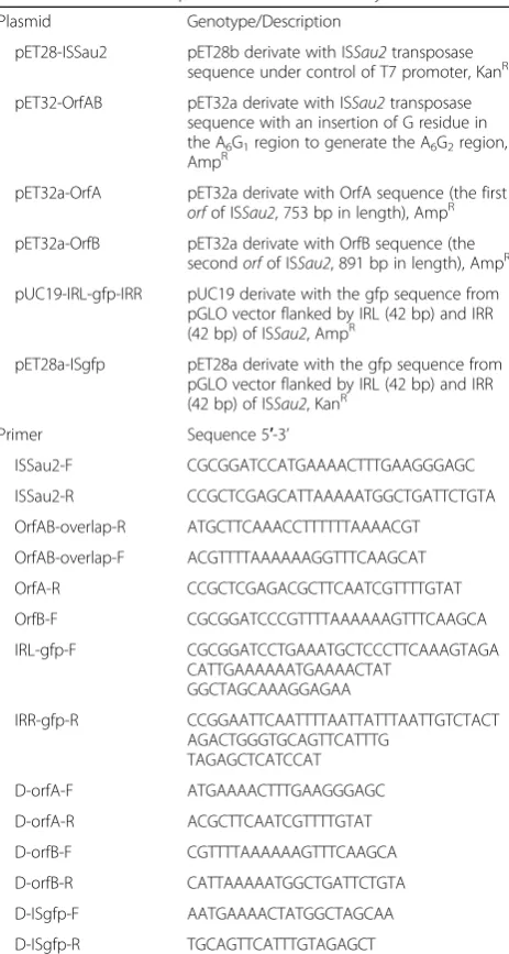
To investigate whether OrfA or OrfB has the activity to inhibit the transposition of ISgfp induced by OrfAB, the pET32a-OrfA or pET32a-OrfB was first introduced Table 1Plasmids and primers used in this studya
Plasmid Genotype/Description
pET28-ISSau2 pET28b derivate with ISSau2transposase sequence under control of T7 promoter, KanR
pET32-OrfAB pET32a derivate with ISSau2transposase sequence with an insertion of G residue in the A6G1region to generate the A6G2region,
AmpR
pET32a-OrfA pET32a derivate with OrfA sequence (the first
orfof ISSau2, 753 bp in length), AmpR
pET32a-OrfB pET32a derivate with OrfB sequence (the secondorfof ISSau2, 891 bp in length), AmpR
pUC19-IRL-gfp-IRR pUC19 derivate with the gfp sequence from pGLO vector flanked by IRL (42 bp) and IRR (42 bp) of ISSau2, AmpR
pET28a-ISgfp pET28a derivate with the gfp sequence from pGLO vector flanked by IRL (42 bp) and IRR (42 bp) of ISSau2, KanR
Primer Sequence 5′-3’
ISSau2-F CGCGGATCCATGAAAACTTTGAAGGGAGC
ISSau2-R CCGCTCGAGCATTAAAAATGGCTGATTCTGTA
OrfAB-overlap-R ATGCTTCAAACCTTTTTTAAAACGT
OrfAB-overlap-F ACGTTTTAAAAAAGGTTTCAAGCAT
OrfA-R CCGCTCGAGACGCTTCAATCGTTTTGTAT
OrfB-F CGCGGATCCCGTTTTAAAAAAGTTTCAAGCA
IRL-gfp-F CGCGGATCCTGAAATGCTCCCTTCAAAGTAGA CATTGAAAAAATGAAAACTAT
GGCTAGCAAAGGAGAA
IRR-gfp-R CCGGAATTCAATTTTAATTATTTAATTGTCTACT AGACTGGGTGCAGTTCATTTG
TAGAGCTCATCCAT
D-orfA-F ATGAAAACTTTGAAGGGAGC
D-orfA-R ACGCTTCAATCGTTTTGTAT
D-orfB-F CGTTTTAAAAAAGTTTCAAGCA
D-orfB-R CATTAAAAATGGCTGATTCTGTA
D-ISgfp-F AATGAAAACTATGGCTAGCAA
D-ISgfp-R TGCAGTTCATTTGTAGAGCT
a
into E. coli DH5α and confirmed by PCR with the pri-mer pairs OrfA-F/OrfA-R or OrfB-F/OrfB-R (Table 1) and then the pET28a-ISgfp and pET32a-OrfAB were in-troduced into E. coliDH5αby electroporation and con-firmed by PCR using the primers ISgfp-F/ISgfp-R and OrfA-F/OrfB-R (Table 1). The GFP fluorescence of the
cells containing pET28a-ISgfp, pET32a-OrfAB and
pET32a-OrfA (or OrfB) were detected by FACSCalibur cytometer.
Abbreviations
DDE:Aspartate-Aspartate-Glutamate;E. coli:Escherichia coli;
EMSA: Electrophoretic Mobility Shift Assays; FACS: Fluorescence Activated Cell Sorting; GFP: Green Fluorescent Protein; HA-MRSA: Healthcare-Associated Methicillin ResistantStaphylococcus aureus; HTH: Helix-Turn-Helix;
IPTG: Isopropyl-β-Dthiogalactopyranoside; IRs: Inverted Repeat sequences; ORF: Open Reading Frame; PBS: Phosphate Buffer solution;S.
aureus:Staphylococcus aureus
Acknowledgements
Not applicable
Funding
National Science Foundation of China (31372282): design of the study and collection, analysis, and interpretation of data; China Thousand Talents program: analysis of data.
Availability of data and materials
Data sharing not applicable to this article as no datasets were generated or analysed during the current study.
Authors’contributions
XZ LW HX conceived and designed the experiments. LW WS performed the experiments. WS LW analyzed the data. HX contributed reagents/materials/ analysis tools. XZ LW WS wrote the paper. All authors read and approved the final manuscript.
Ethics approval and consent to participate
Not applicable
Consent for publication
Not applicable
Competing interests
The authors declare that they have no competing interests.
Publisher’s Note
Springer Nature remains neutral with regard to jurisdictional claims in published maps and institutional affiliations.
Author details 1
College of Animal Science and Technology, Northwest A&F University, No.3 Taicheng Road, Yangling 712100, Shaanxi Province, People’s Republic of China.2School of Pharmaceutical Sciences, Tsinghua University, Beijing, People’s Republic of China.3Tsinghua University-Peking University Joint
Center for Life Sciences, Beijing, People’s Republic of China.4Department of Animal Science, McGill University, Quebec, Canada.
Received: 4 September 2017 Accepted: 8 January 2018
References
1. Wang L, Xue H, Li L, Zhao X. Characterization of insertion sequence ISSau2 in the human and livestock-associatedStaphylococcus aureus. PLoS One. 2015;10:e0127183.
2. Rousseau P, Tardin C, Tolou N, Salome L, Chandler M. A model for the molecular organisation of the IS911transpososome. Mob DNA. 2010;1:16.
3. Izsvák Z, Khare D, Behlke J, Heinemann U, Plasterk RH, Ivics Z. Involvement of a bifunctional, paired-like DNA-binding domain and a transpositional enhancer inSleeping Beautytransposition. J Biol Chem. 2002;277:34581–8. 4. Mahillon J, Chandler M. Insertion sequences. Microbiol Mol Biol Rev.
1998;62:725–74.
5. Siguier P, Perochon J, Lestrade L, Mahillon J, Chandler M. ISfinder: the reference centre for bacterial insertion sequences. Nucleic Acids Res. 2006;34:D32–6.
6. Sekine Y, Eisaki N, Ohtsubo E. Translational control in production of transposase and in transposition of insertion sequence IS3. J Mol Biol. 1994;235:1406–20. 7. Chandler M, Fayet O. Translational frameshifting in the control of transposition
in bacteria. Mol Microbiol. 1993;7:497–503.
8. McGavin MJ, Arsic B, Nickerson NN. Evolutionary blueprint for host- and niche-adaptation inStaphylococcus aureusclonal complex CC30. Front Cell Infect Microbiol. 2012;2:48.
9. Haas M, Rak B.Escherichia coliinsertion sequence IS150: transposition via circular and linear intermediates. J Bacteriol. 2002;184:5833–41. 10. Sekine Y, Aihara K, Ohtsubo E. Linearization and transposition of circular
molecules of insertion sequence IS3. J Mol Biol. 1999;294:21–34.
11. Turlan C, Chandler M. IS1-mediated intramolecular rearrangements: formation of excised transposon circles and replicative deletions. EMBO J. 1995;14:5410–21. 12. Chen CC, Hu ST. Two frameshift products involved in the transposition of
bacterial insertion sequence IS629. J Biol Chem. 2006;281:21617–28. 13. Rubens CE, Heggen LM, Kuypers JM. IS861, a group B streptococcal insertion
sequence related to IS150and IS3ofEscherichia coli. J Bacteriol. 1989;171:5531–5. 14. Smith GP, Ellar DJ, Keeler SJ, Seip CE. Nucleotide sequence and analysis of
an insertion sequence from bacillus thuringiensis related to IS150. Plasmid. 1994;32:10–8.
15. Vögele K, Schwartz E, Welz C, Schiltz E, Rak B. High-level ribosomal frameshifting directs the synthesis of IS150gene products. Nucleic Acids Res. 1991;19:4377–85.
16. Holden MT, Feil EJ, Lindsay JA, Peacock SJ, Day NP, et al. Complete genomes of two clinicalStaphylococcus aureusstrains: evidence for the rapid evolution of virulence and drug resistance. Proc Natl Acad Sci U S A. 2004;101:9786–91. 17. Rousseau P, Gueguen E, Duval-Valentin G, Chandler M. The helix-turn-helix motif of bacterial insertion sequence IS911transposase IS required for DNA binding. Nucleic Acids Res. 2004;32:1335–44.
18. Chandler M, Fayet O, Rousseau P, Ton Hoang B, Duval-Valentin G. Copy-out-paste-in transposition of IS911: a major transposition pathway. Microbiol Spectr. 2015;3:https://doi.org/10.1128/microbiolspec.MDNA3-0031-2014 19. Saito T, Chibazakura T, Takahashi K, Yoshikawa H, Sekine Y. Measurements of
transposition frequency of insertion sequence IS1by GFP hop-on assay. J Gen Appl Microbiol. 2010;56:187–92.
20. Takahashi K, Chibazakura T, Sekine Y, Yoshikawa H. Development of a new "GFP hop-on assay" system for insertion sequence transposition inBacillus subtilis168 using IS4Bsu1fromB-subtilis(natto). Biochem Biophys Res Commun. 2007;355:426–30.
21. Twiss E, Coros AM, Tavakoli NP, Derbyshire KM. Transposition is modulated by a diverse set of host factors inEscherichia coliand is stimulated by nutritional stress. Mol Microbiol. 2005;57:1593–607.
22. Díaz-Maldonado H, Gómez MJ, Moreno-Paz M, San Martín-Úriz P, Amils R, et al. Transposase interaction with theβsliding clamp: effects on insertion sequence proliferation and transposition rate. Sci Rep. 2015;5:13329. 23. Sousa A, Bourgard C, Wahl LM, Gordo I. Rates of transposition inEscherichia
coli. Biol Lett. 2013;9:20130838.
24. Lampe DJ, Akerley BJ, Rubin EJ, Mekalanos JJ, Robertson HM. Hyperactive transposase mutants of the Himar1 mariner transposon. Proc Natl Acad Sci U S A. 1999;96:11428–33.
25. Nagy Z, Chandler M. Regulation of transposition in bacteria. Res Microbiol. 2004;155:387–98.
26. Sekine Y, Izumi K, Mizuno T, Ohtsubo E. Inhibition of transpositional recombination by OrfA and OrfB proteins encoded by insertion sequence IS3. Genes Cells. 1997;2:547–57.
27. Xue H, Lu H, Zhao X. Sequence diversities of serine-aspartate repeat genes amongStaphylococcus aureusisolates from different hosts presumably by horizontal gene transfer. PLoS One. 2011;6:e20332.
28. Polard P, Chandler M. An in vivo transposase-catalyzed single-stranded DNA circularization reaction. Genes Dev. 1995;9:2846–58.