RESEARCH
Variability of serum novel serum peptide
biomarkers correlates with the disease states
of multiple myeloma
Ju Bai
1, Yun Yang
1, Jianli Wang
1, Lei Zhang
2, Fangxia Wang
1and Aili He
1*Abstract
Background: The bone marrow microenvironment provides an optimal substrate for multiple myeloma (MM) initia-tion and progression. The soluble component of MM niche is dynamic with the disease states of MM. We formerly employed proteomic profiling to construct a MM model. Four peptides constituting the model were selected by supervised neural network algorithm (SNN).
Methods: 62 Newly diagnosed (ND) MM and 64 healthy controls (HCs) were picked up for validating the distinguish-ing capability of the SNN model. Nano-liquid chromatography-electrospray ionization-tandem mass spectrometry was used for peptide identification. MM in different disease states and HCs were choosed for peptides relative intensi-ties comparison. Western blot and ELISA were employed to validate the variability.
Results: The sensitivity and specificity of the independent testing data set for blind validation were 93.55% and 92.19%. The relative intensities of three out of the four peptides were increased in ND and refractory and relapse patients but decreased to that level of HCs in complete remission and very good partial remission patients. Rela-tive intensity of the remaining peptide was negaRela-tively associated with MM remission. The peptides sequencing results showed that they were derived from dihydropyrimidinase-like 2, fibrinogen alpha chain, platelet factor 4 and alpha-fetoprotein.
Conclusions: The potential value of the four peptides in monitoring MM treatment response was arised from their close correlation with MM disease states.
Keywords: Biomarkers, Proteomic profiling, Bone marrow microenvironment, Treatment response, Multiple myeloma
© The Author(s) 2019. This article is distributed under the terms of the Creative Commons Attribution 4.0 International License (http://creat iveco mmons .org/licen ses/by/4.0/), which permits unrestricted use, distribution, and reproduction in any medium, provided you give appropriate credit to the original author(s) and the source, provide a link to the Creative Commons license, and indicate if changes were made. The Creative Commons Public Domain Dedication waiver (http://creat iveco mmons .org/ publi cdoma in/zero/1.0/) applies to the data made available in this article, unless otherwise stated.
Background
Multiple myeloma (MM) is a malignant B cell clonal dis-ease with a worldwide incidence of 6–7 cases per 100,000 persons per year [1]. More disturbingly, it has been noted that for some unknown reason MM displays a trend of increasing incidence [2]. Despite the great efforts in the past decades, treatment of MM is not effective and the prognosis of MM is poor. Therefore, MM is still consid-ered largely as an incurable disease.
Cytogenetic abnormalities play a crucial role in MM pathogenesis. The bone marrow microenvironment vides an optimal substrate for MM initiation and pro-gression [3]. MM cells and bone marrow derived stroma cells (BMSCs) profoundly influence each other’s behavior via a variety of molecular mechanisms. Among these, changes in the secretion of cytokines, growth factors, and chemokines play a prominent role in supporting cancer cell growth, survival, trafficking, and resistance to ther-apies [4]. Plasma cells can secrete cytokines (e.g. tumor necrosis factor alpha, transforming growth factor beta, vascular endothelial growth factor (VEGF), fibroblast growth factor-2, etc.) to allow for infiltration, growth, proliferation, adhesion, and migration of MM cells [5– 9]. Moreover, binding of plasma cells to BMSCs triggers
Open Access
*Correspondence: heaili@mail.xjtu.edu.cn
1 Department of Hematology, Second Affiliated Hospital, Medical School of Xi’an Jiaotong University, 157 Xiwu Road, Xincheng District, Xi’an 710004, Shaanxi Province, China
transcription and secretion of cytokines by the latter, such as interleukin-6 (IL-6), insulin-like growth factor-1 (IGF-1), VEGF and CXCL12/stromal cell derived factor-1 (SDF-1) [10–13] that mediate plasma cell growth, sur-vival, and drug resistance, as well as bone marrow (BM) angiogenesis. However, a definite pathogenic role for the BM niche in MM remains under investigation.
The soluble component of MM niche is dynamic with the disease states of MM. Considering that the hetero-geneous nature of MM and the involvement of multiple proteins in BM milieu, a combinatory approach using a panel of biomarkers is more reliable and sensitive than relying on a single protein. Mass spectrometry based pro-teomic profiling holds great promise for the identification of circulating biomarkers useful for a better understand-ing of bone marrow microenvironment of MM. Previ-ously, we applied a comparative peptidomics method combining weak cation exchange beads (MB-WCX) and matrix assisted laser desorption ionization time of flight mass spectrometry (MALDI-TOF-MS) to analyze serum peptide profiles of newly diagnosed (ND) MM and healthy controls. A diagnostic model including four peptides (2660.65, 2900.4, 3315.96, 7763.24 Da) was con-structed based on supervised neural network algorithm
(SNN), which could discriminate newly diagnosed (ND) MM and healthy controls. Appreciable levels of sensitiv-ity and specificsensitiv-ity, 87.5% and 86.36%, respectively, were achieved in a small patient samples [14]. Therefore, to better understand the soluble component of MM niche, we used mass spectrometry based proteomic profiling to characterize the variability of serum proteome in differ-ent MM treatmdiffer-ent durations.
Materials and methods Study population
This study was approved by Ethics Committee of the Sec-ond Affiliated Hospital of Xi’an Jiaotong University. 62 ND patients, 38 complete remission and very good par-tial remission (CR&VGPR) patients and 43 refractory and relapse (RR) patients hospitalized in our hospital were recruited for the study (2010.1–2016.12) (Characteris-tics details were showed in Table 1). Their diagnoses were made according to the International Myeloma Work-ing Group (IMWG) criteria [15]. Treatment response was assessed based on IMWG uniform response crite-ria for MM [16]. All MM cases were treated with VAD (Vinorelbine, Pirarubicin and Dexamethasone), CHOP (Cyclophosphamide, Vinorelbine, Pirarubicin and
Table 1 Clinical features of patients in different MM groups before chemotherapy
This table showed clinical features of MM patients in different groups at the time of diagnosis
ESR erythrocyte sedimentation rate, LDH lactate dehydrogenase
Clinical features ND cases (n = 62) CR&VGPR cases (n = 38) RR cases (n = 43)
Sex
Male 34 21 27
Female 28 17 16
Age (year) 56.5 (44–73) 54.5 (44–71) 57.5 (45–73)
WBC (× 109/L) 5.7 (2.7–14) 5.3 (3.2–11.5) 6.2 (2.3–15.3)
Hb (g/L) 83 (52–101) 82 (63–132) 72 (48–95)
PLT (× 109/L) 190 (23–356) 202 (89–387) 183 (56–361)
Subtype
IgG type 32 20 26
IgA type 12 8 8
IgM type 0 0 0
Light chain type 8 5 5
Non-secretory type 10 5 4
MM cell > 10% 62 38 43
ESR↑ 53 31 36
Glb (> 30 g/L) 52 33 39
Renal failure (serum creatinine ≥ 2 mg/100 mL) 13 6 9
Hypercalcemia 15 6 8
Bence-Jone protein 21 5 9
LDH↑ 35 19 28
Bone lesions (lytic lesions, pathologic fractures or severe
Dexamethasone) regimen and oral thalidomide. 64 HCs (age range 45–70, median age 57.5 years old, male/female 34/30) were enrolled. They had no finding of abnormal symptoms or test results in physical examination. All patients and healthy controls signed the informed con-sent form.
Serum peptides enrichment and MALDI‑TOF data acquisition
The procedure of serum peptides enrichment and MALDI-TOF data acquisition were described previously [14]. The serum peptides were fractionated using weak cation exchange magnetic-beads (MB-WCX) (ClinProt purification reagent sets; Bruker Daltonics, Bremen, Germany) with a magnetic separator. Mixture with the eluted sample and matrix was spot onto a MALDI-TOF mass spectrometry target (AnchorChip™, Bruker Dalton-ics) for peptide profiling acquisition in Microflex mass spectrometer (Bruker Daltonics). Cilnplot standard was used for mass calibration. The scan range was 0.7–10 KD. Peptide profiling by different mass to charge ratio was obtained through FlexControl2.2 software. The coeffi-cient of variability less than 30% indicated that the system was stable and reliable.
Peptides identification
A nano-liquid chromatography-electrospray ionization-tandem mass spectrometry (nano-LC/ESI-MS/MS) system was used for peptide sequencing and peptide identification. The detailed procedure was done accord-ing to previous description [17]. The peptide solutions purified by magnetic beads, were loaded to a C18 trap column (nanoACQUITY) [180 μm × 20 mm × 5 μm (symmetry)]. The flow rate was 15 μL/min. Then the desalted peptides were analyzed by C18 analytical col-umn (nanoACQUITY) [75 μm × 150 mm × 3.5 μm (symmetry)] at a flow rate of 400 nl/min for 60 min. The mobile phases A (5% acetonitrile, 0.1% formic acid) and B (95% acetonitrile, 0.1% formic acid) were used for ana-lytical columns. The gradient elution profile was as fol-lows: 5%B–45%B–80%B–80%B–5%B–5%B in 60 min. The spray voltage was 1.8 kV. MS scan time was 60 min. Experimental mode were data dependent and dynamic exclusion, scilicet MS/MS spectra were limited to two consecutive scans per precursor ion within 10 s followed by 90 s of dynamic exclusion. Mass scan range was from m/z 400 to 2000. MS scan used Obitrap, resolution was set at 100,000. CID and MS/MS scan employed LTQ. In MS spectrogram, we selected single isotope composed of 10 ions with strongest intensity as parent ion for MS/ MS. Single charge was excluded and not as parent ion. We applied data analysis software Bioworks Browser 3.3.1 SP1 for Sequest™ retrieving. Retrieval Database was
International Protein Index (IPI human v3.45 fasta with 71,983 entries). Parent ion error and fragment ions error were set at 100 ppm and 1 Da, respectively. Digested mode was non-digested and variable modification was methionine oxidation. Positive protein identification was accepted for a peptide with Xcorr of greater than or equal to 3.50 for triply charged ions and 2.5 for doubly charged ions, and all with ΔCn ≥ 0.1, peptide probability ≤ 1e−3.
Western blot analysis
Immunoblotting was performed as described previ-ously [17]. Cell pellets lysed in lysis buffer (10 mM Tris-HCl (pH 7.4), 5 mM MgCl2, 1% Triton X-100, 100 mM NaCl, 10 mM NaF, 1 mM Na3VO4) and protease
inhibi-tor cocktail. Then cellular proteins were subjected to sonication. The proteins were separated sodium dodecyl sulfate polyacrylamide gel electrophoresis (SDS-PAGE) and transferred to polyvinylidene fluoride membranes (Roche Diagnostics Corporation, Indianapolis, Indiana United States), which were incubated with the appropri-ate primary antibodies. Immunoreactivity was detected by horseradish peroxidase-labeled secondary anti-bod-ies. Color development was done by chemiluminescence substrates (7seapharmtech, Shanghai, China). Image Reader Tano-5500 (Tano, Shanghai, China) was used to document the results. 2D Densitometry Image Analyzer IPP 7.0 software (Tano, Shanghai, China) was applied to quantify the images. Samples were measured in triplicate and then mean densitometry was calculated.
Enzyme linked immunosorbent assay
Enzyme linked immunosorbent assay (ELISA) was used to detect the level of fibrinogen alpha chain, platelet factor 4 (PF4) and vascular endothelial growth factor (VEGF) in the serum of 62 ND MM patients and 64 HC. The experiments were conducted following the manufac-turers’ instructions of ELISA kit (R&D, USA).
Statistics
Results
Blind validation of MM SNN model
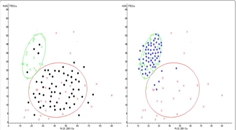
62 newly diagnosed MM and 64 HCs serum specimens were used for blind validation of MM SNN model which was established by our group in previous study. 58 MM were correctly identified as the diseased, so sensitivity of SNN model was 93.55% (Fig. 1a). 59 HCs were correctly assigned to the non-diseased group, 5 cases was misdi-agnosed as MM patients, indicating the specificity was 92.19% (Fig. 1b).
Comparison of the four peptides relative intensities
The multiple comparisons of relative intensities of the four peptides were made in MM with different diease states (ND, CR&VGPR and RR) and HC. The results were
summarized in Table 2. The relative intensities of three peptides (2660.65, 2900.4 and 3315.96 Da) were increased in ND and RR patients but decreased to that level of HCs in CR&VGPR patients (p= 0.0000505; p < 0.000001;
p < 0.000001). The relative intensity of the rest of peptide (7763.24 Da) was negatively associated with MM remis-sion (p < 0.000001).
Identification of peptides
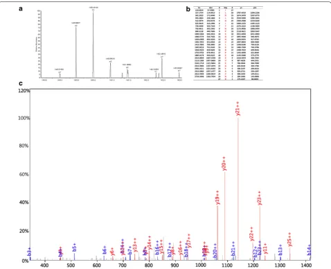
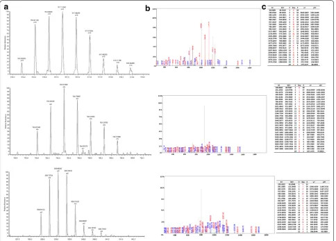
We adopted the HPLC-MS/MS to identify the four pep-tides in the SNN diagnostic model. The four peppep-tides from the diagnostic pattern for MM were derived from dihydropyrimidinase-like 2, fibrinogen alpha chain, plate-let factor 4 and alpha-fetoprotein, respectively (Table 3, Figs. 2, 3, 4, 5).
Fig. 1 Serum specimens from 62 ND MM and 64 HCs were used for SNN model blind validation. 58 cases of MM were correctly assigned to MM group (93.55% sensitivity). 59 HCs were correctly identified as the non-diseased, 5 cases had been wrongly judged as MM (92.19% specificity). Green circle: HCs; Red cross: MM ND patients; Black dot: MM ND patients in validation group; Blue dot: HCs in validation group
Table 2 Relative intensity of the four peptides in MM different groups and healthy control
a “n” stands for the number of patients
Peaks (Da) ND group (n = 62)a HC group (n = 64) CR&VGPR group (n = 38) RR group (n = 43)
2660.65 47.73 ± 28.41 24.58 ± 11.3 26.73 ± 12.43 46.37 ± 27.54
2900.4 4.96 ± 2.12 1.31 ± 0.35 1.44 ± 0.38 5.08 ± 2.58
Validation of protein fragments by western blot
Fibrinogen alpha chain and dihydropyrimidinase-like 2 immunoreactive bands showed that protein expression in MM with different disease states and HC samples were quite heterogeneous (p= 0.0034, p= 0.0067, Fig. 6a, c, d). It is noticeable that PF4 protein levels in ND and RR MM cases were higher than those in CR&VGPR and HC (p= 0.0031, Fig. 6a, b).
Determination of serum PF4, fibrinogen alpha chain and VEGF
ELISA was used to determine the serum PF4, fibrinogen alpha chain and VEGF content in 62 ND MM patients and 64 HC. The PF4 concentration was significantly increased in HC (1.43 ± 0.77 μg/L) when comparing with ND MM (0.67 ± 0.18 μg/L) (p= 1.86 E−6). Higher con-centrations of fibrinogen alpha chain and VEGF were seen in MM than in HC samples (276.78 ± 194.75 μg/L vs. 30.44 ± 22.15 μg/L, p= 1.82E−12; 669.69 ± 137.81 ng/L vs. 120.75 ± 36.96 ng/L, p= 7.28E−29).
We classified 62 ND MM patients into thrombocytope-nia group and platelet normal group in order to exclude the impact of platelet count upon PF4. PF4 concentra-tions were similar in the two groups (0.65 ± 0.18 μg/L vs. 0.69 ± 0.17 μg/L, p= 0.55). Further correlation analysis of platelet count and serum PF4 level revealed the cor-relation coefficient was 0.18 (p= 0.17) (Fig. 7a), indicat-ing no relationship was found between them. However, serum PF4 and serum VEGF appear in an obvious nega-tive correlation. The correlation coefficient was − 0.96 (p= 1.11E−14) (Fig. 7b).
Discussion
The exchange of MM cells with the bone marrow micro-environment also plays a pivotal role in MM pathogen-esis. Since the malignant B-cells produce large amount of proteins and these proteins are readily detectable in circulation, mass spectrometry-based proteomic profil-ing, with its high throughput and sensitivity, holds great promise to identify biomarkers useful for the molecular mechanisms of MM. Such biomarkers may also serve as therapeutic target or indicator for disease state of MM patients. In the present study, an independent testing data
set composed of 62 newly diagnosed MM and 64 healthy control serum specimens was used for blind validation of previously established MM SNN model [14]. The sen-sitivity and specificity reached the encouraging levels of 93.55% and 92.19%, respectively, suggesting that SNN model has a decent power in distinguishing MM patients from healthy individuals. Further efforts for improvement and standardization of the procedure are required for potential clinical application of this SNN model.
Interestingly, the MM candidate biomarkers identified by us and other researchers are distinct. This discrepancy is partially attributable to the different methods used for peptides enrichment and different analytic algorithm and software in data analysis and interpretation [18–22]. The divergent results from different studies suggested that a large pool of differentially expressed proteins is present in MM samples, and the searching of these differences is far from exhausted.
As part of the validation efforts, comparisons of the peptides relative intensity differences were further made in MM with different response states and the healthy control cases. Three peptides with higher relative inten-sities in ND and RR cases were decreased to that level of HCs in CR&VGPR patients. Relative intensity of the remaining peptide was negatively associated with MM remission. All the aforementioned peptides decreased significantly after effective treatment. This is probably the result of the direct cytotoxic effect of the drugs on plasma cells, leading to a reduction in the release of cytokines from the myeloma cells as well as a reduction in their proliferation. Our work shows that, apart from well-identified MM cell-derived soluble factors, the dynamic changes in these peptides along with the progression or remission of the disease raised a possibility for their application to monitoring of disease state and assessment of therapeutic effects.
Fibrinogen alpha chain (FGA), together with beta and gamma chains, is circulating fibrinogens. Numer-ous studies have shown that many malignancies own over expression of fibrinogen, and high fibrinogen is correlated with disease-free, distant disease-free and overall survival [23–28]. In current study, we found an inverse correlation between the relative intensity
Table 3 Peptides sequencing and identification results
Molecular weight (Da) Peptide name International Protein Index Amino acid sequence
2660.65 α-Fibrinogen IPI00021885 DEAGSEADHEGTHSTKRGHAKSRPV
2990.4 Dihydropyrimidinase-like 2 IPI00106642.4 ILDLGITGPEGHVLSRPEEVEAEAVNR 3315.96 Alpha-fetoprotein IPI00022443 FLGDRDFNQFSSGEKNIFLASFVHEYSR 7763.24 Platelet factor 4 IPI00022446
of FGA peptide fragment and the remission of the dis-ease. The relative intensity raised again as MM patients relapsed. This finding was validated by Western blot analysis. Endogenous protein substrates are generated ex vivo by endogenous proteases and these degradation
Fig. 3 MS/MS map of peptide with MW of 2990.4 Da. a The enlarged picture of peptide with MW of 2990.4 Da. b The b and y ions spectra used to identify the peptide with MW of 2990.4 Da. c The sequence of the peptide with MW of 2990.4 Da
proteolytic fragment. Therefore, it is likely that raised enzymatic activity may constantly cleave the FGA C-terminal end to form the over expression of serum peptide fragments (2660.65 Da).
Dihydropyrimidinase-like 2, namely collapsin response mediator protein-2 (CRMP-2), was implicated in the development of nervous system and highly expressed in mouse neuroblastoma Neuro2a cell lines [30]. Wu et al. analyzed the proteome of colorectal carcinoma (CRC) cell lines and found that CRMP-2 can be used as a poten-tial marker for CRC [31]. A research shows that CRMP-2 phosphorylation and mRNA splicing was correlated with carcinoma cell migration and invasion [32]. In current study, the expression of dihydropyrimidinase-like 2 was relevant to the remission of the disease. Thus, CRMP-2 can be used for monitoring response to treatment in MM patients.
The peptide with molecular weight of 7763.24 Da is identified as PF4. ELISA measurement reveals that its levels in serum significantly decreased in ND group
compared to HC group. After comparing the differ-ences between the thrombocytopnia group and the platelet normal group in MM patients and conducting the correlation analysis between the PF4 level in serum and the platelet count, it was found that this difference was not due to alterations in platelet count. Interest-ingly, Cheng’s group reported that Chromosome 4q deletion in clonal plasma cells were present in 70–74% monoclonal gammopathy of undetermined significance (MGUS) and MM cases, suggesting that this region may harbor tumor suppressor gene (TSG). Further analysis found that platelet factor 4 was the candidate TSG and down-regulated in MM due to promoter hypermeth-ylation [33]. Comparing with normal peripheral blood plasma cells, PF4 mRNA expression was not detectable in MM cell lines and a significant down regulation was observed in newly diagnosed MM. Thus, data from this and other studies strongly suggested that PF4 inacti-vation may be involved in MM pathogenesis, and PF4 could be a potential therapeutic target for MM.
VEGF stimulates bone marrow stromal cells to pro-duce IL-6 that can promote the differentiation of nor-mal B cells to plasma cell [34, 35]. VEGF binding to VEGF receptor is mediated by heparin. PF4 also has a strong heparin-binding ability. Thus PF4 can compete with VEGF in binding to heparin, by which PF4 may block the VEGF interaction with VEGFR, and hence, its angiogenic activity [36]. In our study, serum pep-tidome and immunoblotting results showed that PF4
expression was positively related with MM response. Furthermore, serum PF4 and VEGF levels were nega-tively correlated. Physical interaction between PF4 and VEGF provided a molecular mechanism for the tumor suppressive activity of PF4. Further studies along this direction may help our understanding on the tumori-genesis of MM.
Fig. 6 Validation of protein fragment by Western blot analyses. a Expression levels of the fibrinogen alpha chain and dihydropyrimidinase-like 2 differ among the four groups of HC, MM-ND, and MM-RR. PF4 immunoreactive bands show that weak or no band was detected in ND and RR MM cases. b Densitometry comparison of PF4 protein relative to β-actin as determined by Western blot analyses in (a). c Densitometry comparison of dihydropyrimidinase-like 2 protein relative to β-actin as determined by Western blot analyses in (a). d Densitometry comparison of fibrinogen alpha chain protein relative to β-actin as determined by Western blot analyses in (a)
Conclusions
In summary, we showed that the MM SNN model pos-sessed decent efficiency in the differential diagnosis of MM and healthy controls. The four peptides should be considered strong candidate biomarkers of MM. PF4 is down regulated in newly diagnosed MM patients and has great potential of tumour-suppressive function in MM development. These candidate biomarkers have shown dynamic changes along the progression or remission of MM. Their levels may be used for monitoring of disease state and assessment of therapeutic effects.
Abbreviations
MM: multiple myeloma; SNN: supervised neural network algorithm; ND: newly diagnosed; HCs: healthy controls; BMSCs: bone marrow derived stroma cells; VEGF: vascular endothelial growth factor; IL-6: interleukin-6; IGF-1: insulin-like growth factor-1; SDF-1: stromal cell derived factor-1; CR&VGPR: complete remission and very good partial remission; RR: refractory and relapse; MB-WCX: weak cation exchange magnetic-beads; nano-LC/ESI-MS/MS: nano-liquid chromatography-electrospray ionization-tandem mass spectrometry; ELISA: enzyme linked immunosorbent assay; PF4: platelet factor 4; FGA: fibrinogen alpha chain; CRMP-2: collapsin response mediator protein-2; CRC : colorectal carcinoma; TSG: tumor suppressor gene; SDS-PAGE: sodium dodecyl sulfate polyacrylamide gel electrophoresis; BM: bone marrow; IMWG: International Myeloma Working Group; MALDI-TOF-MS: matrix-assisted laser desorption/ ionization time of flight mass spectrometry; MGUS: monoclonal gammopathy of undetermined significance.
Authors’ contributions
JB were involved in serum peptide purification and writing the draft of the manuscript; JB performed serum peptide profiling data acquisition and processing; YY and JLW were involved in serum samples and clinical data col-lection; JB and LZ performed ELISA and western-blot validation; ALH designed the experiments and supervised the research manuscript. All authors read and approved the manuscript.
Author details
1 Department of Hematology, Second Affiliated Hospital, Medical School of Xi’an Jiaotong University, 157 Xiwu Road, Xincheng District, Xi’an 710004, Shaanxi Province, China. 2 Department of Clinical Lab, Second Affiliated Hospi-tal, Xi’an Jiaotong University, 157 Xiwu Road, Xi’an 710004, Shaanxi, China.
Acknowledgements
We are very grateful to Professor Juan Yang and Professor Chen Huang from department of Cell Biology and Genetics of Xi’an Jiaotong University for the help in our experiment.
Competing interests
The authors declare that they have no competing interests.
Availability of data and materials
We wish to share our data.
Consent for publication
Not applicable.
Ethics approval and consent to participate
This study was approved by Ethics Committee of the Second Affiliated Hos-pital of Xi’an Jiaotong University. All patients and healthy controls signed the informed consent form.
Funding
This work was supported by Program for Changjiang Scholars and Innovative Research Team in University (PCSIRT:1171) and Shaanxi Province Science and Technology Research and Development Program (No. 2011K13-02).
Publisher’s Note
Springer Nature remains neutral with regard to jurisdictional claims in pub-lished maps and institutional affiliations.
Received: 13 December 2018 Accepted: 12 April 2019
References
1. Gerecke C, Fuhrmann S, Strifler S, Schmidt-Hieber M, Einsele H, Knop S. The diagnosis and treatment of multiple myeloma. Dtsch Arztebl Int. 2016;113:470–6.
2. Rajkumar SV, Kumar S. Multiple myeloma: diagnosis and treatment. Mayo Clin Proc. 2016;91:101–19.
3. Fairfield H, Falank C, Avery L, Reagan MR. Multiple myeloma in the mar-row: pathogenesis and treatments. Ann N Y Acad Sci. 2016;1364:32–51. 4. Bianchi G, Munshi NC. Pathogenesis beyond the cancer clone(s) in
multi-ple myeloma. Blood. 2015;125:3049–58.
5. Ribatti D, Moschetta M, Vacca A. Microenvironment and multiple myeloma spread. Thromb Res. 2014;133(Suppl 2):S102–6. 6. Hideshima T, Nakamura N, Chauhan D, Anderson HC. The role of
tumor necrosis factor alpha in the pathophysiology of human multiple myeloma: therapeutic applications. Oncogene. 2001;20:5991–6000. 7. Vacca A, Ribatti D, Presta M, Minischetti M, Iurlaro M, Ria R, et al. Bone
marrow neovascularization, plasma cell angiogenic potential, and matrix metalloproteinase-2 secretion parallel progression of human multiple myeloma. Blood. 1999;93:3064–73.
8. Børset M, Hjorth-Hansen H, Seidel C, Sundan A, Waage A. Hepatocyte growth factor and its receptor c-met in multiple myeloma. Blood. 1996;88:3998–4004.
9. Giuliani N, Colla S, Lazzaretti M, Sala R, Roti G, Mancini C, et al. Proangio-genic properties of human myeloma cells: production of angiopoietin-1 and its potential relationship to myeloma-induced angiogenesis. Blood. 2003;102:638–45.
10. Ferlin M, Noraz N, Hertogh C, Brochier J, Taylor N, Klein B. Insulin-like growth factor induces the survival and proliferation of myeloma cells through an interleukin-6 independent transduction pathway. Br J Hae-matol. 2000;111:626–34.
11. Gupta D, Treon SP, Shima Y, Hideshima T, Podar K, Tai YT, et al. Adherence of multiple myeloma cells to bone marrow stromal cells upregulates vascular endothelial growth factor secretion: therapeutic applications. Leukemia. 2001;15:1950–61.
12. Dankbar B, Padro T, Leo R, Feldmann B, Koropff M, Mesters RM, et al. Vascular endothelial growth factor and interleukin-6 in paracrine tumor-stromal cell interactions in multiple myeloma. Blood. 2000;95:2630–6. 13. Pellegrino A, Ria R, Di Pietro G, Cirulli T, Surico G, Pennisi A, et al.
Bone marrow endothelial cells in multiple myeloma secrete CXC-chemokines that mediates interactions with plasma cells. Br J Haematol. 2005;129:248–56.
14. He A, Bai J, Huang C, Yang J, Zhang W, Wang J, et al. Detection of serum tumor markers in multiple myeloma using the CLINPROT system. Int J Hematol. 2012;95:668–74.
15. Rajkumar SV, Dimopoulos MA, Palumbo A, Blade J, Merlini G, Mateos MV, et al. International Myeloma Working Group updated criteria for the diagnosis of multiple myeloma. Lancet Oncol. 2014;15:e538–48. 16. Kyle RA, Rajkumar SV. Criteria for diagnosis, staging, risk stratification and
response assessment of multiple myeloma. Leukemia. 2009;23:3–9. 17. Bai J, He A, Zhang W, Huang C, Yang J, Yang Y, et al. Potential biomarkers
for adult acute myeloid leukemia minimal residual disease assessment searched by serum peptidome profiling. Proteome Sci. 2013;11:39. 18. Wang QT, Li YZ, Liang YF, Hu CJ, Zhai YH, Zhao GF, et al. Construction of
a multiple myeloma diagnostic model by magnetic bead-based MALDI-TOF mass spectrometry of serum and pattern recognition software. Anat Rec. 2009;292:604–10.
•fast, convenient online submission •
thorough peer review by experienced researchers in your field • rapid publication on acceptance
• support for research data, including large and complex data types •
gold Open Access which fosters wider collaboration and increased citations maximum visibility for your research: over 100M website views per year •
At BMC, research is always in progress.
Learn more biomedcentral.com/submissions
Ready to submit your research? Choose BMC and benefit from:
20. Hsieh FY, Tengstrand E, Pekol TM, Guerciolini R, Miwa G. Elucidation of potential bortezomib response markers in mutliple myeloma patients. J Pharm Biomed Anal. 2009;49:115–22.
21. Zhang HT, Tian EB, Chen YL, Deng HT, Wang QT. Proteomic analysis for finding serum pathogenic factors and potential biomarkers in multiple myeloma. Chin Med J. 2015;128:1108–13.
22. Dowling P, Hayes C, Ting KR, Hameed A, Meiller J, Mitsiades C, et al. Identi-fication of proteins found to be significantly altered when comparing the serum proteome from multiple myeloma patients with varying degrees of bone disease. BMC Genom. 2014;15:904.
23. Tao YL, Li Y, Gao J, Liu ZG, Tu ZW, Li G, et al. Identifying FGA peptides as nasopharyngeal carcinoma-associated biomarkers by magnetic beads. J Cell Biochem. 2012;113:2268–78.
24. Yu W, Wang Y, Shen B. An elevated preoperative plasma fibrinogen level is associated with poor overall survival in Chinese gastric cancer patients. Cancer Epidemiol. 2016;42:39–45.
25. Zhang SS, Lei YY, Cai XL, Yang H, Xia X, Luo KJ, et al. Preoperative serum fibrinogen is an independent prognostic factor in operable esophageal cancer. Oncotarget. 2016;7:25461–9.
26. Zhang D, Zhou X, Bao W, Chen Y, Cheng L, Qiu G, et al. Plasma fibrinogen levels are correlated with postoperative distant metastasis and prognosis in esophageal squamous cell carcinoma. Oncotarget. 2015;6:38410–20. 27. Sun ZQ, Han XN, Wang HJ, Tang Y, Zhao ZL, Qu YL, et al. Prognostic
signifi-cance of preoperative fibrinogen in patients with colon signifi-cancer. World J Gastroenterol. 2014;20:8583–91.
28. Matsuda S, Takeuchi H, Fukuda K, Nakamura R, Takahashi T, Wada N, et al. Clinical significance of plasma fibrinogen level as a predictive marker for postoperative recurrence of esophageal squamous cell carcinoma in patients receiving neoadjuvant treatment. Dis Esophagus. 2014;27:654–61.
29. Villanueva J, Shaffer DR, Philip J, Chaparro CA, Erdjument-Bromage H, Olshen AB, et al. Differential exoprotease activities confer tumor-specific serum peptidome patterns. J Clin Invest. 2006;116:271–84.
30. Gu Y, Ihara Y. Evidence that collapsin response mediator pro-tein-2 is involved in the dynamics of microtubules. J Biol Chem. 2000;275:17917–20.
31. Wu CC, Chen HC, Chen SJ, Liu HP, Hsieh YY, Yu CJ, et al. Identification of collapsin response mediator protein-2 as a potential marker of colorectal carcinoma by comparative analysis of cancer cell secretomes. Proteomics. 2008;8:316–32.
32. Morgan-Fisher M, Couchman JR, Yoneda A. Phosphorylation and mRNA splicing of collapsin response mediator protein-2 determine inhibition of rho-associated protein kinase (ROCK) II function in carcinoma cell migra-tion and invasion. J Biol Chem. 2013;288:31229–40.
33. Cheng SH, Ng MH, Lau KM, Liu HS, Chan JC, Hui AB, et al. 4q loss is poten-tially an important genetic event in MM tumorigenesis: identification of a tumor suppressor generegulated by promoter methylation at 4q13.3, platelet factor 4. Blood. 2007;109:2089–99.
34. Bommert K, Bargou RC, Stuhmer T. Signalling and survival pathways in multiple myeloma. Eur J Cancer. 2006;42:1574–80.
35. Manier S, Sacco A, Leleu X, Ghobrial IM, Roccaro AM. Bone marrow micro-environment in multiple myeloma progression. J Biomed Biotechnol. 2012;2012:157496.