I
*
Cor
N. Ar
1Divis Medic Scienc Nadu,2 Divis
Genet Techn Tamil
3 Rena
Resea Scienc Unive 4Divis Schoo Unive
Intr
Uroli on th are North comm wedd stone incre ineffi surge to be Ayur home its le medi is di herb hisut carth curat effec and c ThisIn vitro an
Reshma R
rresponding
runai Nambi R
ion of Photonics cal Physics, Scho
ces, VIT Univers , India.
sion of Bio-molec tics, School of B nology, VIT Univ
Nadu, India. al Research Lab arch Centre, Sch ces and Techno rsity, Vellore, Ta ion of Pharmace ol of Advanced S rsity, Vellore, Ta
oduction
thiasis is a glob he normal metab
more prominen hern Australia, mon types of ur delite etc. [2.3]. e disease is its eases with incre
iciency of variou eries. Hence, an e addressed in tr rvedic treatmen
eopathic treatme ess side effects
icine, there is a uretic and are al plants like Co t [7] , Helichrysu hami [10] were
tive medicine fo ct of Scoparia D curative medicin
work is licens
Intern
nd In vivo
Rajan
1, Mahi
g author:
Raj
s , Nuclear and ool of Advanced sity, Vellore, Tam cules and Bio Sciences and
versity, Vellore, b, Biomedical hool of Bio ology, VIT
amil Nadu, India eutical Chemistry Sciences, VIT amil Nadu, India
bally concerned a bolic activity of ntly detected in India, Europe rinary stones ar
One of the majo recurrence. The easing years a us available che n alternative me reatment of this nt started re ent for the majo and better resu group of plants used to dissolv Costus Igneus [5]
um plicatum [8] successfully pro or urolithiasis. In Dulcis, a Pashan ne for urolithiasis
sed under a C
national J
http
o study on
ma Vedi
2, B
d mil d . y, .
A b s
Urolithia nations been us Western Scopari kidney a The inv techniqu statistic significa In invivo effect o tested f histopat the invit urinary Keywor
ailment due to it a human being. n the areas like
and Pakistan re brushite, struv
or threatening fa e frequency of s and this is main emotherapy and
dicine is the imp painful disease. placing the a rity of diseases lts. In the Ayurv
named ÂPashan ve urinary stone ], Costus spiralis , Tribulus Terre oved as preven
this study, we a nabheda herb, a s. It is a folk me
Creative Comm
Journal o
p://www.arjo
Origin
n the effe
of uri
Badrinathan S
s t r a c t
asis is a very co due to the cha sed as a traditio n Ghats, India. ia Dulcis in disso and liver. vitro testing wa
ue and the wate cal analysis was ance in variation o testing, the uri on the growth of
for all the group thology analysis tro as well as in crystals. rds: Urolithiasis;
ts severe effect . Urinary calculi e British isles,
[1]. The most vite, whewelite, actors in kidney stone formation nly due to the side effects of portant concern
allopathic and in India, due to vedic system of nabhedaÊ which es [4]. Various s[6], Herrniaria resteris[9], Flos ntive as well as are studying the as a preventive edicine, which is
mons Attributio
f Phytom
ournals.org/in
nal Resear
ect of
Sco
inary crys
ridharan
3, Hi
mmon and high ange in lifestyle
onal medicine fo This experimen olving the urinar
as done by dev er extract of the
done for the in .
nary crystals we crystals as wel p of rats and pa s gives the sectio nvivo testing, the
Scoparia Dulcis s abund and lo Diabe consti scopa effecti The in growth vivo e struvit phosp cause tract a statist males applie growth other ability calciu by ind diseas
on 3.0 License
medicine 6
ndex.php/ijpm
rch Article
oparia Du
stals.
maja.M
4, E.P
ly recurring pain and food habits or dissolving ur ntal study gives ry crystals and w
veloping urinary drug is incorpor n vitro study and
ere induced in W l as on nearby arameters analy onal view and re e drug showed
s; Natural crystal
dantly found in h ocally called as Â
tes [11] as w tuents of Scopa adulciol [12,13]
ive medicinal pla nhibitory effect h of struvite crys experiments. Th
te stones which phate hexahydra ed due to the pre and persist due
ical studies, fem s with a ratio of ed to the devel h before and af hand in vivo ex of the plant ex m oxalate urolit ducing kidney s se progression w
e.
6 (2014) 6
m/index
e
ulcis
in inh
P. Sabina
2, N
nful disease both s. Scoparia Dul inary stones by s a scientific aw will confirm the d
y crystals using rated to monitor d has proved 2m
Wistar Rats and t organs. The ur ysis proved the eport of disease a significant eff
l growth; Wester
hilly areas of So KallurukiÊ. It is b well as for hyp paria Dulcis inclu
scopadulin [14] ants.
of the herbal stals was monito
e in vitro study h are also know ate [MAPH] [1 esence of urea s e to alkaline urin males are more
2:1 [19].The he lopment in vitro fter the incorpo xperiments were xtract in treatm hiasis. In vivo e stones in the W was noted after i
617-624
hibiting th
N. Arunai Nam
h in developing a cis is herbal pla the tribal group wareness about drugÊs effect on t
g single diffusio the growth of th ml dosage show
the drug was fed rine and serum significance of ed and treated g fect in inhibiting
rn Ghats; tribal m
uth India, espec being used for th pertension.The p
ude scoparic ac ]which made th
drug Scoparia ored using both y was totally co wn as magnesiu 5]. The struvite splitting bacteria ne[16-18] . Acc
prone to struvit erbal extract of o crystals and oration of the ex e also performed ent of hyperoxa experimental stu Wistar rats and ntervention of ex
he growth
mbi Raj
1*and developed ant which has p of people in t the effect of he organs like
on gel growth he crystal. The ws the highest
d to monitor its samples were the drug. The roups. In both the growth of
medicine.
cially in Kerala he treatment of phytochemical cid [A, B, D], hem as highly
Dulcis in the in vitro and in ncentrated on m ammonium e stones are a in the urinary
cording to the te stones than the plant was monitored its xtract. On the d to check the aluria induced dies are done modulation of xtract.
ISSN: 0975-0185
Rajan
et al
. International Journal of Phytomedicine 6 (4) 617-624 [2014]
PAGE |
618
|
Materials and Methods
Collection of plant material
The herb which is used for the study is Scoparia Duclis which is commonly known as goat-weed. This plant material is collected from South Kerala during the monsoon season. These plant were made as a herbarium (Voucher no: 75151, Jawaharlal Nehru Tropical Botanic Garden and Research Institute) and identity of specimen is also confirmed.
Preparation of herbal extract
The leaves were separated from the branches and the leaves was washed and dried for more than a week. The dried leaves are
powdered [100g] and added distilled water [250ml] and kept in a Soxhlet apparatus for extraction. It was kept in a heating mantle with a temperature of 70 0C for 3 days. The crude extract of color
blackish-brown was obtained and was used for the inhibition studies.
In vitro testing procedure
Preparation of additive solutions
In order to do a comparative study on the inhibition rate of different dosage of the drug in the growth of crystals, various additive solutions were prepared as shown in Table 1. The additive solutions will be used as the supernatant reactant for the growth of crystals.
Table 1: Composition of the supernatant solution
No of additive solutions Volume of 1M magnesium acetate solution Volume of Scoparia Dulcis extract
TTA 20ml 0ml
TTB 19ml 1ml
TTC 18ml 2ml
The test tube A [TTA] will be the control test tube where no drug will be there. In test tube B [TTB] and test tube C [TTC] 1ml and 2ml dosage of the drug is mixed respectively with the supernatant solution.
Growth of Struvite Crystals
The single diffusion gel growth technique was used for the growth of crystals. In this method, the gel was prepared by mixing sodium meta silicate solution of density 1.03 g cm-3 with 0.5M of
ammonium dihydrogen phosphate solution and the pH was adjusted to 7.2 [16] . It took approximately 48 h for the gel to set. After gelling, 1M of magnesium acetate solution is added as a supernatant solution to the test tube. The test tube is kept at room temperature for the crystallization to occur.
Characterization
The crystals fromed in the control tube [TTA] were given for X-Ray Diffraction [XRD] characterization to confirm the crystalline nature of struvite crystal. The characterization was done with the instrument PANalytical Model XÊpert PRO X-ray diffractometer with Cu-K radiation [of wave length 1.54060 Å]. The FT-IR characterization of the grown crystals was done with the instrument Thermo Nicolet model AVATAR 330 Spectrometer which confirms the functional groups present in the developed crystal. The optical images of the grown crystals were taken with the help of the optical microscope to view the morphology of the developed crystal.
Invivo testing procedure
Animals
Twenty four male Wistar rats, weighing approximately 150 - 200 g were used in the present experimental study. All animal experiments and maintenance were carried out according to the ethical guidelines suggested by the Institutional Animal Ethics Committee (Registration Number - 1333/c/10/CPCSEA; Ethical Clearance Number - VIT/IAEC/8th/07). The animals were housed in
polypropylene cages and maintain under the standard conditions of 12 hours dark/light cycle at 27μ1 ÀC.
Study design
The animals were divided into four groups and were kept in four different cages. The normal diet was given to the rats in the first cage, which was the control group, Group I. The drinking water was replaced by the solution of 1% ethylene glycol in water [20] for
the Group II. The Group III animals were ingested with ethylene glycol solution and the extract (50mg/kg) was supplemented simultaneously for 22 days, to study the prophylactic effect of the drug. In Group IV, the animals were supplied with ethylene glycol solution for 22 days to induce the stone and the extract was administered (50mg/kg) from 22nd to 42nd day to observe the
curative effect.
Biochemical Analysis
Hist
At th dislo remo clean cut w moun E). magn
Stat
The term test p< 0
Res
X-R
The patte confi Diffra grow obtai cryst % pe
topathology S
he end of the stu cation under a oved and perfus ned organs were with 4 øm thickn
nted on slides a The sections nification 400x a
tistical Analys
results of the s s of mean μ S. and the results .05.
sults and Di
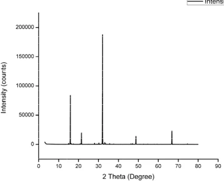
ay Diffraction
crystalline natu ern as given i
irmed by the s action Standard] wn crystals were ined at 350 wh
tals. The next int eak value for the
Study
udy period the ra anesthesia. Kid sed using phosp
e fixed in 10% f nesses using 4 L after staining wi
were focusse and the photogra
sis
serum and urin .D. The results are said to be
iscussion
n [XRD] analys
ure was confirm n figure1. The standard JCPDS
] data [96-900-7 e of struvite type hich is the 100 tensity peak is o e struvite crystals
Ra
ats were euthani ney and liver phate buffer sal
formalin solution Leica RM 2126
th Haematoxylin ed using a m aphs were taken
ne analysis were were done One significant for p
sis
med with the he e peaks in the
S [Joint Commi 7675] and is con e. The highest in % peak value obtained near 15 s.
Fig
ajan
et al
. Inte
ized by cervical were carefully ine (PBS). The n. Section were microtome and n & Eosin (H & microscope of .
e expressed in e- way ANOVA probability value
elp of its XRD e pattern were ttee for Power nfirmed that the ntensity peak is of the struvite 50 which is a 60
gure 2: Optical im
ernational Jou
Optic
The o confirm Figure rectan approx lie in t
mage of the stru
rnal of Phytom
Figure 1: The
cal image ana
optical image o ming the morph e 2. The grown ngular platelet, ximate size of th he range of 0.5
uvite crystals
medicine 6 (4
PA
e XRD pattern o
alysis
f developed str hology of the gro
n crystal exhibi needle type, l he crystals was
cm to 1 cm.
4) 617-624 [2
AGE |
619
|
of the struvite cry
ruvite crystals w own crystals an ts various morp eaf type, dend
found from the
2014]
ystals
In vi
Crys
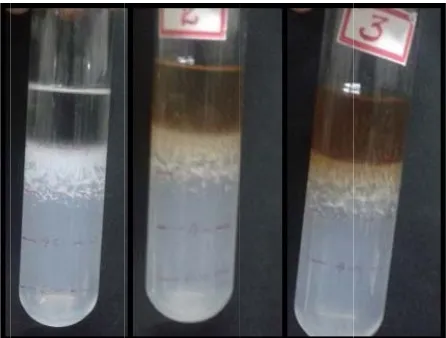
The [TTA inhib show tubes F The secti secti and in th tube liquid Fitro studies on
stal growth ra
crystals were g A], test tube B bitory effect of ws the difference
s according to th
Figure 3: The inv
growth rate of ons of the test on is the margin the drug directly his interface and . The size of the d interface as giv
Figure 4: The va
n the effect of
ate and inhibito
grown in three t [TTB] and test Scoparia Dulcis e in the density o he dosage of a d
vitro growth of st
the crystals w tube for differe nal area where y interacts. Hen d proceeds towa e crystals varies ven in Figure 4.
riation of growth interfac
Ra
f Scoparia Du
ory effect ana
test tubes, nam t tube C [TTC] s Figure 3. The of crystals grown drug applied.
truvite crystals in
was monitored a ent time periods
the gel, outer re ce the crystal g ards the depth
for different tim
h rate of crystals ce
ajan
et al
. Inte
lcis
alysis
ely test tube A ], to study the e figure clearly n in various test
n test tubes
at two different s. The interface eactant solution rowth initializes inside the test me period in
gel-in gel-liquid
ernational Jou
The g dosag and 2 test tu growth measu incorp compa TTC t proves The g interfa the ge of the test tu Fig It was where dissolv fragm After a remov shown shows fragm with th The c due to crysta take p TTB a
rnal of Phytom
growth rate has ge of drug, nam ml dosage of dr ubes the crystals
h rate will decre uring the crysta porated in TTC r
ared to the other that the crystals s that the herbal growth of cryst ace was also me el-liquid interface test tube since ube.
gure 5: The grow
s observed that eas after a time ving in TTB and entation of urina a specific time p ved from all the n in Figure 6. I s more mass c entation and lim he effect of the d crystal collected o the large size o al size decrease place. Hence the and TTC respect
medicine 6 (4
PA
s been compar ely control [TTA ug [TTC]. It sho s will be growing ease in TTB and al size it was p reduces the crys
r two test tubes. s started dissolv l drug used is hig tals at different
easured as sho e, the crystal wil e the gel matrix
wth rate of crysta
the crystal size period the crys d TTC. This pro ary crystals.
period, the crys three tubes and It was observed compared to TT mited crystal grow drug during the t
from TTA show of the crystal. Af es as well as d e mass of the cry
tively.
4) 617-624 [2
AGE |
620
|
ed by incorpora A], 1ml dosage
ws that initially i g rapidly. After 24
d TTC and duri proved that the stal size to the m
After 96 h it wa ving in the gel m
ghly effective. t depth from th own in Figure 5.
ll be grown at di is present all o
als at the depth
e does not vary stals started fra oves the effect
stals from the te their mass was d that the cryst TB and TTC. T wth in test tubes ime of crystal gr ws the higher m fter incorporating dissolution and ystal collected w
2014]
ating different of drug [TTB] in all the three 4 h the crystal ng 68 h while e 2ml dosage maximum than as observed in medium which
he gel- liquid Despite from ifferent depths over inside the
in test tube
much in TTA agmenting and of drug in the
est tubes were s measured as tals from TTA This is due to TTB and TTC rowth. mass or weight
Rajan
et al
. International Journal of Phytomedicine 6 (4) 617-624 [2014]
PAGE |
621
|
Figure 6: The mass of the crystals in various test tubes
Drug Activity
The phytochemical screening was done for the drug using different assays [21] and the qualitative analysis result is given in the Table 2.
Table 2: Qualitative analysis of phytochemicals of the medicinal plant.
Phytoconstituent Present (+) or Absent (-)
Allkaloids +
Saponin + Steroid - Flavanoid +
Tannin +
Statistical Analysis
The One-Way ANOVA test was done to check whether there is a significant difference in the growth rate of crystals in different test
tubes at depth. It gives a probability value 0.0001 [p < 0.05] which proves that there is a highly significant difference in the growth rate of crystals in three test tubes.
In vivo studies on the effect of Scoparia dulcis
Urine Parameters and Analysis
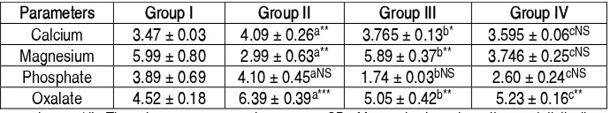
The 24-hr urine has been collected from all the animals of all the groups. The urine parameters were quantitatively analyzed and given in Table 3.
The calcium level has been increased in diseased group II compared to the group I, which occurs due to excessive tubular damage in the kidney, which may lead to excretion of intracellular calcium. However, during the administration of extract the calcium level was managed in Group III and IV, which proves that extract is effective in inhibiting hypercalciuria. Magnesium reduces super saturation and so, it is considered as one of the potent inhibitors of calcium oxalate crystallization [22]. Magnesium level was significantly decreased in Group II animals compared to normal animals, due to metabolic acidosis and super saturation. When the administration of the extract was started, magnesium level was improved in Group III and IV animals in order to reduce the concentration of calcium. The level of magnesium was in preventive group reached almost normal value while the curative therapy showed a mild improvement than the urolithic animals. Hypercalciuria leads to increased phosphate leakage and in the urolithic rats it was only a slight increase in comparison to control rats [23]. In treated rats decreased in level of phosphate was also at par to control rats. Formation of CaOx crystals can be well exhibited from the obvious increase in urinary oxalate level in the ethylene glycol ingested rats compared to control Group I. The effect of the extract can be well demonstrated by its potency in treating hyperoxaluria. This has been proved by the decreased level of oxalate in Group III and IV.
Table 3: The changes in urine parameters in control and experimental animals Parameters Group I Group II Group III Group IV
Calcium 3.47 μ 0.03 4.09 μ 0.26a** 3.765 μ 0.13b* 3.595 μ 0.06cNS
Magnesium 5.99 μ 0.80 2.99 μ 0.63a** 5.89μ 0.37b** 3.746 μ 0.25cNS
Phosphate 3.89 μ 0.69 4.10 μ 0.45aNS 1.74 μ 0.03bNS 2.60 μ 0.24cNS
Oxalate 4.52 μ 0.18 6.39 μ 0.39a*** 5.05 μ 0.42b** 5.23μ 0.16c**
All the parameters were expressed as mg/dL. The values are expressed as mean μ SD of four animals and results are statistically analysed by one way ANOVA
with Bonferroni's multiple comparison post test (n=6). The comparisons are made as follows: ÂaÊ Control Vs Diseased; ÂbÊ Diseased Vs Prevention; ÂcÊ Diseased Vs Treatment. *** p<0.001, ** p<0.01, * P<0.05, NS Not Significant.
Serum Parameters and Analysis
The serum collected from the experimental animals was analyzed and the quantity of various parameters has been calculated and noted in Table 4. The value of urea and creatinine is significantly (p< 0.05) increasing in Group II, III and 1V compared to the control
Rajan
et al
. International Journal of Phytomedicine 6 (4) 617-624 [2014]
PAGE |
622
|
Table 4: The changes in serum parameters in control and experimental animals Parameters Group I Group II Group III Group IV
Urea 29 μ 0.94 53 μ 0.85a*** 31.1 μ 1.12b*** 30.3 μ 0.75c***
Creatinine 0.35 μ 0.06 0.65 μ 0.06a* 0.4 μ 0.07b* 0.35 μ 0.04c*
Calcium 9.6 μ 0.24 8.4 μ 0.38aNS 9 μ 0.40bNS 9.4 μ 0.16cNS
All the parameters were expressed as mg/dL. The values are expressed as mean μ SD of four animals and results are statistically analysed by one way ANOVA
with Bonferroni's multiple comparison post test (n=6). The comparisons are made as follows: ÂaÊ Control Vs Diseased; ÂbÊ Diseased Vs Prevention; ÂcÊ Diseased Vs Treatment. *** p<0.001, ** p<0.01, * P<0.05, NS Not Significant.
Serum calcium level was mildly decreased in the Group II animals compared to the control rats, corresponds to excessive urinary excretion. This was very well improved in group III and IV animals. The calcium level seems to be increasing in the treated groups compared to group II, which proves the effect of inhibition of calcium crystal formation by the extract.
Histopathology studies
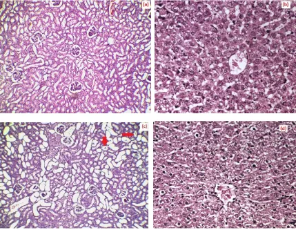
The microscopic images of kidney and liver sections of various groups are given in Figure 7. According to the histopathology observations, the intake of ethylene glycol has induced polymorphic irregular crystals in the kidneys of Group II animals (Figure 7c) with early tubular necrosis.
Rajan
et al
. International Journal of Phytomedicine 6 (4) 617-624 [2014]
PAGE |
623
|
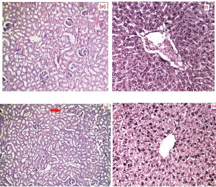
Figure 7: The microscopic images of the histopathological sections of (a) Group I control kidney section (b) Group II control liver section (c) Group II ethylene induced kidney section with crystals marked (d) Group II diseased liver section (e) Group III prophylactic drug effect kidney section (f) Group III liver section (g) Group IV curative effect of drug tested kidney section (h) Group IV liver section.
While the liver section of the diseased animal shown in Figure 7 (d) , demonstrates sinusoidal and venular dilation. In prophylactic treatment, kidney section Figure 7(e) shows significantly reduced tubular vacoulization and crystals deposition compared to the diseased group due to the intake of drug whereas the liver section Figure 7(f) shows a central venular congestion. The curative effect study shows that there is a reduction on number of crystals in the kidney section as shown in Figure 7 (g) and a mild liver degeneration is being detected in the curative treated liver section. The pathological observations in the liver and kidney show the efficacy of the extract in restoring the normal function of liver and kidney, which can be one of the signs in management of hyperoxaluria and urolithiasis. The significant contribution of the bioactive compounds from the plant can be the sole reason for reduction in the disease progression [14].
Conclusion
Rajan
et al
. International Journal of Phytomedicine 6 (4) 617-624 [2014]
PAGE |
624
|
Conflict of Interest
There is no conflict of interests between the authors in submitting and publishing this research article.
References
[1]. Menon MD, Parulkar BG, Drach GW. CampbellÊs Urology, seventh ed.W.B. Saunders Company. New York; 1988. [2]. Pak CYC. Kidney stones. Lancet 1998;
351: 1797-1801.
[3]. Kok DJ. Modulation of calcium oxalate monohydrate crystallization kinetics in vitro. Endocrin Metab Clin 2002; 31:855-867.
[4]. Chow SY, Chen SM, Yang CM, Hsu H. Pharmacological studies on china herbs [I] Hypotensive effect of 30 chinese herbs. J Formosan Med Assoc 1974; 73: 729-739.
[5]. Manjula K, Rajendran K, Eevera T, Kumaran S. Effect of Costus igneus stem extract on calcium oxalate urolithiasis in albino rats. Urol Res 2012; 40: 499 510.
[6]. Viel TA, Domingos CD, Monteiro APS, Lima-Landman MTR, Lapa AJ, Souccar C. Evaluation of the antiurolithiatic activity of the extract of Costus Spiralis Roscoe in rats. J Ethnopharmacol 1999; 66: 193 198. [7]. Grases F, Ramis M, Costa-Bauz A,
March J. G. Effect of Herniaria hirsuta and Agropyron repens on calcium oxalate urolithiasis risk in rats. J Ethnopharmacol 1995; 45: 211-214. [8]. Bayir Y, Halici Y, Kelesc M. S, Colakd
S, Cakire A, Kayad Y, Akcay F. Helichrysum plicatum DC. subsp. plicatum extract as a preventive agent in experimentally induced urolithiasis
model. J Ethnopharmacol 2011;138: 408 414.
[9]. Aggarwal A, Tandon S, Singla S. K, Tandon C. Diminution of Oxalate Induced Renal Tubular Epithelial Cell Injury and Inhibition of Calcium Oxalate Crystallization in vitro by Aqueous Extract of Tribulus terrestris. Int Brazil J Urol 2010; 36: 480-489.
[10].Lin WC, Lai MT, Chen HT, Ho CY, Man KM, Shen JL, Lee YJ, Tsai FJ, Chen YH, Chen WC. Protective effect of Flos carthami extract against ethylene glycol-induced urolithiasis in rats. Urol Res 2012; 40: 665- 661.
[11].Freire SMF, Emim AJS, Lapa AJ, Souccar C, Torres LMB. Analgesic and anti-inflammatory properties of Scoparia dulcis L. extract and glutinol in rodents. Phytother Res 1993; 7: 408-414.
[12].Hayashi T, Kawaski M, Miwa Y, Taga T, Morita N. Antiviral agents of plant origin III. Scopadulin, a novel tetracyclic diterpene from Scoparia dulcis L. Chemical and Pharmaceutical Bulletin 1990; 38: 945-947.
[13].Hayashi T, Asano S, Mizutani M, Takeguchi N, Okamura K, Morita N. Scopadulciol, an inhibitor of gastric H+, K+ ATPase from Scoparia dulcis and its structure activity relationships. J Nat Prod 1991; 54: 802-809.
[14].Hayashi T, Okamura K, Tamada Y, Iida A., Fujita T, Morita N. A new chemotype of Scoparia dulcis. Phytochemistry 1993; 33: 349-352.
[15].Chauhan CK, Joshi MJ, Vaidya ADB. Growth inhibition of struvite crystals by the aqueous root extract of Rotula aquatic. Indian J Biochem Bio 2011; 48: 201-207.
[16].Griffith DP. Struvite. Kidney Int 1978; 13: 372-382.
[17].Coe FL, Parks JH, Asplin JR. Medical process: The pathogenesis and the treatment of kidney stones.The New Eng J Med 1992; 327: 1141-1152. [18].Hesse A and Heimbach D. Causes of
phosphate stone formation and the importance of metaphylaxis by urinary acidification: a review. World J Urol 1999; 17: 308-315.
[19].Takasake E. The neurotoxicity of drugs. Urology Int 1975; 30: 585- 587. [20].Lyon ES, Borden TA, Vermeulen CW.
Experimental oxalate lithiosis produced with ethylene glycol. Invest Urol 1966; 4: 143-151.
[21].Ahmed M, Shikha H.A, Sadhu S.K, Rahman M.T, Datta B.K. Analgesic, diuretic and anti-inflammatory principle from Scoparia Dulcis. Die Pharmazie 2001; 56: 57-60.
[22].Rushton HG and Spector M. Effects of magnesium deficiency on intratubular calcium oalate formation and crystalluria in Hyperoxaluric rats. J Urology. 1982; 127: 598-602.