R E S E A R C H
Open Access
Evolution of retinoic acid receptors in chordates:
insights from three lamprey species,
Lampetra
fluviatilis
,
Petromyzon marinus
, and
Lethenteron
japonicum
Florent Campo-Paysaa
1,2, David Jandzik
3,4, Yoko Takio-Ogawa
5, Maria V Cattell
3,6, Haley C Neef
7,8, James A Langeland
7,
Shigeru Kuratani
5, Daniel M Medeiros
3, Sylvie Mazan
9,10, Shigehiro Kuraku
11,12, Vincent Laudet
1and Michael Schubert
13,14*Abstract
Background:Retinoic acid (RA) signaling controls many developmental processes in chordates, from early axis specification to late organogenesis. The functions of RA are chiefly mediated by a subfamily of nuclear hormone receptors, the retinoic acid receptors (RARs), that act as ligand-activated transcription factors. While RARs have been extensively studied in jawed vertebrates (that is, gnathostomes) and invertebrate chordates, very little is known about the repertoire and developmental roles of RARs in cyclostomes, which are extant jawless vertebrates. Here, we present the first extensive study of cyclostome RARs focusing on three different lamprey species: the European freshwater lamprey,Lampetra fluviatilis, the sea lamprey,Petromyzon marinus, and the Japanese lamprey,Lethenteron japonicum.
Results:We identified fourrarparalogs (rar1,rar2,rar3, andrar4) in each of the three lamprey species, and phylogenetic analyses indicate a complex evolutionary history of lampreyrargenes including the origin ofrar1andrar4by lineage-specific duplication after the lamprey-hagfish split. We further assessed their expression patterns during embryonic development byin situhybridization. The results show that lampreyrargenes are generally characterized by dynamic and highly specific expression domains in different embryonic tissues. In particular, lampreyrargenes exhibit combinatorial expression domains in the anterior central nervous system (CNS) and the pharyngeal region. Conclusions:Our results indicate that the genome of lampreys encodes at least fourrargenes and suggest that the lampreyrarcomplement arose from vertebrate-specific whole genome duplications followed by a lamprey-specific duplication event. Moreover, we describe a combinatorial code of lampreyrarexpression in both anterior CNS and pharynx resulting from dynamic and highly specific expression patterns during embryonic development. This‘RAR code’might function in regionalization and patterning of these two tissues by differentially modulating the expression of downstream effector genes during development.
Keywords:Agnathan, Cyclostome, Developmental patterning, Gene duplication, Gnathostome, RAR code, Retinoid signaling, Vertebrate
* Correspondence:michael.schubert@obs-vlfr.fr
13Sorbonne Universités, UPMC Université Paris 06, UMR 7009, Laboratoire de
Biologie du Développement de Villefranche-sur-Mer, Observatoire Océanologique de Villefranche-sur-Mer, 181 Chemin du Lazaret, 06230 Villefranche-sur-Mer, France
14CNRS, UMR 7009, Laboratoire de Biologie du Développement de
Villefranche-sur-Mer, Observatoire Océanologique de Villefranche-sur-Mer, 181 Chemin du Lazaret, 06230 Villefranche-sur-Mer, France
Full list of author information is available at the end of the article
Background
Developmental functions of vitamin A derivatives (also called retinoids) have been described in great detail in various vertebrate species since the first half of the twen-tieth century [1]. Although a variety of retinoids are de-tectable in vertebrates [2], all-transretinoic acid (RA) is the main biologically active vitamin A derivative during embryogenesis [3-5]. RA signaling is involved in the control of a wide range of biological processes. In the course of vertebrate development, for example, RA con-trols cell proliferation, cell differentiation, apoptosis, and cell survival, acting at different developmental stages, from early gastrulation to late organogenesis, and in all embry-onic tissue layers. Roles for RA signaling during develop-ment have also been characterized in invertebrate chordates, that is, in cephalochordates and tunicates, and it has been shown that RA functions, in particular in de-velopmental patterning, are well conserved, at least be-tween cephalochordates and vertebrates [3].
The molecular response to RA is controlled by heterodi-mers of two members of the nuclear hormone receptor superfamily: the retinoic acid receptor (RAR) and the ret-inoid X receptor (RXR) [6-9]. According to the classical model of RAR/RXR heterodimer function, unliganded heterodimers exert a repressive action on the expression of their targets, while, upon RA binding, the heterodimers recruit co-activators and promote the transcription of tar-get genes [6,9]. RAR/RXR tartar-get gene specificity is medi-ated by the binding of the heterodimer to specific DNA elements in the regulatory regions of target genes, the so-called retinoic acid response elements (RAREs) [10-13].
In vertebrates, whole genome duplication (WGD)
events led to the expansion of the repertoire of rarand
rxr genes [14]. Thus, while the cephalochordate
amphi-oxus possesses only onerarand onerxr, the mouse
gen-ome encodes threerargenes and three rxrgenes, called
rarα, rarβ, and rarγ, and rxrα, rxrβ, and rxrγ,
respect-ively. The rar and rxr repertoires have been expanded
further in some bony fish (the teleosts), whose genomes have undergone an additional round of WGD [15-17].
Thus, the zebrafish genome encodes four rar genes
(rarαa,rarαb,rarγa, andrarγb) and sixrxrgenes (rxrαa,
rxrαb, rxrβa, rxrβb, rxrγa, and rxrγb). During chordate
development, expression of rar and rxr genes is
gener-ally dynamic and detectable in most embryonic tissues
[16-21]. Interestingly, paralogous rar and rxr genes in
jawed vertebrates (that is, gnathostomes) show highly di-verse expression patterns as well as divergent functions during development, indicating that vertebrate-specific genome duplications have mediated lineage-specific di-versification of the developmental processes controlled
by specificrarandrxrgenes [17,21].
Surprisingly, while developmental roles of RA signal-ing have been extensively studied in gnathostomes and
invertebrate chordates, much less is known about RA functions in cyclostomes [22-24], a group of jawless ver-tebrates comprising lampreys and hagfish and represent-ing the phylogenetic sister group of the gnathostomes [25]. Cyclostomes are particularly appealing models for
comparative studies, because they possess many
vertebrate-specific features, such as neural crest deriva-tives, but lack key characters that are present in other vertebrates, such as the jaws [26-28]. In addition to the overall morphology, cyclostome genomes, when com-pared to those of gnathostomes, also exhibit both simi-larities and differences [29]. For instance, while lamprey genomes have very likely experienced the two rounds of WGDs characteristic of vertebrates [30-32], their ge-nomes undergo dramatic remodeling during develop-ment, resulting in the elimination of hundreds of millions of base pairs (bp), including hundreds of genes, from somatic cell lineages [33,34].
In lampreys, some preliminary studies have been car-ried out to investigate the roles of RA signaling during embryonic development and have provided insights into the evolution of RA functions in the vertebrate lineage [22-24]. For example, it has been shown that RA treat-ments during gastrulation induce rostral truncations of both the brain and the pharynx, leading, in the severest cases, to embryos that consist only of trunk segments [22]. Previous work has also suggested that the genomes
of the sea lamprey, Petromyzon marinus, the Japanese
lamprey, Lethenteron japonicum, the Australian lamprey,
Mordacia mordax, as well as of the inshore hagfish, Epta-tretus burgeri, encode at least threerar genes [24,31], al-though phylogenetic analyses have failed to unambiguously assign orthologies between the cyclostome and
gnathos-tome rars [31]. Furthermore, the expression patterns of
lampreyrars have so far only been described for a single
developmental stage, and the functions of these genes in the lamprey embryo still remain elusive [24].
Given the lack of data about the developmental
expres-sion and the functions of lamprey rargenes, we decided
to isolate and characterize therargenes from a third
lam-prey species, the European river lamlam-prey (Lampetra
flu-viatilis). We identified and cloned cDNAs of four L. fluviatilis rar genes and investigated their phylogenetic
relationship to rars of other vertebrates, including two
additional lamprey species (P. marinusandL. japonicum).
Furthermore, we carefully characterized the expression of
the fourL. fluviatilis rargenes in the course of
embryo-genesis and compared the obtained patterns to those of the P. marinus and L. japonicum rar genes. Our results
indicate that lamprey genomes encode at least four rar
overlapping gene expression patterns in both central
ner-vous system (CNS) and pharynx. Therarexpression
do-mains in these embryonic tissues seem to establish a
combinatorial ‘RAR code,’ which might function in
pat-terning and regionalization of the lamprey CNS and phar-ynx. Taken together, this work provides the first detailed
description ofrarexpression during cyclostome
develop-ment and reveals fundadevelop-mental information on the elabor-ation of RA signaling functions following duplicelabor-ation of an ancestralrargene early in vertebrate evolution. Methods
Embryos
Adult male and female L. fluviatilis, P. marinus, and L.
japonicum were collected, respectively, from the Loire river (France), from tributaries of Lake Huron and Lake Michigan (USA), and from the Miomote and Shiribetsu rivers (Japan). Collection and handling of animals was carried out in full compliance of institutional, national, and international guidelines and did not require ap-proval by an ethics committee. After stripping the adults, eggs were artificially fertilized and incubated in filtered water at 12°C (for L. fluviatilis), in 0.1× Marc’s
modified Ringer’s (MMR) buffer [24] at 18°C (for P.
marinus), or in 10% Steinberg’s solution [35] at 16°C to
23°C (forL. japonicum). Embryonic stages were assessed
morphologically according to the developmental table forL. reissneri [36]. Forin situhybridization and immu-nohistochemistry analyses, the embryos were fixed in 4% paraformaldehyde (PFA) in phosphate-buffered saline (PBS), dehydrated in a methanol dilution series, and
stored in 100% methanol at−20°C.
Gene isolation, cloning, and sequencing
The clones ofL. fluviatilis rars were isolated from cDNA
libraries and by polymerase chain reaction (PCR) amplifi-cation. Sequences of the primers used for PCR experiments are available from the authors. Template cDNA was
pre-pared from total RNA extracted from pooledL. fluviatilis
embryos of stages 22 through 26 using the First-Strand cDNA Synthesis Kit (GE Healthcare, Velizy-Villacoublay, France). PCR products were purified with the QIAquick PCR Purification Kit (Qiagen, Courtaboeuf, France) and then cloned into the pCRII-TOPO vector (Invitrogen,
Cergy Pontoise, France). After cloning, the obtainedL.
flu-viatilis rar gene fragments were sequenced on both
strands (Cogenics, Meylan, France). Amplification of 5′
and 3′regions of the original cDNA clones was carried
out by 5′ and 3′ rapid amplification of cDNA ends
(RACE) using the GeneRacer Advanced RACE Kit (Invi-trogen, Cergy Pontoise, France). After cloning, the
ob-tained 5′ and 3′ RACE fragments were sequenced on
both strands (Cogenics, Meylan, France). Altogether, the cloning yielded the followingL. fluviatilis rargene pieces:
rar1 (length 1,116 bp, GenBank accession number
KJ948416), rar2 (length 1,104 bp, GenBank accession
number KJ948417),rar3(length 1,475 bp, GenBank
acces-sion number KJ948418), andrar4(length 1,071 bp,
Gen-Bank accession number KJ948419). Additionally, the
genome sequences of bothP. marinus[32] andL.
japoni-cum[37] were systematically screened for the presence of
rargenes, which led to the identification of theP. marinus
andL. japonicum rar1,rar2,rar3, andrar4genes, which,
with the exception ofL. japonicum rar4, were subsequently
validated by cloning and sequencing, as previously
de-scribed [24,31]. The GenBank accession numbers of theP.
marinusandL. japonicum rar1,rar2,rar3, andrar4genes
are as follows:P. marinus rar1, LC019144 (length 576 bp);
P. marinus rar2, LC019145 (length 1,378 bp); P. marinus
rar3, LC019146 (length 1,247 bp); P. marinus rar4,
LC019147 (length 1,050 bp);L. japonicum rar1, AB292622
(length 1,842 bp); L. japonicum rar2, AB292623 (length
2,753 bp);L. japonicum rar3, AB292624 (length 2,307 bp);
L. japonicum rar4, LC019148 (length 1,077 bp). Sequence analysis and phylogenetic reconstruction
The deduced protein sequences encoded by therargenes
from various animals, including the three lamprey species analyzed in this study, were aligned using MAFFT [38] followed by manual refinement (the alignment is available from the authors upon request), and the phylogenetic tree was inferred with the maximum likelihood (ML) method using PhyML version 3.0 [39] based on 144 gap-free amino acid sites in the alignment. The phylogenetic
infer-ence employed the LG + I +Γ4model of amino acid
sub-stitution and bootstrap resampling (1,000 replicates).
Whole mountin situhybridization
Digoxigenin (DIG)-labeled antisense and sense
ribo-probes of the fourL. fluviatilisandP. marinus rargenes
as well as of the three cloned L. japonicum rar genes
were transcribed using DIG-11-UTP (Roche,
Boulogne-Billancourt, France) according to the manufacturer’s
in-structions. The in situ hybridization experiments for P.
marinusandL. japonicumwere performed as previously
described [24,40]. For L. fluviatilis, fixed embryos were
rehydrated in PBS containing 0.1% Tween 20 (PBT). After rehydration, the embryos were digested for 30 min
in 10μg/ml proteinase K (Sigma-Aldrich, Saint-Quentin
Fallavier, France) in PBT. Subsequently, the embryos were post-fixed for 15 min with PFA/PBT containing 0.2% glutaraldehyde, then washed with PBT and pre-hybridized in hybridization buffer (50% formamide, 1.3×
SSC, 0.2% Tween 20, 50 μg/ml total yeast RNA, 100 μg/
ml heparin sulfate, 5 mM ethylenediaminetetraacetic acid
(EDTA)-Na2, 0.5% CHAPS) for 2 h at 72°C. The
After hybridization, the embryos were washed four times in hybridization buffer for 30 min at 72°C. Subsequently, the solution was substituted gradually with 10 mM
Tris–HCl (pH 8) containing 0.5 M NaCl and 1 mM
EDTA (NTE). RNase A was added to a final
concentra-tion of 100μg/ml, and the specimens were incubated for
30 min at 37°C. The samples were then washed twice with hybridization buffer for 30 min at 65°C and once in 50% hybridization buffer and 50% maleic acid buffer (pH 7.5) with 0.1% Tween 20 (MABT) for 30 min at 65°C. The embryos were blocked with MABT containing 0.5% blocking reagent (Roche, Boulogne-Billancourt, France) and 20% sheep serum for 2 h and developed at 4°C over-night with alkaline phosphatase (AP)-conjugated anti-digoxigenin Fab fragments, diluted 1:2,000 (Roche, Boulogne-Billancourt, France). The embryos were washed ten times for 30 min each in MABT at room temperature and then overnight at 4°C. Subsequently, the embryos were washed twice in 100 mM NaCl, 100 mM Tris
(pH 9.6), 50 mM MgCl2, and 0.1% Tween 20 for 15 min at
room temperature. Alkaline phosphatase activity was de-tected with BM Purple (Roche, Boulogne-Billancourt, France). Stained specimens were fixed in 4% PFA in PBS, dehydrated with a methanol series and clarified with a 1:2 mixture of benzyl alcohol and benzyl benzoate.
Histology
After in situ hybridization, embryos were post-fixed in
4% PFA/PBS (at 4°C, overnight), rinsed in PBS, dehy-drated in methanol (25%, 50%, 75%, 100%, and 100%),
and incubated in methylcyclohexane. L. fluviatilis
em-bryos were subsequently embedded in paraplast and
sec-tioned at 10μm with a microtome, whileP. marinusand
L. japonicum embryos were embedded in gelatin and
sectioned at 10μm using a cryostat [24,41].
Whole mount immunohistochemistry
The fixed embryos stored in 100% methanol were placed in dimethylsulfoxide (DMSO) and methanol (1:1). After washes with Tris-buffered saline (pH 7.6) with 0.1% Tween 20 (TST) containing 5% DMSO (TSTd), the em-bryos were blocked with aqueous 1% periodic acid and 5% nonfat dry milk in TSTd (TSTM). The specimens were incubated for 4 days at room temperature with antibodies directed against acetylated tubulin (Sigma-Al-drich, Saint-Quentin Fallavier, France) diluted 1:1,000 in TSTM. After incubation, the samples were washed with TST and incubated with HRP-conjugated anti-mouse IgG antibodies, diluted 1:200 in TSTM (Invitrogen, Cergy Pontoise, France). After the final wash in TSTd, the embryos were developed with the peroxidase
sub-strate 3,3′-diaminobenzidine (DAB) at 100 mg/ml in
TST for 1 h and subsequently with DAB at 100 mg/ml in TST in the presence of 0.01% hydrogen peroxide.
Results and discussion
Lamprey genomes encode at least fourrargenes
Previous analyses have identified three rar genes (called
rar1,rar2, andrar3) in the genomes of three different
lam-prey species, P. marinus, L. japonicum, and M. mordax
[24,31]. Lamprey rar3 was shown to group with the
gnathostome rarα genes, whilerar2 andrar1could
tenta-tively be associated, respectenta-tively, with the gnathostomerarβ
andrarγgenes [31]. However, the statistical support for this phylogenetic arrangement was not very strong [31]. In this
study, we assessed the rar complement of the European
river lamprey,L. fluviatilis, and successfully identified and
cloned fourrargenes from this lamprey species. The newly
characterized fourth rar gene was subsequently identified
in the genomes of bothP. marinus[32] and L. japonicum
[37] and validated in P. marinus by PCR-based cloning
from cDNA. Following the logic of the nomenclature used in previous studies on lampreyrargenes, this novel, fourth,
lampreyrargene was namedrar4.
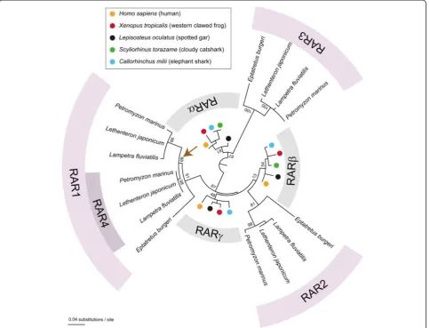
We subsequently carried out a phylogenetic analysis to
address the relationships between the four RARs of L.
fluviatilis,P. marinus, andL. japonicumand those from other animals. Consistent with previously published data [31], the resulting phylogeny recovered the proximities
of gnathostome RARα and cyclostome RAR3 as well as
of gnathostome RARβand cyclostome RAR2 (Figure 1).
In contrast, our analysis did not retrieve the relationship
of gnathostome RARγand cyclostome RAR1 (Figure 1).
More importantly, in our phylogenetic tree, the lamprey RAR1 and RAR4 sequences grouped together at the
ex-clusion of the RAR1 from the inshore hagfish (E.
bur-geri), strongly suggesting that rar1 and rar4 arose by
gene duplication in the lineage leading to extant lam-preys after the hagfish-lamprey split (Figure 1).
Taken together, our phylogenetic data are compatible
with the scenario that lampreyrargenes have undergone
both pan-vertebrate and lamprey-specific gene
duplica-tions. The lamprey rars thus likely belong to the three
gnathostome subtypes (that is,rarα,rarβ, andrarγ), but further bioinformatic analyses will be required to valid-ate the proposed associations of lamprey and
gnathos-tomerarparalogs. Although it was initially believed that
cyclostomes (that is, lampreys and hagfish) might have diverged from other vertebrates (that is, gnathostomes) before the second round of WGD in the vertebrate lineage [42], more recent publications propose instead that cyclostomes may have also undergone the two rounds of WGD [30-32]. Our data on the phylogenetic
relationships of lampreyrargenes are in agreement with
hox gene repertoires have identified lineage-specifichox
cluster duplications in lampreys [37,43-45]. With the
de-scription of two rarγ subtype genes in lampreys, rar1
and rar4, our work adds another example to the list of
genes that underwent a lineage-specific duplication in lampreys.
The lampreyrars are expressed in specific spatiotemporal domains during embryonic development
Following the phylogenetic analysis of lampreyrargenes,
we assessed the temporal and spatial expression patterns of these genes during development. We thus characterized the expression of the four L. fluviatilis rars (rar1, rar2,
rar3, andrar4) byin situhybridization at various stages of embryonic development and carefully mapped the ob-tained signals using as landmarks the expression of marker genes in specific brain regions as well as the immunohisto-chemical signature of neurons in the developing head
(Additional file 1: Figure S1 and Additional file 2: Figure S2). Additionally, we assessed the developmental expres-sion patterns of the fourP. marinus rars (rar1,rar2,rar3, andrar4) as well as of three of the fourL. japonicum rars (rar1,rar2, andrar3) to allow comparisons ofrar expres-sion between the different lamprey species. For studying the developmental patterns of the fourL. fluviatilisandP. marinus rargenes, we focused on embryos from stages 19 (that is, neurula) through 26 (that is, body elongation),
and, for analysis of the domains of the threeL. japonicum
rargenes, we used embryos at body elongation stages 24
through 26 [36].
Expression of lampreyrar1andrar4genes during development
Based on the results of previous phylogenetic analyses
[31], the lamprey rar1 and rar4 genes probably belong
to the gnathostome rarγ subtype and may have arisen
from a lamprey-specific duplication that, according to the results of our phylogenetic inference (Figure 1), probably occurred after the divergence from the hagfish lineage. Despite their relative phylogenetic proximity,
rar1 and rar4 exhibit very different expression patterns
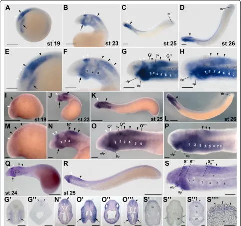
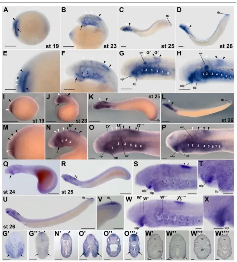
during lamprey development (Figures 2 and 3). InL.
flu-viatilis embryos at stage 19, rar1expression is detected in the hindbrain and anterior spinal cord, with expression in the hindbrain being less conspicuous than in the spinal cord (Figure 2A,E). In the course of development (that is, at stages 23 and 25),L. fluviatilis rar1expression splits into an anterior domain, comprising rhombomeres 1 and 2, and a posterior domain, which includes the anterior spinal cord (Figure 2B,C,F,G). Interestingly, at least the
posterior-most neural expression ofrar1 seems to be slightly more
conspicuous dorsally (Figure 2G”). At stage 26, expression
in the anterior hindbrain is lost, whilerar1transcripts are still detectable in the spinal cord (Figure 2D, H).
Outside the CNS,rar1is dynamically expressed in
sev-eral structures of the developing L. fluviatilis embryo.
For example, at stage 19, expression of rar1 is detected
in the presumptive pharyngeal region (Figure 2A,E).
Pharyngeal expression of rar1 becomes more
conspicu-ous at stage 23, especially in the mandibular region (that is, in the upper and lower lips). Later on, at stages 25
and 26, rar1 is prominently expressed in the pharynx
(Figure 2G’) as well as in the upper and lower lips of the
mouth (Figure 2C,D,G,H). This widespread expression
of rar1 in the pharyngeal region is suggestive of a role
for RA signaling in the patterning and specification of the developing lamprey pharynx, as has recently been
proposed [24]. At stages 25 and 26, rar1 is further
expressed in the tail bud (Figure 2C,D) and, at least at stage 25, in the otic vesicle (Figure 2C,D,G,H).
Comparisons of rar1 expression between the three
different lamprey species, that is, between L. fluviatilis
(Figure 2A-H,G,G’ ”), P. marinus (Figure 2I-P,N’,O’,O”,O”’), and L. japonicum(Figure 2Q-S,S’,S”,S”’,S””), reveal that the expression patterns are generally well conserved. At corre-sponding developmental stages, the main differences are discernable in the otic vesicle, whererar1expression is con-spicuous inL. fluviatilisandP. marinusand likely absent in
L. japonicum. Furthermore, rar1 is also abundantly
expressed in the pharynx of bothL. fluviatilisandP.
mari-nus embryos, with the notable difference of L. fluviatilis
turning on the pharyngeal expression of this gene earlier than P. marinus. Interestingly, in L. japonicum the pharyngeal signal seems to be restricted exclusively to the buccal cavity. Finally, tail bud-associated expression ofrar1
is detectable inL. fluviatilis, inconspicuous inP. marinus,
and likely absent inL. japonicum.
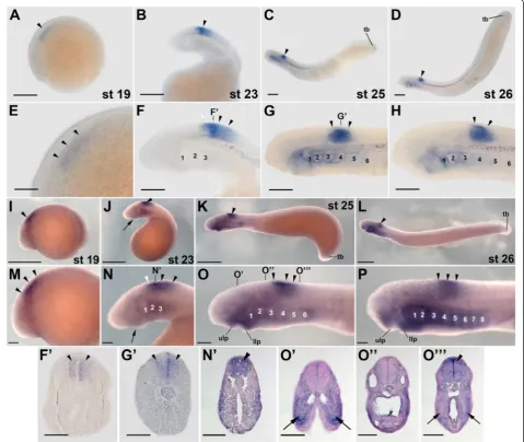
In contrast to the other lampreyrargenes, expression
of rar4 has only been assessed in L. fluviatilis and P.
marinus (Figure 3). In L. fluviatilis, rar4 expression,
from stage 19 through 26, is almost exclusively restricted to the developing CNS, conspicuously in the anterior spinal cord and transiently, at stage 23, in the
posterior-most hindbrain (Figure 3A-H,F’,G’). Cross sections
indi-cate that the gene is homogeneously expressed along the dorsoventral axis of the spinal cord, at least from stage 23
through 26 (Figure 3F’,G’). The only other tissues that
might expressrar4at the assayed stages are the most
pos-terior hindbrain at stages 19 and 23 as well as the anpos-terior pharyngeal gill pouches and the tail bud at stages 25 and 26 (Figure 3C,D,G,H). Of note, the expression domains of
rar4do not seem to change significantly during lamprey
development and are thus remarkably stable over time
(Figure 3A-D). The expression of rar4 in P. marinus
(Figure 3I-P,N’,O’,O”,O”’) generally resembles that ofL. flu-viatilisand suggests that the expression patterns between both lamprey species are well conserved. Slight differences can be observed in the onset of pharyngeal expression,
which seems to be advanced in P. marinus. Thus, in this
species, very weak pharyngeal expression can already be observed at stage 23 (Figure 3J,N), while it only appears inL. fluviatilisat stage 25 (Figure 3C,G).
Taken together, although rar1 and rar4 exhibit
par-tially overlapping expression domains during lamprey development, most conspicuously in the anterior CNS,
rar1 expression is dynamic, whereas rar4 expression is
stable through embryogenesis. This difference suggests that, following the lineage-specific gene duplication, the
regulatory regions ofrar1andrar4have evolved distinct
sets of transcriptional control elements.
In other vertebrates, genes of the rarγ subtype are
expressed in various tissues in the course of development.
For example, in frog and mouse embryos,rarγis expressed
in the tail bud and the pharynx as well as in specific regions of the developing brain [19]. In contrast, developmental ex-pression ofrarγaandrarγbin zebrafish is strikingly
differ-ent fromrarγexpression in other gnathostomes as well as
from that described here for lampreys. Thus, zebrafish
rarγais expressed in a dynamic rhombomere-specific
pat-tern in the anterior CNS, whereas rarγb expression is not
at all detected in hindbrain structures [17,46]. Although certainly not the result of the same gene duplication event, the divergence of therarγaandrarγb expression patterns in zebrafish is nonetheless reminiscent of the situation of
therar1andrar4expression patterns in lampreys.
Follow-ing the gene duplication, the regulatory regions of
zebra-fish rarγa and rarγb thus very likely accumulated
mutations that led to the acquisition of distinct sets of de-velopmental expression domains, which in turn resulted in the preservation of both duplicates in the genome [47].
Expression of lampreyrar2genes during development
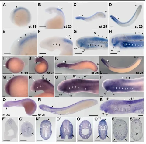
Transcripts of L. fluviatilis rar2, a possible ortholog of
situ hybridization from stage 19 onwards (Figure 4A,E).
Interestingly, the domains of rar2 expression in the L.
fluviatilis CNS do not significantly change between
stages 19 and 26. Thus, rar2 is expressed all along the
CNS, with an anterior limit of expression in the poster-ior hindbrain at the level of rhombomere 6. In addition,
the neural expression of rar2 is homogenous along the
dorsoventral axis, as shown in cross sections at stage 23
(Figure 4B,F,F’). These characteristics of the expression
of the lamprey rar2 are reminiscent of rarβ expression
in frogs and mice, where rarβ is conspicuously
detect-able in the spinal cord and characterized by an anterior limit of expression in the posterior hindbrain [19]. In addition to the spinal cord and the posterior hindbrain,
at stages 25 and 26, L. fluviatilis rar2 is also
promin-ently expressed in the upper and very weakly in the
lower lip of the mouth as well as in the pharyngeal pouches (Figure 4C,D,G,H), which is also comparable to
the expression of rarβin frogs and mice [19]. While, in
stage 25 embryos, the pharyngeal expression ofrar2is
ini-tially separated into two distinct domains, one located
an-teriorly and one posan-teriorly in the pharynx, therar2signal
subsequently expands and becomes detectable throughout
the pharynx at stage 26 (Figure 4C,D,G,H,G’). This
con-spicuous expression of rar2 in the pharynx further
sup-ports the notion that RA signaling might be required for pharyngeal patterning and specification in the lamprey
embryo [24]. Finally, at stages 25 and 26, rar2 is also
expressed inconspicuously in the L. fluviatilis tail bud
(Figure 4C,D).
Comparisons of rar2 expression between L. fluviatilis
(Figure 4A-H,F’,G’), P. marinus(Figure 4I-P,N’,O’,O”,O”’), and L. japonicum (Figure 4Q-S,S’,S”) indicate that the overall patterns are very well conserved, with the main differences being detectable in the pharynx, where both
L. fluviatilis and P. marinus rar2 are expressed in the
pharyngeal pouches, while the L. japonicum rar2 signal
seems to be limited to the pharyngeal territory just an-terior to the heart. Intriguingly, contrary to the situation of L. fluviatilis and P. marinus rar1, but similar to the
situation of L. fluviatilis and P. marinus rar4, the
pharyngeal expression ofL. fluviatilis rar2turns on only
after that of P. marinus rar2. Further rar2 expression
differences between the three lamprey species include an inconspicuous domain in the anterior hindbrain
detect-able exclusively inP. marinusat stages 19 and 23, a
stron-ger mandibular signal in P. marinus, as well as the tail
bud-associated expression domain of this gene, which is obvious inP. marinus, weak inL. fluviatilis, and possibly absent inL. japonicum.
It has previously been proposed that members of the
rarβsubtype have retained expression patterns that
resem-ble the ancestral vertebrate condition [19]. This hypothesis is based on the observation that the domains of both frog
and mouserarβexpression are comparable to those of the
single amphioxus rar. Furthermore, the amphioxus RAR
ligand-binding pocket exhibits a ligand-binding selectivity
that is of a RARβtype, which was thus proposed to be a
chordate synapomorphy [19]. The patterns of lampreyrar2
expression, reported here, closely resemble those of both
frog and mouserarβas well as those of the single
amphi-oxusrar. These data thus seem to confirm the hypothesis
that, of the three gnathostome rar subtypes, the
develop-mental expression, and possibly function, of rarβ genes
most closely approximate the ancestral vertebrate condi-tion. Along these lines, it is interesting to note that the
zeb-rafish genome does not encode any rarβ ortholog, while
the medaka fish genome contains tworarβgenes [48],
in-dicating that the rarβsubtype was specifically lost in the lineage leading to extant zebrafish [16].
Expression of lampreyrar3genes during development
The lamprey rar3 gene is a likely ortholog of the
gnathostome rarαsubtype [31], and its expression in L.
fluviatilis, like the one of rar2, is already detectable by
in situ hybridization in stage 19 embryos. Thus, in stage 19L. fluviatilisembryos,rar3 is expressed in neural tis-sues, more specifically in the presumptive hindbrain and anterior spinal cord, as well as in the future pharynx (Figure 5A,E). Later in development, at stage 23, two
distinct domains of L. fluviatilis rar3expression are
ob-servable in the anterior CNS: the first one in the hind-brain, in rhombomeres 4 and 5, and the second one in the anterior spinal cord (Figure 5B,F). These separate domains likely arose from the unique signal observed earlier in development, through partitioning during elongation of the embryo. In addition to its expression in neural tissues, at stage 23,L. fluviatilis rar3is also de-tectable in the upper and lower lips as well as in the pharynx, both anteriorly in differentiated and posteriorly in presumptive pharyngeal pouches (Figure 5B,F).
At stages 25 and 26, expression of L. fluviatilis rar3in
the anterior CNS does not significantly change from its ex-pression at stage 23 (Figure 5C,D,G,H). Thus, both the do-mains in rhombomeres 4 and 5 and in the anterior spinal
cord are maintained dorsally in the CNS (Figure 5G”), even
though expression in the hindbrain is much less conspicu-ous at stages 25 and 26, when compared to the signal at
stage 23. Furthermore, L. fluviatilis rar3 expression is
strongly induced in the newly formed otic vesicle at stage 25, which closely correlates with the differentiation of this anatomical structure derived from the ectoderm, and the expression is maintained at stage 26 (Figure 5C,D,G,H).
The observation of rar expression in the lamprey otic
vesicle is in accordance with results obtained in other
vertebrate models, where rar genes are specifically
expressed in the developing ear and contribute to a time- and space-dependent activation of the RA (See figure on previous page.)
signaling cascade [20]. Consistent with the signals de-tected at earlier stages, at stages 25 and 26,L. fluviatilis
rar3is still strongly expressed in the pharyngeal region,
in all pharyngeal pouches (Figure 5C,D,G,H). This pharyngeal expression is clearly visible in cross sections of stage 25 larvae (Figure 5G’,G”). Moreover,L. fluviati-lis rar3is also detectable in the mouth region at stages
25 and 26, both in the upper and lower lips (Figure 5C,
D, G, H). As discussed above for rar1 and rar2, the
pharyngeal expression of rar3 is highly suggestive of a
direct implication of RA signaling in patterning and specification of the developing lamprey pharynx [24].
The expression of rar3 is quite well conserved
be-tween L. fluviatilis (Figure 5A-H,G’,G”), P. marinus
(Figure 5I-P,N’,O’,O”,O”’), and L. japonicum (Figure 5Q-X,W’,W”,W”’,W””). As for the other lampreyrargenes, the main differences are detectable in the pharynx. Thus, whilerar3is clearly detectable in the pharyngeal region of bothL. fluviatilisandP. marinus, theL. japonicumpharynx
does not seem to express this gene. InP. marinus,rar3was
shown to be the onlyrargene expressed in the pharyngeal
endoderm [24], which is consistent with our results from the three lamprey species. In addition to the pharynx,
lam-preyrar3genes also seem to be differentially expressed in
the developing otic vesicle. Thus, while the gene is con-spicuously expressed in the otic vesicle ofL. fluviatilis em-bryos, its expression in this structure is only weak in
P. marinusand undetectable byin situhybridization in de-velopingL. japonicum.
When compared to rarα expression in other
verte-brates, the lamprey rar3 pattern shows both similarities
and significant differences. For example, in the frog and
the mouse, rarα is broadly expressed in various
embry-onic tissues during development, including the neuroec-toderm and the mesenchyme of the head [19]. The
expression ofrar3in lampreys, with specific domains in
CNS, pharynx, and otic vesicle, more closely resembles
the situation in zebrafish, where both rarαa and rarαb
are expressed in distinct regions of the developing em-bryo [15,17,46]. Even if only the expression domains of
gnathostome rarα subtype genes in the CNS are
com-pared, despite some conserved domains of expression (for example, in the posterior rhombomeres and the an-terior spinal cord), the overall spatiotemporal dynamics of gene expression seem to vary significantly between different species [17,19,20]. Taken together, these com-parisons suggest that, in the course of vertebrate
evolution, the developmental expression ofrarαparalogs
has been subjected to lineage-specific modifications.
Conclusions
In this study, we have identified four rar genes (rar1,
rar2,rar3, andrar4) in three lamprey species, the
Euro-pean river lamprey, L. fluviatilis, the sea lamprey, P.
marinus, and the Japanese lamprey, L. japonicum, and subsequently analyzed their phylogenetic position in a tree of the RAR subfamily of the nuclear hormone re-ceptors. We further assessed the developmental
expres-sion of therargenes in these three lamprey species.
Our results are compatible with the notion that lam-prey genomes encode orthologs of each of the three gnathostome rar subtypes (rarα, rarβ, and rarγ), hence supporting the hypothesis that lampreys have also undergone the two rounds of WGD that occurred early in vertebrate evolution [31]. Furthermore, the work
re-ported here suggests that lampreys possess two rar
genes (rar1 and rar4) that duplicated after the split of
the lamprey and hagfish lineages.
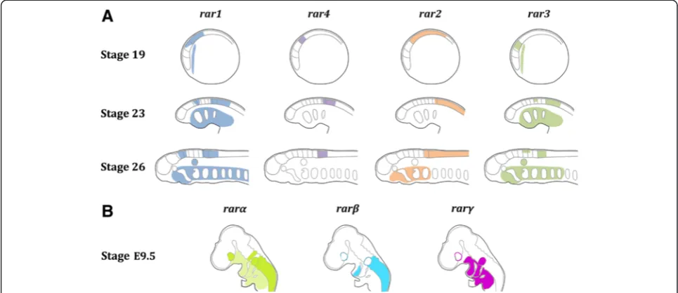
The in situ hybridization experiments indicate that
lamprey rar genes are expressed in very specific
spatio-temporal patterns during development (Figure 6). In
particular, the expression domains of the different rar
genes in the anterior CNS are highly regionalized, and
partially overlapping expression of one or several rar
genes are observable in different regions of the CNS, such as the hindbrain and the anterior spinal cord. This
com-binatorial expression ofrargenes in different domains of
the anterior CNS during neurulation hence creates a‘RAR
code’ defining specific regions of the developing lamprey
brain (Figure 6). In addition to the CNS, a ‘RAR code’
(See figure on previous page.)
might also be observable in the developing pharyngeal re-gion (Figure 6), although characterized by a different
spa-tiotemporal combination ofrarexpression.
Given that RA treatments of developing lamprey em-bryos induce severe rostral truncations, in particular in CNS and pharynx [22], it is very appealing to speculate
that the‘RAR codes’in these two tissues are functionally
required to ensure proper regional patterning and tissue specification. Indeed, in zebrafish, rarαa, rarαb, rarγa,
and rarγb operate combinatorially to pattern the
hind-brain, pharyngeal arches, and pectoral fins [46]. Further-more, several studies have shown that RA signaling is required for the establishment of another gene
expression ‘code’ during chordate development: the
so-called ‘Hox code’ that is crucial for anteroposterior
pat-terning of the embryo [3-5,49] and that has also been
described in the lamprey embryo [50,51]. Given thathox
genes figure prominently amongst the described direct targets of RAR-dependent signaling [3-5], it is very tempting to speculate that at least some of the
func-tional readout of the lamprey ‘RAR code’ is directly
translated into the lamprey‘Hox code’and that this
rela-tionship between the‘RAR code’and the‘Hox code’has
evolved in the last common ancestor of extant verte-brates following the two rounds of WGD.
Comparisons of the developmental expression of rar
genes between vertebrates (Figure 6) reveal that, while
the spatiotemporal patterns of rarα and rarγ subtype
genes display a relatively high degree of diversity
be-tween species, the developmental expression of rarβ
subtype genes is more conserved, at least between lam-preys, frogs, and mice [19]. Previous studies have
pro-posed that vertebraterarβgenes have retained ancestral
chordate characters, both in terms of developmental gene expression and ligand-binding properties of the
re-ceptor [19]. Our analyses of the expression of rargenes
during lamprey development support this hypothesis (Figure 6), which was initially elaborated based on the
characterization of the rar gene of the invertebrate
chordate amphioxus. It is thus likely that, following the two rounds of WGD, which occurred before the cyclostome-gnathostome split, the members of the three
rar subtypes diversified their developmental expression
and functions, with one subtype (rarβ) retaining
ances-tral characteristics and the other two subtypes (rarαand
rarγ) acquiring novel features. In this context, the
emer-gence of ‘RAR codes’in different embryonic tissues may
have resulted in the elaboration of new regulatory circuit-ries supporting the evolution of novel developmental features and hence of morphological innovations.
Figure 6Diagrammatic summary of retinoic acid receptor (rar) expression during development of the European river lamprey,Lampetra fluviatilis,
Additional files
Additional file 1: Figure S1.Precise mapping of retinoic acid receptor (rar) expression in the European river lamprey,Lampetra fluviatilis, by one color doublein situhybridization. The top panel shows expression of each one of the fourrargenes inL. fluviatilisembryos at stage 23. The middle panel displays expression ofkrox20in rhombomeres 3 and 5 in a L. fluviatilisembryo at stage 23. The bottom panel shows expression of each one of the fourrargenes together with that ofkrox20inL. fluviatilis embryos at stage 23. Embryos are oriented with anterior to the left. Scale bars: 50μm.
Additional file 2: Figure S2.Precise mapping of retinoic acid receptor (rar) expression in the European river lamprey,Lampetra fluviatilis, by immunohistochemistry. The top panel shows the expression ofrar4in a L. fluviatilisembryo at stage 25. The bottom panel displays aL. fluviatilis embryo at stage 25 labeled with an antibody directed against acetylated tubulin (Ac-tubulin). The fluorescent signal is detectable in cranial nerves and specific regions of the brain. Embryos are oriented with anterior to the left. Scale bars: 50μm.
Abbreviations
AP:alkaline phosphatase; bp: base pairs; cDNA: complementary
deoxyribonucleic acid; CHAPS: 3-[(3-cholamidopropyl)dimethylammonio]-1-propanesulfonate; CNS: central nervous system; DAB: 3,3′-diaminobenzidine; DIG: digoxigenin; DMSO: dimethylsulfoxide; EDTA: ethylenediaminetetraacetic acid; Fab: fragment antigen-binding; hox: homeobox; HRP: horseradish peroxidase; IgG: immunoglobulin G; MABT: maleic acid buffer (pH 7.5), 0.1% Tween 20; MgCl2: magnesium chloride; ML: maximum likelihood; MMR: Marc’s modified Ringer’s; NaCl: sodium chloride; NTE: 10 mM Tris–HCl (pH 8), 0.5 M NaCl, 1 mM EDTA; PBS: phosphate-buffered saline; PBT: PBS, 0.1% Tween 20; PCR: polymerase chain reaction; PFA: paraformaldehyde; RA: retinoic acid; RACE: rapid amplification of cDNA ends; RAR: retinoic acid receptor; RARE: retinoic acid response element; RNA: ribonucleic acid; RXR: retinoid X receptor; SSC: saline sodium citrate; Tris–HCl: 2-amino-2-(hydroxymethyl)-1,3-propanediol hydrochloride; TST: Tris-buffered saline (pH 7.6), 0.1% Tween 20; TSTd: TST, 5% DMSO; TSTM: TST, 5% DMSO, 1% periodic acid, 5% nonfat dry milk; UTP: uridine triphosphate; WGD: whole genome duplication.
Competing interests
The authors declare that they have no competing interests.
Authors’contributions
FCP performed the experiments onLampetra fluviatilis, compiled figures, and co-wrote the manuscript. DJ and YTO carried out gene expression analyses in Petromyzon marinusandLethenteron japonicum, respectively, compiled figures, and edited the manuscript. MVC, HCN, and JAL contributed to gene cloning and gene expression analyses. SK (Shigeru Kuratani) and SM supported the collection of embryonic material. DMM and VL assisted in the writing of the manuscript. SK (Shigehiro Kuraku) carried out the bioinformatic analyses and edited the manuscript. MS conceived the study, compiled figures, and co-wrote the manuscript. All authors read and approved the final manuscript.
Acknowledgements
This work was supported by research grants from the Agence Nationale de la Recherche to MS (ANR-09-BLAN-0262-02 and ANR-11-JSV2-002-01). The authors are indebted to Dr. Thomas Butts from the Queen Mary University of London (United Kingdom) for critical reading and commenting of the manuscript.
Author details
1Molecular Zoology Team, Institut de Génomique Fonctionnelle de Lyon,
Université de Lyon, Université Lyon 1, CNRS, INRA, Ecole Normale Supérieure de Lyon, 69364 Lyon Cedex 07, France.2MRC Centre for Developmental
Neurobiology, New Hunt’s House, King’s College London, Guy’s Campus, London SE1 1UL, UK.3Department of Ecology and Evolutionary Biology,
University of Colorado Boulder, Ramaley Biology, 1800 Colorado Avenue, Boulder, CO 80309, USA.4Department of Zoology, Comenius University in
Bratislava, Mlynska Dolina B-1, 84215 Bratislava, Slovakia.5Laboratory for Evolutionary Morphology, RIKEN, 2-2-3 Minatojima-minamimachi, Chuo-ku,
Kobe, Hyogo 650-0047, Japan.6Department of Pediatrics, University of
Colorado, Children’s Hospital, 13065 East 17th Avenue, Aurora, CO 80045, USA.7Department of Biology, Kalamazoo College, 1200 Academy Street,
Kalamazoo, Michigan 49008, USA.8Division of Pediatric Gastroenterology, Department of Pediatrics and Communicable Diseases, University of Michigan, C.S. Mott Children’s Hospital, 1540 East Hospital Drive SPC 4259, Ann Arbor, Michigan 48109, USA.9Sorbonne Universités, UPMC Université
Paris 06, FR2424, Station Biologique de Roscoff, Place Georges Teissier, 29680 Roscoff, France.10CNRS, FR2424, Station Biologique de Roscoff, Place Georges
Teissier, 29680 Roscoff, France.11Genome Resource and Analysis Unit, RIKEN Center for Developmental Biology, 2-2-3 Minatojima-minamimachi, Chuo-ku, Kobe, Hyogo 650-0047, Japan.12Phyloinformatics Unit, RIKEN Center for Life Science Technologies, 2-2-3 Minatojima-minamimachi, Chuo-ku, Kobe, Hyogo 650-0047, Japan.13Sorbonne Universités, UPMC Université Paris 06, UMR 7009, Laboratoire de Biologie du Développement de Villefranche-sur-Mer, Observatoire Océanologique de Villefranche-sur-Mer, 181 Chemin du Lazaret, 06230 Villefranche-sur-Mer, France.14CNRS, UMR 7009, Laboratoire de Biologie
du Développement de Villefranche-sur-Mer, Observatoire Océanologique de Villefranche-sur-Mer, 181 Chemin du Lazaret, 06230 Villefranche-sur-Mer, France.
Received: 30 January 2015 Accepted: 20 April 2015
References
1. Gutierrez-Mazariegos J, Theodosiou M, Campo-Paysaa F, Schubert M. Vitamin A: a multifunctional tool for development. Semin Cell Dev Biol. 2011;22:603–10.
2. Kane MA, Napoli JL. Quantification of endogenous retinoids. Methods Mol Biol. 2010;652:1–54.
3. Campo-Paysaa F, Marlétaz F, Laudet V, Schubert M. Retinoic acid signaling in development: tissue-specific functions and evolutionary origins. Genesis. 2008;46:640–56.
4. Duester G. Retinoic acid synthesis and signaling during early organogenesis. Cell. 2008;134:921–31.
5. Rhinn M, Dollé P. Retinoic acid signalling during development. Development. 2012;139:843–58.
6. Gronemeyer H, Gustafsson JA, Laudet V. Principles for modulation of the nuclear receptor superfamily. Nat Rev Drug Discov. 2004;3:950–64. 7. Xu F, Li K, Tian M, Hu P, Song W, Chen J, et al. N-CoR is required for patterning
the anterior-posterior axis of zebrafish hindbrain by actively repressing retinoid signaling. Mech Dev. 2009;126:771–80.
8. Koide T, Downes M, Chandraratna RA, Blumberg B, Umesono K. Active repression of RAR signaling is required for head formation. Genes Dev. 2001;15:2111–21.
9. Evans RM, Mangelsdorf DJ. Nuclear receptors, RXR, and the Big Bang. Cell. 2014;157:255–66.
10. Chambon P. A decade of molecular biology of retinoic acid receptors. FASEB J. 1996;10:940–54.
11. Ross SA, McCaffery PJ, Drager UC, De Luca LM. Retinoids in embryonal development. Physiol Rev. 2000;80:1021–54.
12. Balmer JE, Blomhoff R. A robust characterization of retinoic acid response elements based on a comparison of sites in three species. J Steroid Biochem Mol Biol. 2005;96:347–54.
13. Moutier E, Ye T, Choukrallah MA, Urban S, Osz J, Chatagnon A, et al. Retinoic acid receptors recognize the mouse genome through binding elements with diverse spacing and topology. J Biol Chem. 2012;287:26328–41.
14. Escriva H, Delaunay F, Laudet V. Ligand binding and nuclear receptor evolution. Bioessays. 2000;22:717–27.
15. Hale LA, Tallafuss A, Yan YL, Dudley L, Eisen JS, Postlethwait JH. Characterization of the retinoic acid receptor genesraraa,rarabandrargduring zebrafish development. Gene Expr Patterns. 2006;6:546–55.
16. Bertrand S, Thisse B, Tavares R, Sachs L, Chaumot A, Bardet PL, et al. Unexpected novel relational links uncovered by extensive developmental profiling of nuclear receptor expression. PLoS Genet. 2007;3, e188. 17. Waxman JS, Yelon D. Comparison of the expression patterns of newly identified
zebrafish retinoic acid and retinoid X receptors. Dev Dyn. 2007;236:587–95. 18. Hisata K, Fujiwara S, Tsuchida Y, Ohashi M, Kawamura K. Expression and
19. Escriva H, Bertrand S, Germain P, Robinson-Rechavi M, Umbhauer M, Cartry J, et al. Neofunctionalization in vertebrates: the example of retinoic acid receptors. PLoS Genet. 2006;2, e102.
20. Dollé P. Developmental expression of retinoic acid receptors (RARs). Nucl Recept Signal. 2009;7, e006.
21. Mark M, Ghyselinck NB, Chambon P. Function of retinoic acid receptors during embryonic development. Nucl Recept Signal. 2009;7, e002. 22. Kuratani S, Ueki T, Hirano S, Aizawa S. Rostral truncation of a cyclostome,
Lampetra japonica, induced by all-transretinoic acid defines the head/trunk interface of the vertebrate body. Dev Dyn. 1998;211:35–51.
23. Murakami Y, Pasqualetti M, Takio Y, Hirano S, Rijli FM, Kuratani S. Segmental development of reticulospinal and branchiomotor neurons in lamprey: insights into the evolution of the vertebrate hindbrain. Development. 2004;131:983–95.
24. Jandzik D, Hawkins MB, Cattell MV, Cerny R, Square TA, Medeiros DM. Roles for FGF in lamprey pharyngeal pouch formation and skeletogenesis highlight ancestral functions in the vertebrate head. Development. 2014;141:629–38.
25. Heimberg AM, Cowper-Sal-lari R, Sémon M, Donoghue PC, Peterson KJ. microRNAs reveal the interrelationships of hagfish, lampreys, and gnathostomes and the nature of the ancestral vertebrate. Proc Natl Acad Sci U S A. 2010;107:19379–83.
26. Kuratani S, Kuraku S, Murakami Y. Lamprey as an evo-devo model: lessons from comparative embryology and molecular phylogenetics. Genesis. 2002;34:175–83.
27. Kuratani S, Ota KG. Hagfish (Cyclostomata, Vertebrata): searching for the ancestral developmental plan of vertebrates. Bioessays. 2008;30:167–72. 28. McCauley DW, Kuratani S. Cyclostome studies in the context of vertebrate
evolution. Zoolog Sci. 2008;25:953–4.
29. Smith JJ, Saha NR, Amemiya CT. Genome biology of the cyclostomes and insights into the evolutionary biology of vertebrate genomes. Integr Comp Biol. 2010;50:130–7.
30. Kuraku S. Insights into cyclostome phylogenomics: pre-2R or post-2R. Zoolog Sci. 2008;25:960–8.
31. Kuraku S, Meyer A, Kuratani S. Timing of genome duplications relative to the origin of the vertebrates: did cyclostomes diverge before or after? Mol Biol Evol. 2009;26:47–59.
32. Smith JJ, Kuraku S, Holt C, Sauka-Spengler T, Jiang N, Campbell MS, et al. Sequencing of the sea lamprey (Petromyzon marinus) genome provides insights into vertebrate evolution. Nat Genet. 2013;45:415–21.
33. Smith JJ, Antonacci F, Eichler EE, Amemiya CT. Programmed loss of millions of base pairs from a vertebrate genome. Proc Natl Acad Sci U S A. 2009;106:11212–7.
34. Smith JJ, Baker C, Eichler EE, Amemiya CT. Genetic consequences of programmed genome rearrangement. Curr Biol. 2012;22:1524–9.
35. Steinberg M. A non-nutrient culture medium for amphibian embryonic tissues. Carnegie Inst Wash Year B. 1957;56:347–8.
36. Tahara Y. Normal stages of development in the lamprey,Lampetra reissneri (Dybowski). Zool Sci. 1988;5:109–18.
37. Mehta TK, Ravi V, Yamasaki S, Lee AP, Lian MM, Tay BH, et al. Evidence for at least six Hox clusters in the Japanese lamprey (Lethenteron japonicum). Proc Natl Acad Sci U S A. 2013;110:16044–9.
38. Katoh K, Standley DM. MAFFT multiple sequence alignment software version 7: improvements in performance and usability. Mol Biol Evol. 2013;30:772–80.
39. Guindon S, Dufayard JF, Lefort V, Anisimova M, Hordijk W, Gascuel O. New algorithms and methods to estimate maximum-likelihood phylogenies: assessing the performance of PhyML 3.0. Syst Biol. 2010;59:307–21. 40. Murakami Y, Ogasawara M, Sugahara F, Hirano S, Satoh N, Kuratani S.
Identification and expression of the lampreyPax6gene: evolutionary origin of the segmented brain of vertebrates. Development. 2001;128:3521–31. 41. Kokubo N, Matsuura M, Onimaru K, Tiecke E, Kuraku S, Kuratani S, et al.
Mechanisms of heart development in the Japanese lamprey,Lethenteron japonicum. Evol Dev. 2010;12:34–44.
42. Escriva H, Manzon L, Youson J, Laudet V. Analysis of lamprey and hagfish genes reveals a complex history of gene duplications during early vertebrate evolution. Mol Biol Evol. 2002;19:1440–50.
43. Force A, Amores A, Postlethwait JH.Hoxcluster organization in the jawless vertebratePetromyzon marinus. J Exp Zool. 2002;294:30–46.
44. Irvine SQ, Carr JL, Bailey WJ, Kawasaki K, Shimizu N, Amemiya CT, et al. Genomic analysis of Hox clusters in the sea lampreyPetromyzon marinus. J Exp Zool. 2002;294:47–62.
45. Fried C, Prohaska SJ, Stadler PF. Independent Hox-cluster duplications in lampreys. J Exp Zool B. 2003;299:18–25.
46. Linville A, Radtke K, Waxman JS, Yelon D, Schilling TF. Combinatorial roles for zebrafish retinoic acid receptors in the hindbrain, limbs and pharyngeal arches. Dev Biol. 2009;325:60–70.
47. Force A, Lynch M, Pickett FB, Amores A, Yan YL, Postlethwait J. Preservation of duplicate genes by complementary, degenerative mutations. Genetics. 1999;151:1531–45.
48. Muffato M, Louis A, Poisnel CE, Roest CH. Genomicus: a database and a browser to study gene synteny in modern and ancestral genomes. Bioinformatics. 2010;26:1119–21.
49. Hunt P, Whiting J, Muchamore I, Marshall H, Krumlauf R. Homeobox genes and models for patterning the hindbrain and branchial arches. Dev Suppl. 1991;1:187–96.
50. Takio Y, Kuraku S, Murakami Y, Pasqualetti M, Rijli FM, Narita Y, et al.Hox gene expression patterns inLethenteron japonicumembryos - insights into the evolution of the vertebrate Hox code. Dev Biol. 2007;308:606–20. 51. Parker HJ, Bronner ME, Krumlauf R. AHoxregulatory network of hindbrain
segmentation is conserved to the base of vertebrates. Nature. 2014;514:490–3.
Submit your next manuscript to BioMed Central and take full advantage of:
• Convenient online submission
• Thorough peer review
• No space constraints or color figure charges
• Immediate publication on acceptance
• Inclusion in PubMed, CAS, Scopus and Google Scholar
• Research which is freely available for redistribution