Molecular Targets
Differential Phosphoprofiles of EGF and EGFR
Kinase Inhibitor-Treated Human Tumor Cells
and Mouse Xenografts
David R. Stover,*
,1,3Jennifer Caldwell,
1Jarrod Marto,
1Karen Root,
1Juergan Mestan,
2Michael Stumm,
2Olga Ornatsky,
1Chris Orsi,
1Nina Radosevic,
1Linda Liao,
4Doriano Fabbro,
2and Michael F. Moran
41MDS Proteomics Inc., 1670 Discovery Dr., Charlottesville,VA and 251 Attwell Drive, Toronto M9Q 7H4, Canada
2Oncology Research, Novartis Pharma AG, CH-4002, Basel, Switzerland
3Oncology Research, Novartis Institutes for Biomedical Research, Cambridge, MA 02139
4Banting and Best Department of Medical Research, University of Toronto, 112 College St., Toronto, ON, M5G 1L6, Canada
Copyright ©Humana Press Inc.
All rights of any nature whatsoever are reserved. ISSN 1542-6416/04/01:69–80/$25.00
*Author to whom all correspondence and reprint requests should be addressed:
Abstract
The purpose of this phospho-proteomics study was to demonstrate the broad analysis of cellular protein phosphorylation in cells and tissue as a means to monitor changes in cellular states. As a cancer model, human tumor-derived A431 cells known to express the epidermal growth factor receptor (EGFR) were grown as cell cultures or xenograft tumors in mice. The cells and tumor-bearing animals were subjected to treatments including the EGFR-directed pro-tein kinase inhibitor PK166 and/or EGF stimu-lation. Whole cell/tissue protein extracts were converted to peptides by using trypsin, and phosphorylated peptides were purified by an affinity capture method. Peptides and phospho-rylation sites were characterized and quantified
ier transform mass spectrometry (FTMS); Epider-mal Growth Factor Receptor (EGFR); PKI166.
Introduction
The wide range of cellular processes that are known to involve protein phosphorylation as central elements of their regulation suggest that a clearer picture of these events would be valuable in understanding cellular physiology. Recent reports have highlighted several tech-niques demonstrating, to various degrees, an ability to broadly catalog many protein
phos-phorylation sites (1–3). We have employed a
modified immobilized metal affinity chro-matography (IMAC) approach first reported by Ficarro et al. (1) in a series of differential analyses to identify phosphorylation events that occur following treatment of A431 cells with epidermal growth factor (EGF) or the EGF receptor (EGFR)-directed protein kinase inhibitor, PKI166. By analyzing sample aliquots
representing roughly 106 cell equivalents, 780
unique phosphopeptides from approx 450 dif-ferent proteins were characterized. In these phosphoproteins, all three phospho-amino acids were observed in their expected physio-logical ratios—the great majority being phos-phoserines. Only a small number of these phosphorylation sites have been described pre-viously in the literature. Whereas a targeted analysis of the EGFR pathway was not a spe-cific aim of this study, 22 proteins from the EGFR pathway were identified as being phos-phorylated, including six sites of phosphoryla-tion on the EGFR itself. Three novel sites of phosphorylation were identified on the EGFR, two of which were differentially phosphory-lated in response to EGF and PKI166 treat-ment. Using a custom-built Fourier transform mass spectrometer (FTMS), approx 350 phos-phopeptides were quantified in terms of their relative abundance as a function of growth
factor and drug treatment. Fifty of these pep-tides were found to be differentially phospho-rylated by a significant amount, including differential phosphorylation on the EGFR as noted above. Overall, the results that we report provide a representative subset of the phos-phoproteome that responds to PKI166 treat-ment and is involved in EGFR signaling and further validates the utility of this approach for identifying phosphorylation sites and their involvement in both physiological and patho-physiological processes.
The general schema for differential phos-phoprofiling is depicted in Fig. 1. Protein is extracted from cell or tissue lysates and digested with trypsin. Acidic residues are derivatized to form d0 or d3 labeled methyl esters. For protein identification the individual samples are sub-jected to IMAC to isolate phosphopeptides and analyzed with a 4 h liquid chromatography (LC) gradient on a Q-Star Pulsar using a data dependent tandem mass spectrometry (MS/ MS) algorithm. The data are then searched against public databases using Mascot to iden-tify peptide sequences, sites of phosphorylation, and tentative protein IDs. These data are then manually checked for added veracity.
For determining relative abundance between two samples, differentially labeled samples are pooled prior to IMAC isolation of phospho-peptides and subsequent analysis is performed on a custom-built 7T FTMS. Differentially labeled pairs are identified based on their mass difference of a multiple of three and relative abundance is expressed as a ratio. Phospho-peptide identities are then assigned to specific differential ratios based on alignment of the elution times of abundant marker proteins.
breast, and ovarian carcinomas (4–8). Deregu-lation (constitutive activation) of EGFR is also associated with tumorigenesis and progression
(9). ErbB2, most closely related to EGF-R, is
overexpressed in approx 25% of primary breast and ovarian tumors; ErbB2 overexpres-sion in these two tumor types correlates with
earlier relapse and poor prognosis (10,11). A
number of therapeutic strategies aimed at blocking EGFR activity have been pursued, the most advanced of which include extracel-lularly directed antibodies that block ligand binding, Cetuximab (Erbitux) and ABX-EGF, and small molecule ATP-competitive antago-nists, Iressa (12), and Tarceva (13).
PKI166 is a substituted pyrrolo[2,3-d ]pyrim-idine and is another ATP-competitive EGFR inhibitor, which exhibits dual inhibition of
EGFR and erbB-2 (IC50 = 1 and 11 nM,
respec-tively) (14–16). PKI-166 has shown significant
and dose-dependent in vivo anti-tumor activ-ity in several EGFR expressing xenograft models in nude mice following oral
adminis-tration of 10–100 mg/kg/d(17). Complete and
long-lasting inhibition of EGF-stimulated EGFR autophosphorylation in tumors was observed following administration of a single
100 mg/kg oral dose (18). This study
exam-ines the effect of EGF and PKI166 on the phos-phoprofile of the epidermoid tumor-derived A431 cell line in culture and in xenograft tumors grown in nude mice.
Results
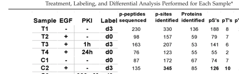
Four samples were prepared from both cul-tured cells and dissected tumors, treated and analyzed as indicated in Table 1.
The results from the phosphopeptide identi-fication analyses are summarized in Table 1. It is important to note that these statistics are based solely on those peptides that were selected (by the MS software) for sequencing and found to have a confirmed sequence from the Mascot search. Roughly equal numbers of phosphory-lation sites were identified from in vivo samples as were found the cell culture samples.
The ratio of the different phosphoamino acids (pS to pT to pY) is very similar between all samples and is consistent with expected physiological ratios. In total, 780 unique phos-phopeptides were sequenced and identified from 441 different proteins. Quantitation ratios for 354 unique phosphopeptides from 117 dif-ferent proteins were generated by matching differentially labeled pairs from the FTMS analysis to corresponding MS/MS data from the Q-Star analysis.
Routinely, greater than 95% of the peptides identified after the modified IMAC enrich-ment procedure are determined to be phos-phorylated. Many more differentially labeled pairs are observed in the FTMS analysis than can be matched to Q-Star identifications, sug-gesting that these numbers still represent just a small percentage of the phosphopeptides.
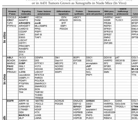
A diverse array of proteins were determined to be phosphorylated, including: signaling molecules, kinase, phosphatases, transcription factors, proteins involved in differentiation and apoptosis, RNA processing proteins, and metabolic enzymes (Table 2). Each protein on this list was identified by MS/MS sequenc-ing of at least one phosphopeptide with a sequence that matches a tryptic peptide within the protein. In many cases multiple phospho-rylation sites were identified for each protein. For example, six sites of phosphorylation were identified within the EGFR itself, including three novel phosphoserine sites.
Whereas the actual identification of any specific phosphoprotein is a statistical event dependent upon selection by the MS software algorithm for sequencing, the numbers of pro-teins identified per class should be roughly proportional to their diversity, abundance, and phosphorylation status. Thus, the representa-tion of phosphoproteins uniquely identified in the cell line samples compared to the tumor samples (Table 2) highlights the greater rep-resentation of desmosomal and gap junc-tion phosphoproteins in the tumor cells. This observed difference can be postulated to reflect the morphology of a solid tumor
com-Treatment, Labeling, and Differential Analysis Performed for Each Sample*
pared to a monolayer of cultured cells, and provides an example to support the notion that broad phosphoprotein analysis offers the potential of an unbiased view of the physio-logical status of a cell or tissue.
Because we compared cell states ±EGF or ±PKI166, we cataloged the phosphoproteins identified that have a known connection to the EGFR signaling pathway (Table 2).
Approxi-mately 22 proteins that are known to be asso-ciated with EGFR signaling were determined to be phosphorylated in at least one sample (shown in bold). This includes EGFR itself and several downstream kinases such as Abl, Pak2, PDK1, and RSK. Eight of these proteins were determined to be differentially phos-phorylated in response to EGF and/or PKI166 treatment.
Table 2
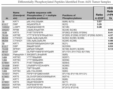
Peptides that were determined to be signif-icantly differentially phosphorylated in at least one of the cell line comparisons are listed in Table 3. A number of phosphopeptides are observed to be more abundant in EGF treated (+) vs untreated (–) A431 cells. Specifically, an EGFR peptide containing two novel phospho-serines is eightfold more abundant in EGF
treated cells, while 300 nM PKI166 (cell 2)
inhibited formation of this phosphopeptide by a factor of 4, suggesting that regulation of S967 and S971 phosphorylation is stimulated by
EGF and inhibited by PKI166. Because PKI166 is a relatively selective EGFR inhibitor, the observed inhibition of phosphorylation of these sites is probably a result of inhibition of EGFR pathways leading to activation of serine kinases and/or serine phosphatases, rather than direct effects on those enzymes.
This same peptide was found to be phos-phorylated on tyrosine, corresponding to EGFR Y974, after treatment with EGF. No signal was detected for this phosphopeptide in the absence of EGF treatment; therefore, the
differential ratio is recorded as greater than the difference between the signal observed in ‘+’ (d3 label) and the background level.
The largest finite difference measured was that of ribosomal protein S6. With almost a 10-fold increase in EGF treated vs nontreated and over a 400-fold increase in cells treated with EGF alone compared to those treated with
EGF and 300 nM PKI166 (Table 3),
phospho-rylation of this peptide is clearly affected by the status of EGFR signaling and modulation by PKI166. The three phosphorylation sites identified on this peptide correspond to phos-phorylation sites on ribosomal protein S6 known to correlate with activation of protein synthesis (19,20).
To highlight one complication of quantify-ing phosphopeptides, it should be pointed out that a related phosphopeptide from RPS6 was determined to be >10-fold more abundant in PKI166 treated cells than in cells treated with EGF alone (differential ratio of 0.07). Because this peptide contains two of the same phosphoserines as the peptide described above (pS236 and pS240), this data would appear to be contradictory. However, upon careful inspection this data is consistent with the expected pattern of EGF stimulation of RPS6 phosphorylation and inhibition of RPS6 phosphorylation by PKI166. Briefly, the RPS6 phosphopeptide carrying two phosphosites is decreased because of increased phosphoryla-tion of a third serine (pS235). This third phos-phorylation also results in a change in the trypsin cleavage site.
ADAM 17 (A disintegrin and
metallopro-teinase domain 17) also known as TNF-α
con-verting enzyme (TACE) has multiple splice variants. One splice variant lacks residues 656...827, we found S786 to be phosphorylated, thereby identifying the presence of the long splice variant. TACE regulates EGFR ligand
availability in vivo (21). We found
phosphory-lation of S786 to be inhibited in the presence of EGF. This suggests the possibility of a novel mechanism regulating TACE mediated pro-duction of EGFR ligands through a feedback mechanism.
Cdc2-related kinase 7 (Crk7) also called CDC2 related kinase with an arginine/serine-rich (RS) domain (CrkRS), is a 1490 amino acid protein that contains RS domains similar to that of RNA splicing factors and co-localizes with a hyperphosphorylated form of RNA Polymerase II in cells and phosphorylates RNA
Polymerase II in vitro (22). Thus Crk7 may
be a link between transcription and splicing machinery. In this study, phosphorylation of Crk 7 on S477, was inhibited by a factor of 3.84
by 300 nMPKI166 treatment. S477 is located
within a PEST-like sequence suggesting a pos-sible role for this phosphorylation in regulat-ing Crk7 degradation in response to EGF.
CDC2-Like kinase 5 (CDC2L5) or CDC2 Related Kinase 5 (Crk5) is the full-length amino acid sequence of the cholinesterase-related cell division controller (CHED) kinase, a previously published partial coding sequence23. This ubiquitous nuclear protein, shares a PITAVRE and PITAIRE motif with other Cdc2-related proteins. Antisense-CHED treatment impairs growth and/or differentiation of hematopoi-etic cells (megakaryocytes), indicating that CHED is a positive growth/differentiation
reg-ulator (24). The kinase domain of CDC2L5 is
89% identical to Crk7 and it also contains a Ser-Arg-rich (RS) domain that may play a role in nuclear localization of the protein. Serines 395, 397, and 400 are localized in the middle of the RS-rich sequence in CDC2L5. Increased phosphorylation of these serines, upon stimu-lation with EGF, may affect the nuclear target-ing necessary for execution of CDC2L5’s role in mitotic regulation.
(liprin), α-1(PPFIA1), is a member of a recep-tor PTPase interacting family that binds to type f receptors such as leukocyte common antigen-related protein (LAR). PPFIA1 binds to the LAR membrane-distal D2 protein tyro-sine phosphatase domain and appears to localize LAR to focal adhesions. Both LAR and PPFIA1 decorate the ends of focal adhesions most proximal to the cell nucleus and are excluded from the distal ends of focal sions, thus localizing to regions of focal adhe-sions presumably undergoing disassembly. Therefore, LAR and PPFIA1 may regulate the disassembly of focal adhesions and thus help
orchestrate cell-matrix interactions. Increased phosphorylation of PPFIA1 in the presence of
300 nM PKI166 is consistent with the large
body of literature claiming a role for EGF sig-naling in down regulating focal adhesions and
thereby stimulating tumor invasion (25,26).
The two phosphopeptides from the tumor extracts determined to be increased with EGF treatment are from Keratin (Krt1) and desmo-plakin (DSP) (Table 4). Significantly, the tumor T3:T4 comparison demonstrated higher lev-els of phosphorylation of many desmosomal and cytoskeletal proteins (DSP, Keratins, and Plakophilins) in tumors from mice treated with
PKI166 for 1 h (T3) as compared to those treated for 24 h (T4). Similarly EMS1 (Cortactin) and catenin D1 (CTNND1) were shown to have higher levels of phosphorylation at the earlier time point. These data suggest that while EGFR itself may be maximally inhibited at 1 h (data not shown), the inhibition of downstream events affecting the cytoskeleton and gap junctions/ desmosomes lags behind. Maximal inhibition of these events may require transcriptional regula-tion, protein turnover, and/or other processes that would follow a different time course than EGFR autophosphorylation.
Cortactin plays a role in regulating cortical actin assembly, transmembrane receptor orga-nization and membrane dynamics. Aberrant regulation of cortactin levels contributes to
tumor cell invasion and metastasis (27).
EGF-stimulation increases phosphorylation of cor-tactin two-fold on Ser/Thr residues. Mek activation is necessary and sufficient for
EGF-induced phosphorylation of Cortactin (28). In
this study, we found increased phosphoryla-tion of Cortactin on S368 upon EGF stimula-tion and reduced phosphorylastimula-tion following treatment with PKI166 in vitro (Table 4). These data are consistent with disruption of the EGFR-MEK-Erk pathway leading to cortical actin rearrangements by PKI166. In vivo phos-phorylation of S380 was elevated in 1 h PKI166-treated mice compared to 24 h post-PKI166 delivery. The time course of phospho-regulation of this protein mirrors that of the cytoskeletal and desmosomal proteins identi-fied consistent with its involvement in regulat-ing actin assembly and membrane dynamics.
P120 Catenin delta 1 (p120ctn) is implicated both in cell transformation by Src and in ligand-induced receptor signaling through the
EGF, PDGF, CSF-1 and Erbb2 receptors (29).
The association of catenins to cadherins pro-duces a complex which is linked to the actin filament network, and which seems to be of primary importance for cadherins
cell-adhe-EGF-induced signaling, three phosphoserines in p120ctn were found to be differentially
reg-ulated in the T3T4 comparison.
This study has further highlighted the util-ity of the IMAC phosphoprofiling method to catalog large numbers of cellular phosphory-lation events. Furthermore, with the addition of the d0/d3 methyl ester labeling step, this method can be used to analyze relative abun-dance of phosphorylations in different cell states. Significantly, this report is the first to demonstrate the utility of this approach to assess in vivo phosphorylation events.
Overall, this approach offers a novel means to analyze protein function and/or drug inter-actions in an unbiased way. By taking a global snapshot of a large number of phosphory-lation events, effects of protein function or drugs can be observed without guessing which pathways you expect might be altered. Once data from such a global analysis has been achieved then more targeted approaches, such as co-immunoprecipitations, can be done to obtain more detailed information within affected pathways.
Experimental Protocol
In Vitro A431 Samples
1 × 10⁷ A431 cells (confluent monolayer in
75 mm dishes: approx 2 ×10⁶ cells per 75 mm
dish) were starved for 16 h in medium con-taining 1% fetal bovine serum (FBS), treated for 60 min with EGF (50 ng/mL) in the
pres-ence or abspres-ence of PKI166 (300 nM, 3000 nM).
Cells were preincubated for 30 min with PKI166 prior to treatment with EGF.
In Vivo A431 Tumor Xenografts
were grown to between 400 and 600 mg in mass. Based upon previous pharmacokinetic determinations PKI166-treated mice received 100 mg/kg po whereas control mice received vehicle (four mice per group). Mice were then sacrificed at 0 h, 1 h, and 24 h after cessation of treatment. Five minutes before sacrifice, the
mice were administered 0.5 µg EGF/g body
weight iv; a group of vehicle-treated control mice received 0.2 mL 0.9% w/v NaCl instead of EGF. The tumors were dissected free of necrotic material and then divided into halves, snap-frozen in liquid nitrogen and stored at –80°C.
Lysis and Protein Extraction
Cells or tumors were lysed and total protein extracted by using 1 mL of TRIzol Reagent
(Invitrogen) per 107 cells or 5:1 vol:wt in the
case of tumors. RNA and DNA were removed by ethanol precipitation and protein was col-lected by isopropanol precipitation. Protein
pellets were washed with 0.3Mguanidine-HCl
in ethanol prior to protein solubilization and analysis. Tryptic digests of protein samples were derivatized in the presence of either d0 or d3 methanol as a means to differentially mass-label peptides, as indicated in Table 1. By this approach, labeled peptide mixtures were combined prior to mass spec analysis, and identical peptides differing by 3 daltons (per esterified methyl group) were identified and quantified for relative abundance.
Sample Preparation
Sample preparation followed the procedure of Ficarro et al. (1). Briefly, the Trizol protein pellets were solubulized in 1% sodium dode-cyl sulfate (SDS) and diluted to 0.2% SDS in digest buffer for digestion overnight with trypsin. The digests were taken to dryness and methyl esters were made using d0 or d3 methanol, as listed in Tables 1 and 2. After esterification the samples were aliquoted and
taken to dryness. Aliquoted samples were stored at –80°C for future use.
IMAC Enrichment
IMAC enrichment followed the procedure of Ficarro et al. (1). Each sample was subjected to IMAC phosphopeptide enrichment. A metal chelating microcapillary column was washed with ethylene diamine tetraaccetic acid (EDTA) and then charged with iron. The column was acidified and the digested sample was loaded to allow the phosphopeptides to remain on the column. Organic washes were used to remove non-phosphorylated peptides and the phosphopeptides were eluted onto a C-18 precolumn with a phosphate wash.
Qualitative MS analysis
The precolumn was attached to a C-18 ana-lytical column with an emitter tip. The whole column is connected to an Agilent 1100 HPLC such that peptides were eluted in to the Sciex Qstar Pulsar mass spectrometer. For a qualita-tive look at the phosphopeptides present in a sample we used a 4 h HPLC gradient where
solvent A was 0.2M acetic acid (HOAc) and
solvent B was 0.2MHOAc in 70% acetonitrile
(MeCN). The mass spectrometer is set to acquire MS/MS spectra on the top three most abundant ions in a MS scan. Once a mass is subjected to MS/MS it is placed on an exclu-sion list for 1 min to allow lesser abundant ions to be selected for MS/MS analysis.
Qualitative Data Analysis
Quantitative MS Analysis
For quantitative analysis another IMAC enrichment was performed, except that two samples were mixed, one d0 and one d3 labeled sample, and then loaded onto the IMAC column for phospho enrichment. All of the peptides were eluted onto a precolumn and eluted into the mass spectrometer. Either the FTMS or the QStar Pulsar was used for this experiment. A custom-built 7T FTMS was used for this study. During gradient elution of the peptides the ions are accumulated in an exter-nal quadrupole and then are pulsed into the trap. Ions are detected for approx 100 ms/s scan event. The current FTMS only produces MS information during the 1 h HPLC gradient. The QStar Pulsar was set to acquire one MS and one MS/MS spectra during a 4 h gradient. All quantitative data is analyzed manually and with software developed at MDS Proteomics.
Acknowledgments
The authors thank Daniel Figeys and Forest White for invaluable technical contributions, and Don Hunt and Jeff Shabanowitz at the University of Virginia for their cooperation and advice related to this study.
References
1. Ficarro SB, McCleland ML, Stukenberg PT, et al. Phosphoproteome analysis by mass spectrometry and its application to Saccha-romyces cerevisae. Nat Biotechnol 2002;19: 01-305.
2. Oda Y, Nagasu T, Chait BT. Enrichment analy-sis of phosphorylated proteins as a tool for probing the phosphoproteome. Nat Biotechnol 2001;19:379-382.
3. Zhou H, Watts JD,Aebersold R. A systematic approach to the analysis of protein phospho-rylation. Nat Biotechnol 2001;19:375-378. 4. Yarden Y. The EGFR family and its ligands in
human cancer. signalling mechanisms and therapeutic opportunities. Eur J Cancer 2001; 37:S3-S8.
5. Mendelsohn J, Baselga J. The EGF receptor family as targets for cancer therapy Oncogene 2000;19,6550-6565.
6. Voldborg B R, Damstrup L, Spang-Thomsen, M. & Poulsen, H. S. Epidermal growth factor receptor (EGFR) and EGFR mutations, func-tion and possible role in clinical trials. Ann Oncol 1997;12:1197-1206.
7. Moscatello DK, Holgado-Madruga M, God-win, AK, et al. Frequent expression of a mutant epidermal growth factor receptor in multiple human tumors. Cancer Res 1995;55:5536-5539. 8. Salomon DS, Brandt R, Ciardiello F, Normanno N. Epidermal growth factor-related peptides and their receptors in human malignancies. Crit Rev Oncol Hematol 1995;19:183-232. 9. Scambia G, Benedetti-Panici P, Ferrandina G,
et al. Epidermal growth factor, oestrogen and progesterone receptor expression in primary ovarian cancer: correlation with clinical out-come and response to chemotherapy. Br J Cancer 1995;72:361-366.
10. Simpson BJ, Phillips HA., Lessels AM, Lang-don SP, Miller WR. c-erbB growth-factor-receptor proteins in ovarian tumours. Int J Cancer 1995;64:202-206.
11. Slamon, D. J. et al. Studies of the HER-2/neu proto-oncogene in human breast and ovarian cancer. Science 1987;235:177-182.
12. Herbst RS. ZD1839: targeting the epidermal growth factor receptor in cancer therapy. Expert Opin. Invest Drugs 2002;11:837-849. 13. Moyer JD, Barbacci EG, Iwata KK, et al.
Induction of apoptosis and cell cycle arrest by CP-358,774, an inhibitor of epidermal growth factor receptor tyrosine kinase. Cancer Res 1997;57:4838-4848.
14. Traxler P, Bold G, Buchdunger E, et al. Tyro-sine kinase inhibitors: from rational design to clinical trials. Med Res Rev 2001;21,499-512. 15. Caravatti G, Bruggen J, Buchdunger E, et al.
Pyrrolo[2,3-d]Pyrimidine and Pyrazolo[3,4-d] Pyrimidine Derivatives as Selective Inhibitors of the EGF Receptor Tyrosine Kinase. ACS Symposium Series, ed. 796: Anticancer Agents. Oxford University Press, USA: 2001;231–244. 16. Bruns CJ, Solorzano CC, Harbison MT, et al.
Blockade of the epidermal growth factor recep-tor signaling by a novel tyrosine kinase inhibitor leads to apoptosis of endothelial cells and therapy of human pancreatic carcinoma. Cancer Res 2000;60,2926-2935.
glands reconstituted with Neu/ErbB2 trans-formed HC11 cells provide a novel orthotopic tumor model for testing anti-cancer agents. Oncogene 2001;20:5459-5465.
18. Baker CH, Solorzano CC, Fidler IJ. Blockade of vascular endothelial growth factor receptor and epidermal growth factor receptor signal-ing for therapy of metastatic human pancre-atic cancer. Cancer Res 2002;62:1996-2003. 19. Ferrari S, Bandi HR, Hofsteenge J, Bussian
BM, Thomas G. Mitogen-activated 70K S6 kinase. Identification of in vitro 40 S riboso-mal S6 phosphorylation sites. J Biol Chem 1991;266(33):22770-22775.
20. Ferrari S, Pearson RB, Siegmann M, Kozma SC, Thomas G. The immunosuppressant rapa-mycin induces inactivation of p70s6k through dephosphorylation of a novel set of sites. J Biol Chem 1993;268(6):4530-4533.
21. Sunnarborg SW, Hinkle CL, Stevenson M, et al. Tumor necrosis factor-αconverting enzyme (TACE) regulates epidermal growth factor receptor ligand availability. J Biol Chem 2002; 277(15):12838-12845 (2002).
22. Ko TK, Kelly E, Pines J. CrkRS: a novel con-served Cdc2-related protein kinase that colo-calises with SC35 speckles. J Cell Sci 2001; 114:2591-2603 (2001).
23. Marques F, Moreau JL, Peaucellier G, et al. A new subfamily of high molecular mass CDC2-related kinases with PITAI/VRE motifs.
832-837 (2000).
24. Lapidot-Lifson Y, Patinkin D, Prody CA, et al. Cloning and antisense oligodeoxynucleo-tide inhibition of a human homolog of cdc2 required in hematopoiesis. Proc Natl Acad Sci USA 1992;89(2):579-583.
25. Hauck CR, Sieg DJ, Hsia DA, Loftus JC, Gaarde WA, Monia BP, Schlaepfer DD. Inhibition of focal adhesion kinase expression or activity dis-rupts epidermal growth factor-stimulated sig-naling promoting the migration of invasive human carcinoma cells. Cancer Res 2001; 61(19):7079-7090.
26. Lu L, Han AP, Chen JJ. Translation initiation control by heme-regulated eukaryotic initia-tion factor 2alpha kinase in erythroid cells under cytoplasmic stresses. Mol Cell Biol 2001; 12:4016-4031.
27. Weed SA, Parsons JD. Cortactin: coupling membrane dynamics to cortical actin assem-bly. Oncogene 2001;20:6418-6434.
28. Campbell DH, Sutherland RL, Daly RJ. Sig-naling pathways and structural domains required for phosphorylation of EMS1/ cortactin. Cancer Res 1999;59:5376-5385. 29. Mariner DJ. Identification of Src
phosphoryla-tion sites in the catenin p120ctn. J Biol Chem 2001;276:28006-28013 .
30. Lu Q. δ-catenin, an adhesive junction-associated protein which promotes cell scattering. J Cell Biol 1999;144:519-532.
19
20
24
25
26
27
28