ORIGINAL ARTICLE
Can centralised care of complex laparoscopic procedures
prevent urinary tract injuries?
H. Morsi&G. Phillips
Received: 2 November 2008 / Accepted: 29 December 2008 / Published online: 15 January 2009
#Springer-Verlag 2009
Abstract Centralised care of complex laparoscopic proce-dures offers expertise and multidisciplinary care. The objective was to identify if centralised care makes urinary tract injuries less likely or avoidable. This Retrospective Audit was performed at a Tertiary Referral Centre for Advanced Laparoscopic Surgery in North East England. The incidence of injury to bladder/ureter, time of diagnosis, instrument, location, side and mode of repair were evaluated in 105 consecutive complex laparoscopic proce-dures. Injuries were identified in three (2.8%) cases. There was one bladder injury which was unavoidable. The bladder dome was opened to allow excision of bladder endometriosis. There were two ureter injuries. The first injury involved the ureter being locked in a vaginal vault stitch. The second injury had stage IV endometriosis with peri-ureteric endometriosis where the anatomy was distorted, with medial displacement and kinking of the ureter secondary to fibrosis at the level of the ureter crossing below the uterine artery, with resultant accidental transection of the ureter close to the uterine artery. The first ureter injury was not a laparoscopic injury but due to vaginal vault closure. Arguably, vault closure in any vaginal hysterectomy could carry the same theoretical risk. The only direct laparoscopic injury was the ureteral
transection. Such cases present a challenge due to a higher chance of anatomical distortion and predisposition to urinary tract injury. Noteworthy here is the fact that the ureter injury occurred where there was probable distortion of the anatomy, due to endometriosis, of the ureter at the level of the uterine artery. These cases are tackled by experienced laparoscopic surgeons in tertiary centres, yet injuries still occur. Is it possible then that those injuries represent a minimum unavoidable injury rate, and are they injuries or in fact unavoidable consequences of such inherently dangerous and difficult surgery?
Keywords Complex laparoscopic procedures . Advanced laparoscopic surgery . Complications . Injury . Ureter . Bladder
Introduction
Gynaecologic laparoscopic surgery (GLS) is becoming more popular. A prime concern with GLS is its safety and the expertise needed to undertake it. There is still the perception that many complications of GLS can or could have been prevented by open surgery. It is unfortunate that the main drive for this perception is historical concerns rather than closer scrutiny and evidence-based analysis.
The urinary tract is vulnerable to injury because of its subtle appearance, its retroperitoneal nature and its long course from the renal pelvis to the bladder. There is a wide variation in reported rates of injury to the urinary tract as a result of GLS [1], mainly due to differences in study design, surgeons’ experience, technique and case complexity.
Centralised care of complex laparoscopic procedures offers the advantages of established advanced laparoscopic H. Morsi (*)
Obstetrics & Gynaecology Department, Russell’s Hall Hospital, Dudley Group of Hospitals NHS Foundation Trust,
Pensnett Road, Dudley, West Midlands DY1 2HQ, UK e-mail: [email protected]
G. Phillips
Obstetrics & Gynaecology Department, The James Cook University Hospital, Marton Road,
skills, advanced multidisciplinary care and an ideal theatre setup. It has been shown that the risk of complications in GLS is inversely proportionate to the experience of the operator [2]. The theatre setup and hospital structure must be capable of adapting to efficient practice of laparoscopic surgery. This keeps risks of complications to a minimum [3] and reduces cost [4]. It is thus reasonable to assume that established operators at minimal access surgery centres already have the experience and the setup to provide safe, specialised advanced laparoscopic surgery.
The aim was to identify the risk of urinary tract injury in complex laparoscopic procedures performed by skilled laparoscopic operators in an advanced laparoscopy centre, and to compare this centre’s practice to published param-eters for prevention/identification of urinary tract injury [5].
Methods
All the procedures were performed at the James Cook University Hospital (JCUH) in Middlesbrough. This is a large teaching hospital with a well-known (nationally and internationally) referral centre for advanced laparoscopic surgery as well as a regional training centre for minimal access surgery. Until the year 2000, the JCUH was the venue of the ‘The WEL Foundation’, a multidisciplinary renowned centre for treatment of endometriosis, which was a registered charity whose aim was to provide safe endoscopic surgery for women.
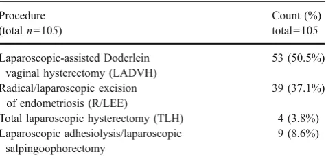
The procedures were identified retrospectively, using the codes of advanced laparoscopic procedures, by searching the operating theatre database. One hundred and five operative procedures were examined. All procedures were performed by experienced laparoscopic surgeons. The notes were examined individually. The individual counts of each procedure are shown in Table1.
Laparoscopic entry and operative techniques were performed using established published techniques [6–9]. All the operations were performed for benign pathology (except for one case with primary peritoneal carcinoma who received four cycles of chemotherapy preoperatively
followed by laparoscopic bilateral salpingoophorectomy). One case had laparoscopic-assisted Doderlein vaginal hysterectomy (LADVH) for atypical endometrial hyperpla-sia and was then found to have FIGO 1B G2 endometrioid adenocarcinoma of the uterus.
The vast majority of the procedures was performed by one operator (93.3%), 1.9% for each of three other operators and 0.9% for one operator. The patients were assessed in terms of surgical risk in view of risk factors; previous surgery, known endometriosis (previous confir-mation by histology), benign ovarian masses, fibroids, pelvic inflammatory disease, previous irradiation or known urogenital congenital anomalies (one case underwent LADVH and was known to have uterus didelphys with a single right kidney with a history of partial nephrectomy for a right duplex system). The documentation during the clinic consultation was assessed in terms of discussion of planned surgery, alternatives offered, risks of surgery, leaflets provided and GP (general practitioner) letter documenta-tion. The operative notes were then analysed for safety rules for entry as per the Middlesbrough consensus document, meticulous technique, documentation of diffi-culties and of identification of the ureters at the start, during and at the end of the procedure, method of ureter identification, time of diagnosis of injury, side, type, site of injury, causative instrument, multidisciplinary input, mode and route of repair, follow-up and sequelae. Where necessary, perioperative illuminated ureteric stents were used with cystoscopy with or without indigo carmine dye, which was included in the preoperative discussion and consenting process.
Results
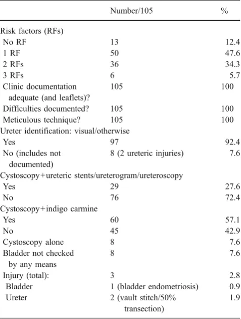
Table 2 shows all the cases needing perioperative ureteric identification. Almost 88% of the procedures had one or more pre-existing risk factors (Table 3), either endome-triosis or adhesions from chronic pelvic inflammatory disease (five cases), previous pelvic surgery (examples include caesarean section, CLAM cystoplasty, colposus-pension, abdominal hysterectomy, previous midline laparotomy for left borderline mucinous ovarian mass) or from advanced endometriosis, large (>5 cm) adherent ovarian endometriomas, benign ovarian masses or uterine fibroids. In 93% of cases, the ureters were identified visually or by an invasive means. There were eight cases where the ureters were not identified or documented presumably due to the surgeon being relatively reassured that the pathology was away from clearly visible, peristaltic ureters. Interestingly, in those cases, there were two ureteric injuries details of which are described below. Eight cases had no form of bladder integrity Table 1 Distribution of procedures
Procedure (totaln=105)
Count (%) total=105
Laparoscopic-assisted Doderlein vaginal hysterectomy (LADVH)
53 (50.5%)
Radical/laparoscopic excision of endometriosis (R/LEE)
39 (37.1%)
Total laparoscopic hysterectomy (TLH) 4 (3.8%) Laparoscopic adhesiolysis/laparoscopic
salpingoophorectomy
check most likely due to uneventful surgery and a reassured surgeon. There were no injuries in this group. Details of the procedures with bladder injury and ureteric injuries are as follows:
Bladder injury
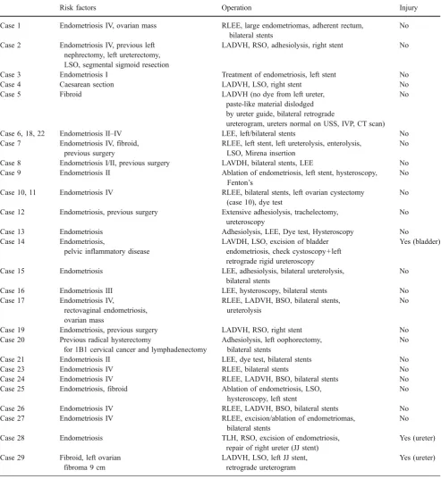
This case was undergoing LADVH and LSO with excision of bladder endometriosis. This patient was known to have Table 2 This table shows the details of all the 29 cases needing preoperative or intraoperative ureteric stenting, ureteroscopy or retrograde ureterogram
Risk factors Operation Injury
Case 1 Endometriosis IV, ovarian mass RLEE, large endometriomas, adherent rectum, bilateral stents
No
Case 2 Endometriosis IV, previous left nephrectomy, left ureterectomy, LSO, segmental sigmoid resection
LADVH, RSO, adhesiolysis, right stent No
Case 3 Endometriosis I Treatment of endometriosis, left stent No
Case 4 Caesarean section LADVH, LSO, right stent No
Case 5 Fibroid LADVH (no dye from left ureter,
paste-like material dislodged by ureter guide, bilateral retrograde
ureterogram, ureters normal on USS, IVP, CT scan) No
Case 6, 18, 22 Endometriosis II–IV LEE, left/bilateral stents No Case 7 Endometriosis IV, fibroid,
previous surgery
RLEE, left stent, left ureterolysis, enterolysis, LSO, Mirena insertion
No
Case 8 Endometriosis I/II, previous surgery LAVDH, bilateral stents, LEE No Case 9 Endometriosis II Ablation of endometriosis, left stent, hysteroscopy,
Fenton’s
No
Case 10, 11 Endometriosis IV RLEE, bilateral stents, left ovarian cystectomy (case 10), dye test
No
Case 12 Endometriosis, previous surgery Extensive adhesiolysis, trachelectomy, ureteroscopy
No
Case 13 Endometriosis Adhesiolysis, LEE, Dye test, Hysteroscopy No Case 14 Endometriosis,
pelvic inflammatory disease
LAVDH, LSO, excision of bladder endometriosis, check cystoscopy+left retrograde rigid ureteroscopy
Yes (bladder)
Case 15 Endometriosis LEE, adhesiolysis, bilateral ureterolysis, bilateral stents
No
Case 16 Endometriosis III LEE, hysteroscopy, bilateral stents No Case 17 Endometriosis IV,
rectovaginal endometriosis, ovarian mass
RLEE, LADVH, BSO, bilateral stents, ureterolysis
No
Case 19 Endometriosis, previous surgery LADVH, RSO, right stent No Case 20 Previous radical hysterectomy
for 1B1 cervical cancer and lymphadenectomy
Adhesiolysis, left oophorectomy, bilateral stents
No
Case 21 Endometriosis II LEE, dye test, bilateral stents No
Case 23 Endometriosis IV RLEE, bilateral stents No
Case 24 Endometriosis IV RLEE, LADVH, BSO, bilateral stents No
Case 25 Endometriosis, fibroid Ablation of endometriosis, LSO, hysteroscopy, left stent
No
Case 26 Endometriosis IV RLEE, LADVH, BSO, bilateral stents No
Case 27 Endometriosis IV RLEE, excision/ablation of endometriomas, bilateral stents
No
Case 28 Endometriosis TLH, RSO, excision of endometriosis, repair of right ureter (JJ stent)
Yes (ureter)
Case 29 Fibroid, left ovarian fibroma 9 cm
LADVH, LSO, left JJ stent, retrograde ureterogram
Yes (ureter)
endometriosis and pelvic inflammatory disease from previ-ous laparoscopies. The patient was counselled appropriately in the clinic. The endometriotic nodule was noted to involve the bladder wall. An intentional cystotomy was performed to allow complete excision of this bladder nodule. It was repaired laparoscopically with the urological surgeon in attendance. The urological surgeon then performed check cystoscopy to verify the integrity of the repair with a left retrograde rigid ureteroscopy which revealed no abnormalities. This patient has suffered no long-term morbidity.
Ureteric injury: case 1
This case was undergoing TLH, RSO and LEE. This patient was nulliparous with narrow vaginal access and known to have stage IV endometriosis. Preoperative counselling was adequate. In the operative notes, the surgeon wrote;‘there was thick nodular endometriosis around the right ureter pulling it medially’. The injury of the right ureter was noted immediately and was caused by the bipolar diathermy and scissors leading to 50% transection. It was erroneously believed to be a blood vessel as the injury of the right ureter
was close to the site of the right uterine artery. The urology team were asked to attend who inserted a JJ-shaped ureteric stent and laparoscopic repair of the right ureter followed. A 10-mm portion of the coagulated ureter ends (till healthy ureter) was excised and tension-free reanastomosis of healthy ureter ends followed using full thickness 3/0 polyglactin sutures. An intravenous urogram 10 days later showed no abnormalities. The stent was removed at 8 weeks and intravenous pyelograms (IVP) at 6 and 12 months were normal. To date, this patient has suffered no long-term morbidity.
Ureteric injury: case 2
This case was undergoing LADVH and LSO. This patient was known to have a fibroid uterus and a 9-cm left ovarian fibroma. Preoperative counselling was adequate. The ureters were identified visually transperitoneally at the start of the operation. At the end of the operation, the ureter integrity was checked by cystoscopy and indigo carmine. It was then noted that there was no dye coming from the left ureteric orifice. The urology team were called who then performed a retrograde ureterogram. The left ureter was blocked possibly due to a uterine pedicle or vaginal vault stitch that had perforated the ureter and locked it. A JJ-shaped stent was placed and the stitch released. Eight weeks later, a retrograde ureterogram revealed no problems and the left ureteric stent was removed. Table 4 shows a summary of the cases of ureteric injury.
Discussion
There is now accumulating evidence for the comparable safety of GLS. A recent publication by the National Institute for Health and Clinical Excellence (NICE) institute on laparoscopic hysterectomy [10] has said that there is now adequate evidence to support the safety, efficacy of laparoscopic techniques for hysterectomy. It has been shown in a meta-analysis that GLS is not inherently dangerous for patients presenting with benign gynae-cological pathologies and that the complication risk should no longer be advanced as an argument against laparoscopic surgery [11].
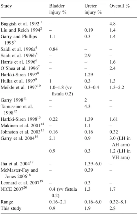
The total rate of injury to the urinary tract in this study was 2.8%. If considered separately, the risk of bladder injury was 0.9% and that of ureter injury was 1.9%. These rates are somewhat higher than published figures (Table5). We, however, have to take into consideration the complex-ity of the cases being studied, most of whom have been referred to this tertiary centre following unsuccessful treatment elsewhere and their pre-existing risk factors. They are therefore more ‘challenging’ cases. Ou et al. Table 3 Adherence to recommendations to prevent/diagnose urinary
tract injury
Number/105 %
Risk factors (RFs)
No RF 13 12.4
1 RF 50 47.6
2 RFs 36 34.3
3 RFs 6 5.7
Clinic documentation adequate (and leaflets)?
105 100
Difficulties documented? 105 100
Meticulous technique? 105 100
Ureter identification: visual/otherwise
Yes 97 92.4
No (includes not documented)
8 (2 ureteric injuries) 7.6
Cystoscopy+ureteric stents/ureterogram/ureteroscopy
Yes 29 27.6
No 76 72.4
Cystoscopy+indigo carmine
Yes 60 57.1
No 45 42.9
Cystoscopy alone 8 7.6
Bladder not checked by any means
8 7.6
Injury (total): 3 2.8
Bladder 1 (bladder endometriosis) 0.9 Ureter 2 (vault stitch/50%
transection)
[12] questioned the existence of a learning curve as they could not find a reduction in operating time in 839 LAVHs undertaken by four surgeons. The explanation for that was that as the surgeon accrues experience, more difficult cases are undertaken. The results of an American survey suggested that complication rates increase with more complex laparoscopic procedures [13]. Furthermore, a noteworthy fact is that the ureter injured in Dorderlein’s hysterectomy was associated with distortion of the pelvic anatomy and probable kinking and displacement of the ureter at the uterine vessel complex. Therefore, based on the above arguments, those cases were more challenging and more prone to injury, despite being operated upon by expert laparoscopic surgeons.
It could be argued that the bladder hole was unavoidable if complete excision of the bladder endometriotic nodule was to be achieved. The alternatives were to avoid surgery or to avoid removing the nodule or to try and remove it without causing a bladder hole. Certainly, avoidance of surgery or leaving the nodule behind would not have treated the patient’s cyclical hematuria. A similar challenge would have been encountered if this procedure was done by open surgery, with the surgeon losing the added benefit of magnification and clear views of organs and anatomical
landmarks with laparoscopy compared to the crude, gross views offered by laparotomy. Relevant disadvantages of laparoscopy in this case are the loss of depth perception and tactile sensation. Nevertheless, the bladder hole was detected immediately and repaired laparoscopically with no added long-term morbidity. A further benefit of this surgery being done in this tertiary centre is the ability to repair this complication laparoscopically.
Both ureters were injured in their distal segment, one on the right side and one on the left side. One case of ureteric injury had stage IV endometriosis. It is known that 38% of ureter injuries occur during treatment of endometriosis [14]. There was clear documentation of the presence of thick nodular endometriosis around the right ureter causing anatomical distortion and pulling the ureter medially. Studies have indicated that remaining strictly within the boundaries of the lateral edge of the uterus medially and a sectioned uterine artery laterally would make injury of the ureter theoretically impossible as it is located outside the uterine artery [5]. In this instance, however, the ureter was
Table 5 Urinary tract complications with laparoscopic surgery reported in different studies
Study Bladder
injury %
Ureter injury %
Overall %
Baggish et al. 19921 – – 4.8 Liu and Reich 19942 – 0.19 1.4 Garry and Phillips
19953
1.1 0.3 1.4
Saidi et al. 1996a4 0.84 – – Saidi et al. 1996b5 – 2.9 – Harris et al. 19966 – – 1.6 O’Shea et al. 19967 – – 2.4 Harkki-Siren 19978 – 1.29 – Hulka et al. 19979 1 0.3 1.3 Meikle et al. 199710 1.0–1.8 (vv
fistula 0.2)
0.3–0.4 1.3–2.2
Garry 199811 – 2 –
Tamussino et al.
199812 –
4.3 –
Harkki-Siren 199813 0.22 1.39 1.61 Makinen et al. 200114 – 1.1 – Johnston et al. 200315 0.16 0.16 0.32 Garry et al. 200416 2.1 0.9 3.0 (LH in
AH arm)
0.9 0.3 1.2 (LH in
VH arm) Jha et al. 200417 – 1.39–6.0 – McMaster-Fay and
Jones 200618
– 0.39 –
Leonard et al. 200719 – 0.3 – NICE 200720 0.4 (vv fistula
0.2)
1.3 1.7
Range 0.16–2.1 0.16–6.0 0.32–8.1
This study 0.9 1.9 2.8
Table 4 Details of the two cases with ureteric injury
Ureteric injury cases
Case 1 Case 2
Surgeon Senior Senior
Procedure LADVH, LSO TLH, RSO, RLEE stage IV Risk factors Fibroid, 9-cm
ovarian fibroma
Stage IV endometriosis
Diagnosis Intraoperative; suspected at check cystoscopy
Intraoperative: transected, ?? blood vessel
Side Left Right
Type Perforation thru and thru/locking
50% transaction
Site Distal ureter close to left vaginal vault
Close to right uterine artery, thick nodular endometriosis
Instrument Vaginal vault stitch Bipolar diathermy, scissors Urology input Yes (intraoperative) Yes (intraoperative) Mode of
repair
Retrograde ureterogram, left JJ stent, vaginal vault stitch release
Right JJ ureteric stent, full thickness repair of ureter, excision, tension-free reanastomosis Route of
repair
Vaginal Laparoscopic
Follow-up 8 weeks 10 days, 8 weeks, 6 months, 12 months Investigations/
sequelae
Cystoscopy, stent removal, left retrograde ureterogram/none
pulled medially, away from its normal position. Indeed, it was mistaken for a blood vessel and was injured close to the uterine artery. The injury, as documented, was 50% transection using bipolar diathermy and scissors. The injury was identified immediately and repaired laparoscopically with the urology team. The immediate identification of the injury, multidisciplinary input and laparoscopic repair all help reduce the impact of the injury on morbidity, recovery time, kidney and ureter function.
Using a retrospective critical analysis of this complica-tion, it is possible that preoperative ureter stent in this particular case might have prevented the ureter injury. The policy in this unit regarding usage of ureteric stents is that of selective use. This depends on the ability to visualise, dissect the ureter, the proximity of the disease to the ureter or if there is significant pelvic sidewall disease at the time of the procedure. Therefore, it is usually a retrospective decision during the laparoscopy. In this case, a problem in ureteric identification was not anticipated and therefore this complication was not expected. The ureter was completely surrounded by disease and its location was distorted as it was pulled medially at the level of the uterine artery. The authors feel that in this particular case, had the ureter been identified, safe removal of disease would have involved leaving residual disease due to the extensive nature of this disease at this critical area. This selective policy of stent usage is based on the surgeon’s expertise, judgement at the time and it is a process that is undergoing continuous modification and review depending on complication rates.
From the authors’point of view, this complication shows that the judgement made at the time was incorrect and that preoperative stent usage in that particular instance might have averted the injury. The learning point for the authors is to have a lower threshold for stent usage in the future.
The other ureteric injury was completely unexpected as the procedure was straightforward. It is possible that the ureteric anatomy was distorted by the fibroid uterus and the ovarian fibroma. The left ureter injury was suspected when indigo carmine dye failed to show at the left ureteric orifice at the time of cystocopy. The left ureter was locked by a uterine pedicle or vaginal vault stitch. Arguably, this complication is not a direct laparoscopic injury and, theoretically speaking, could occur in any vaginal hyster-ectomy. Complications of laparoscopic surgery have been classified as either approach or technique related [15]; this complication does not fit in either category. Had it not been for the check cystoscopy and indigo carmine at the end, this complication would have been missed with disastrous consequences.
The major limitation of this study is that it is retrospective in nature with underestimation of the potential risk (reporting bias) due to subclinical injury or inadvertent omission if the identification mechanism fails. Furthermore,
the sample size was relatively small. Extrapolation of the findings in this study would be difficult for other units as the caseload in this study represents complicated cases referred to skilled laparoscopic surgeons for tertiary care.
Conclusion
Injuries to the urinary tract, albeit some cases have anatomical distortion with increased likelihood of urinary tract injury, still occur despite skilled laparoscopic surgeons undertaking those advanced laparoscopic procedures in a well-established theatre setting. It has already been said that surgery adjacent to the ureter will continue to result in occasional iatrogenic injury [16]. Is it then fair to say that those injuries represent a minimum unavoidable injury rate, and are they injuries or in fact unavoidable consequences of such inherently dangerous surgery? The important issue primarily should be the early recognition and management of urinary tract injuries, yet undoubtedly avoidance, if at all possible, remains the most attractive option.
Acknowledgements The authors are grateful to the theatre database team and the central clinical audit team who provided valuable help in case identification and patients’notes retrieval.
Disclosure of interests None declared
Contribution to authorship Hassan Morsi designed the study, collected and analysed the data and prepared the draft paper. Graham Phillips reviewed the paper and provided corrections until the final draft was achieved.
Ethics approval This was a retrospective audit conducted, within the Trust’s Clinical Audit program, with Trust approval through the Audit Lead and the Clinical Audit department and registered (audit design and results) on the Trust’s Clinical Audit Database. In view of this not being within the confines of research, independent research ethics committee approval was not sought.
Funding None
Declaration The abstract of this paper has been presented as an oral presentation at the 7th annual congress of the RCOG in Montreal Canada on 18.09.2008
References
2. Chapron C, Querleu D, Bruhat M-A, Madelenat P, Fernandez H, Pierre F et al (1998) Surgical complications of diagnostic and operative gynaecological laparoscopy: a series of 29,966 cases. Hum Reprod 13:867–872
3. Pierre F, de Poncheville L, Chapron C (1998) Laparoscopic surgery complication rate should be evaluated in an unselected population of operators: a French survey on gynaecological laparoscopy. Hum Reprod 13:1761
4. Bachmann GA, Trattler B, Ko T, Tweddel G (1998) Operational improvement of gynecologic laparoscopic operating room services: an internal review. Obstet Gynecol 92:142–144 5. Leonard F, Fotso A, Borghese B, Chopin N, Foulot H, Chapron
C (2007) Ureteral complications from laparoscopic hysterectomy indicated for benign uterine pathologies: a 13-year experience in a continuous series of 1300 patients. Hum Reprod 22(7):2006– 2011
6. Garry R (1999) A consensus document concerning laparoscopic entry techniques: Middlesbrough. Gynaecol Endosc 8:403–406 7. The Royal College of Obstetricians and Gynaecologists. Preventing
Entry related gynaecological laparoscopic injury. RCOG Green top guideline No. 49. May 2008. Available from:http://www.rcog.org. uk/resources/Public/pdf/green_top49_PreventingLaparoscopicInjury. pdf
8. Garry R (1994) The evolution of a technique for laparoscopic hysterectomy: laparoscopic-assisted Doderlein’s hysterectomy. Gynaecol Endosc 3:123–128
9. El Bishry G, Phillips G (2006) A new technique for dissecting the bladder laparoscopically. Gynaecol Surg 3:259–263
10. National Institute for Health and Clinical Excellence. Laparoscop-ic techniques for hysterectomy. Interventional procedure guidance 239. November 2007. Available from: http://www.nice.org.uk/ nicemedia/pdf/IPG239Guidance.pdf
11. Chapron C, Fauconnier A, Goffinet F, Bréart G, Dubuisson JB (2002) Laparoscopic surgery is not inherently dangerous for patients presenting with benign gynaecologic pathology. Results of a meta-analysis. Hum Reprod 17:1334–1342
12. Ou CS, Beadle E, Presthus J, Smith M (1994) A multicenter review of 839 laparoscopic-assisted vaginal hysterectomies. J Am Assoc Gynecol Laparosc 1:417–422
13. Hulka JF, Levy BS, Parker WH, Phillips JM (1997) Laparoscopic-assisted vaginal hysterectomy: American Association of Gynecol-ogists’1995 membership survey. J Am Assoc Gynecol Laparosc 4:167–171
14. Weingertner A, Rodriguez B, Ziane A, Gibon E, Thoma V, Osario F et al (2008) The use of JJ stent in the management of deep endometriosis lesion, affecting or potentially affecting the ureter: a review of our practice. BJOG 115:1159–1164
15. Jansen FW, Kapiteyn K, Trimbos-Kemper T, Hermans J, Trimbos JB (1997) Complications of laparoscopy: a prospective multi-centre observational study. BJOG 104:595–600