Iranian Biomedical Journal 11 (3): 161-167 (July 2007)
Distribution of Enterococcal Species and Detection of Vancomycin
Resistance Genes by Multiplex PCR in Tehran Sewage
Fateh Rahimi, Malihe Talebi, Mahnaz Saifi
and Mohammad R. Pourshafie
*Dept. of Microbiology, Pasteur Institute of Iran, Tehran, Iran
Received 24 July 2006; revised 4 December 2006; accepted 2 January 2007
ABSTRACT
Background: Enterococci are important because of their role as the leadingcause of nosocomial infections
which havea significant role in the dissemination and persistence of antimicrobialresistance genes. Methods:
In this study, we determined the distribution of enterococcal species in the sewage treatment plants in Iran. Furthermore, we improved a rapid and specific PCR method using primers (sodA and ddl genes) for
identification of enterococci spp. Results and Conclusion: A total number of 712 enterococci spp. were
isolated and the results showed that 56%, 24%, 12%, 4%, 2%, 1% and 1% isolates were E. faecium, E. hirae, E. faecalis, E. gallinarum, E. casseliflavus, E. mundtii and other enterococcal spp., respectively. The use of
species-specific PCR was in agreement with the biochemical tests. Furthermore, multiplex PCR was developed to study the presence of vancomycin resistant genes in E. faecium or E. faecalis. The multiplex
PCR appeared to be a useful, rapid and specific method for detecting and discriminating genotypes for vancomycin-resistant Enterococcus. Iran. Biomed. J. 11 (3): 161-167, 2007
Keywords: Enterococcus, Vancomycin, PCR, Multiplex PCR, sodA
INTRODUCTION
he major niche of enterococci is the gastrointestinal tract of human and animal where they make up a significant portion of the normal flora [1]. They are released into the environment via the sewage where they can survive for a long period of time [2, 3].
Enterococci, nowadays, represent the second leading cause of nosocomial urinary tract infections and the third leading cause of nosocomial bacteremia [4, 5]. They include 20 species, but most human enterococcal infections are caused by E. faecalis and E. faecium [4, 5]. A few cases of
human infections caused by other enterococci spp. such as E. durans, E. gallinarum and E. casseliflavus have also been reported [4].
Biochemical identification tests to the species level of enterococci are not routinely performed in the clinical laboratories which are very laborious and require time-consuming steps. This, therefore, explain a possible underestimation of the frequency of various enterococcal species in the infections. In order to overcome the problems associated with the biochemical testing, the use of molecular methods
for the identification of enterococcal species has been suggested [6]. Several genes coding heat shock protein 60, elongation factor EF-Tu, D-Ala: D-Ala ligase and manganese-dependent superoxide dismutase have been recommended for the molecular identification of the enterococcal species [6-8]. The use of PCR for identification of genus and species of enterococci has been reported previously [6-8]. However, we report for the first time the distribution of enterococci in Tehran (Iran) sewage followed by a multiplex PCR for rapid identification of Enterococcus spp. and vancomycin
resistant genes, simultaneously.
MATERIALS AND METHODS
Sample collection. Samples were collected ten
times from 2004 to 2005 from four different urban sewagetreatment plants located in different parts of Tehran including Shoosh (south), Jonoob (south), Sahebgharanyeh (north) and Ekbatan (west). At every sewage treatment plant, sampling was carried out on incoming raw sewage (n = 282), outgoing treated sewage (n = 221) and sludge (n = 209). All
T
162 Rahimi et al. Iran. Biomed. J., July 2007
of the samples were collected in the sterile 250 ml bottles and were kept refrigerated and analyzed within 3 h.
Isolation of enterococci. For achieving isolated
colonies, various sample dilutions were filtered with 0.45 µm membrane (Millipore Corporation, Bedford, MA, USA). Outgoing treated sewage was vortexed and filtered directly. The samples taken from sludge and incoming raw sewage were diluted 5-folds with phosphate-buffered saline before filtration as described before by others [2, 3]. The membranes were subsequently transferred in the mEnterococcus agar (Becton Dickinson and Co.,
Sparks, MD, USA) and incubated at 37°C for 48 h. The membranes with well isolated colonies were transferred to bile esculin agar plates and then incubated at 44°C for 2 h. The black isolated colonies were then selected for Gram-staining, growth at 6.5% NaCl, Pyrrolidonyl aminopeptidase (PYR) [9] and catalase tests [10, 11]. The total number of enterococci was recorded and the presumed Enterococcus was defined as the isolates
that grew at 44°C and in 6.5% NaCl, esculin and PYR positive and catalase negative.
Phenotyping of enterococcal isolates. Isolates
exhibiting Enterococcus characteristics were
identified to the species level using the following biochemical tests: acid production of L-arabinose,
lactose, D-sorbitol, D-mannitol, L-sorbose, glucose, methyl-α-D-glucopyranoside, arginine dihydrolase, motility, hippurate hydrolysis, haemolysis, pigmentation, tetrazolium 0.01% and tellurite 0.04% reduction [10, 11].
Antimicrobial susceptibility testing. The
susceptibility tests were performed with disk diffusion method and interpreted according to the guidelines from the Clinical and Laboratory Standards Institute [12]. The following antibiotics were purchased from BD BBL (Becton, Dickinson and Company, Sparks, MD, USA); vancomycin (30 µg), tetracycline (30 µg), gentamicin (120 µg), erythromycin (15 µg), ciprofloxacin (5 µg) and chloramphenicol (30 µg). Minimum inhibitory concentration (MIC) of the vancomycin resistant enterococci (VRE) isolates was determined by using Etest (AB Biodisk, Solna, Sweden). E. faecalis
(ATCC 29212) and E. faecalis (ATCC 51299) were
used as the quality control strains.
PCR and multiplex PCR. The genes encoding
D-alanine-D-alanine ligases specific for E. faecium
(ddl E. faecium) and for E. faecalis (ddl E. faecalis) and the vancomycin resistance genes, vanA and vanB, were
detected by a modified multiplex PCR assay using the primers listed in Table 1. The classical PCR was done using primers (superoxide dismutase genes) specific for E. faecium (sodAE.faecium), E. faecalis
Table 1. Primers used in this study.
Product size (bp) Reference
Sequence (5’-3’) species
658 14
TTGAGGCAGACCAGATTGACG TATGACAGCGACTCCGATTCC
E. faecium
941 14
ATCAAGTACAGTTAGTCT ACGATTCAAAGCTAACTG
E. faecalis
1030 14
CATGAATAGAATAAAAGTTGCAATA CCCCTTTAACGCTAATACGATCAA
vanA
433 14
GTGACAAACCGGAGGCGAGGA CCGCCATCCTCCTGCAAAAAA
vanB
359 This report
CGAATTTAAATTCAGCAATTGA CTTTCCTTCCATCAATGGAG
E. faecium
347 This report
ATGTGACTAACTTAAACGCAG AATCTTGGTTTGGTGTTGAA
E. faecalis
189 6
TTACTTGCTGATTTTGATTCG TGAATTCTTCTTTGAAATCAG
E. gallinarum
186 6
This report TAAATTCTTCCTTAAATGTTG
CTTTCTGATATGGATGCTGT
E. hirae
253 6
This report GCTAGTTTACCGTCTTTAACG
TTAGCAGACTTTTCTTCTGTAC
E. casseliflavus
301 6
This report CAGACATGGATGCTATTCCATCT
AGGTTTCTTGCCTTCCATCAAT
E. mundtii
Iran. Biomed. J., July 2007 Distribution of Enterococal Species 163
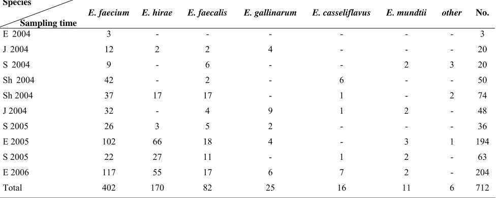
Table 2. Prevalence of enterococci species according to the site of collection.
No. other E. mundtii E. casseliflavus E. gallinarum E. faecalis E. hirae E. faecium Species Sampling time 3 - - - - - -3 E 2004
20 - - - 4 2 2 12 J 2004
20 3 2 - - 6 - 9 S 2004
50 - - 6 - 2 - 42 Sh 2004
74 2 - 1 - 17 17 37 Sh 2004 48 - 2 1 9 4 - 32 J 2004 36 - - - 2 5 3 26 S 2005 194 1 3 - 4 18 66 102 E 2005 63 - 2 1 - 11 27 22 S 2005 204 - 2 7 6 17 55 117 E 2006 712 6 11 16 25 82 170 402 Total
E,Ekbatan; J, Jonoob; Sh, Shoosh; S, Sahebghranyeh sewage treatment plants.
(sodA E. faecalis), E. hirae (sodA E. hira), E. casseliflavus (sodA E. casseliflavus), E. gallinarum (sodA E. gallinarum) and E. mundtii (sodA E. mundtii) listed in Table 1. The following VRE strains carrying vanA and vanB
genes were used as quality control strains; E. faecalis V583 (vanB) and E. faecium BM4147
(vanA). Species identification of the isolates was
done using PCR with genus and species-specific primers (Table 1). For DNA extraction, one isolated colony from each plate was transferred into 200 µl distilled water and boiled at 100°C for 15 min [13]. The mixture was centrifuged and 10 µl of supernatant was used as the DNA template in the PCR mix (for PCR and multiplex PCR) containing 10× PCR buffer, taq DNA polymerase (0.5 U) (HT
Biotechnology, Cambridge, United Kingdom), each primer (1.6 µM), MgCl2 (1.2 µM) and each dNTP (0.64 µM). The PCR cycles for the isolates were as follow: an initial denaturation at 95°C for 4 min, with 30 cycles of denaturation at 95°C for 30 s, annealing at 52°C for 1 min and elongation at 72°C for 1 min and final extension at 72°C for 7 min [14]. PCR products were electrophoresed on a 1.5% agarose gel in a 0.5 X Tris-borate-EDTA buffer and stained in ethidium bromide.
RESULTS
Prevalence and antibiotic resistance. A total of
712 isolates from four different sewage treatment plants in Tehran were identified to the species level using the standard biochemical tests and PCR. Six enterococcal species: E. faecium, E. hirae, E. faecalis, E. gallinarum, E. casseliflavus and E.
mundtii, were isolated from Tehran sewage using
PCR (Fig. 1 and Table 2). The classical PCR using conserved sodA gene which catalyzes the
dismutation of superoxide showed that in some cases distinct bands were not evident for E. hirae and E. gallinarum. On the other hand, a distinct DNA band
for E. mundtii was observed (Fig. 1). These
organisms covered 99% of the total isolated enterococci. Other rare isolates such as E. raffinosus, E. dispar and E. avium with low frequency (1%)
were also obtained. E. faecium were the most
frequently identified Enterococcus spp. (53%)
followed by E. hirae (24%) and E. faecalis (12%). E. faecium was isolated from sewage in every
Fig. 1. PCR for identification of different enterococcal spp.
PCR products were loaded on 1.5% agarose gel. 100 bp DNA ladder (lanes 1 and 8), E. hirae (lane 2), E. gallinarum (lane 3), E. faecalis (lane 4), E. mundtii (lane 5), E. faecium (lane 6) and E. casseliflavus (lane 7).
3000 bp
500 bp
200 bp
1 2 3 4 5 6 7 8
164 Rahimi et al. Iran. Biomed. J., July 2007
Table 3. Antibiotic resistance according to the every sample in E. faecium, E. hirae and E. faecalis isolates (%) no isolates
was detected. Species
Sampling time
E. faecium E. hirae E. faecalis
V Tet Gm E Cip C V Tet Gm E Cip C V Tet Gm E Cip C
E 2004 0 10 0 0 67 33 - - - - - - -
J2004 0 50 0 0 17 42 0 0 0 0 0 0 0 50 0 0 0 0
S2004 0 0 0 67 29 0 - - - 0 83 0 50 0 0
Sh2004 0 19 2 64 10 17 - - - 0 50 0 0 50 0
Sh 2004 8 16 5 32 30 5 0 0 0 0 0 0 0 36 0 0 0 0
J 2004 0 41 0 34 28 19 - - - 0 25 0 25 25 0
S 2005 0 27 0 38 12 12 0 0 0 0 0 0 0 20 0 0 20 0
E 2005 11 16 10 70 38 9 0 5 0 5 2 0 0 6 6 6 17 0
S 2005 18 18 18 55 41 14 0 0 0 0 7 0 0 73 0 0 18 9
E 2006 1 22 1 57 15 0 0 0 0 2 4 0 0 59 6 12 12 6
Species
Sampling time
E. gallinarum E. casseliflavus E. mundtii
V Tet Gm E Cip C V Tet Gm E Cip C V Tet Gm E Cip C
E 2004 - - - - - - - - -
J2004 0 0 0 0 25 0 - - - - - -
S2004 - - - - - - 0 0 0 0 0 0
Sh2004 - - - - - - 0 17 0 67 17 0 - - - - - -
Sh 2004 - - - - - - 0 0 0 0 0 0 - - - - - -
J 2004 0 0 0 0 0 0 0 0 0 0 0 0 0 50 0 0 0 0
S 2005 0 50 0 50 0 0 - - - - - - - - - -
E 2005 0 25 0 25 0 0 - - - 0 67 33 67 67 33
S 2005 - - - - - - 0 0 0 100 0 0 - - - - - -
E 2006 0 33 0 17 33 0 0 43 0 43 14 14 0 0 0 0 50 0
V, vancomycin; Te, tetracycline; Gm, gentamicin; E, erythromycin; Cip, ciprofloxacin; C, chloramphenicol; The symbol (-) denotes no isolates was detected. E, Ekbatan; J, Jonoob; Sh, Shoosh; S, Sahebghranyeh treatment plants.
sampling, E. faecalis in 9, E. hirae in 6, E. gallinarum in 5, E. mundtii and E. casseliflavus were
isolated in 4 out of 10 sampling.
Antimicrobial susceptibility tests showed that the highest level of resistance was observed with erythromycin, tetracycline and ciprofloxacin (Tables 3). E. avium was susceptible to all of the antibiotics
tested here (data not shown). The antibiotic resistance to three major isolates is shown in Table 3. E. faecium, E. faecalis and E. hirae, showed
resistance to more than one antibiotic examined. The level of antibiotic resistance to the least common isolates, E. gallinarum, E. casseliflavus and E. mundtii, was less evident (Table 3).
Antibiotic resistance study showed that all VRE were E. faecium. All 19 VRE isolates were
simultaneously resistant to erythromycin, amikacin and ciprofloxacin. The highest level of resistance was observed with ampicillin (95%), gentamicin (89%) and chloramphenicol (53%). Sixteen percent of the VRE isolates were resistant to all of the antibiotics examined here. The frequency of VRE strains isolated from the three sewage treatment plants was similar to an average of 3% VRE from the total enterococcal isolations.
Multiplex PCR. Since distinct DNA bands were
not observed following amplification of sodA gene,
the gene coding for D-Ala:D-Ala ligase (ddl) was
used in the multiplex PCR. The multiplex PCR was performed and vanA gene was detected in all of the
19 VRE strains. These isolates were highly resistant to vancomycin with MIC≥256 mg/l. The vanB gene
was also detected in 6 of 19 isolates (32%) (Fig. 2). VRE strains (36%) were isolated from Ekbatan treatment plant (west part of Tehran), 21% and 16% were isolated from Sahebgharanyeh (north) and Shoosh (south) treatment plants, respectively.
DISCUSSION
For the first time we have determined the prevalence of enterococci population in the sewage in Iran. Similar to the report by the European study group [3], it was shown here that E. faecium was the
most common enterococcal isolate followed by E. hirae. The presence of E. faecium in every sampling
and sewage treatment plant indicated the capability of this organism to survive and resist in the sewage. Furthermore, it was shown that all of the VRE strains were E. faecium, that have important role to
disseminate and establish antibiotic resistance within the enterococcal populations.
Iran. Biomed. J., July 2007 Distribution of Enterococal Species 165
Fig. 2. Multiplex PCR for detection of vanA and/or vanB genes, and E. faecalis or E. faecium. PCR products were loaded on 1.5%
agarose gel. 100 bp DNA ladder (lane 1), positive control vanB gene alone (lane 2), positive control vanA gene alone (lane 3), E. faecium control without van genes (lane 4), E. faecalis control without van genes (lane 5), E. faecium BM4147 carrying vanB gene
(lane 6), E. faecalis V583carrying vanA gene (lane 7), and E. faecium sample carrying both vanA and vanB genes (lane 8).
Contrary to the report by others [15], we couldn’t isolate certain rare species of enterococci such as E. durans and E. cecorum in the sewage [15]. This
could be due to i) climate differences and ii) bacterial composition in the sewage treatment plants in Tehran, that could justify the differences observed in our study and the studies done in Europe [15].
No other strains beside E. faecium showed high
level of resistance to vancomycin. E. faecalis and E. gallinarum strains only showed intermediate
resistance to vancomycin as determined by disk diffusion assay. However, upon performance of MIC, it was shown that these strains were vancomycin susceptible. The frequency of vancomycin resistance E. faecium isolated in our
study was about 3% from the total isolates. The prevalence of the VRE obtained from the sewage treatment plants varies in different countries. In Spain, for example, much lower frequency VRE (0.4%) has been observed in the waste water [16]; in New Zealand [17], France [13] and USA [1], the percent of VRE isolation from broilers, human fecal and farm wastewater has been 6%, 4% and 6%, respectively. Up to 20% of VRE, however, has been reported in Portugal from hospital sewage samples [18]. Kuhn and her colleagues [3] showed that the frequency of VRE isolated from animals, humans and the environment in different European regions was in the range of 8 to 11%. Collectively, the data suggest that the frequency of VRE varies in different geographical regions and the site of sample collection.
Several investigators have reported the isolation of vancomycin resistant E. faecium, E. faecalis and E. hirae [3, 15]. We, however, could only isolate
vancomycin resistant E. faecium in our samples
which may suggest that the transfer of vancomycin resistant genetic elements to other enterococcal spp., which has been reported extensively elsewhere [19], is uncommon in Iran. Furthermore, the antibiotic resistance pattern of the VRE was very similar among the isolates obtained from the different sewage treatment plants. For example, E. hirae was
almost susceptible to all of the antibiotics examined. Identification of enterococci by using biochemical tests is a tedious process that requires a numerous tests and long times. The use of molecular techniques could, therefore, enhance the identification process. A multiplex PCR assay was developed in this study to enable the identification of vancomycin resistant genes in E. faecium and E. faecalis.
Variations in the sequencesof sodA genes among
enterococcal spp. have been reported [6]. The sod
gene coding for superoxide dismutase was selected as the target for the classical PCR. The results showed that for enterococcal spp. distinct bands could not be observed; therefore, the gene coding for D-Ala:D-Ala ligase (ddl) was used for further
analysis in our multiplex PCR. The result showed that distinct DNA bands could be viewed when multiple primers for ddl genes for E. faecium/E. faecalis and vanA/vanB genes were included in a
single PCR mixture. 3000 bp
1200 bp
500 bp
vanA E. faecalis
E. faecium
vanB
1 2 3 4 5 6 7 8
166 Rahimi et al. Iran. Biomed. J., July 2007
VanA and vanB genes are two of the most common
vancomycin resistant genes which are transferable and are of significant importance in the pathogenicity of the enterococci. VanA confers
high-level resistance to vancomycin and teicoplanin and vanB confers low-level resistance to
vancomycin and no resistance to teicoplanin [2]. Using the multiplex PCR in a single assay we observed that all VRE strains carry vanA gene. We
also found that vanB gene was more prevalent in our
samples than reported by in other countries [1, 2, 4, 18].
Jackson et al. [6] reported the use of 7 different
mixtures of primers for identification of over 20 species of enterococci. It has been suggested that the reason they were not able to perform a single multiplex PCR is because of the inhibitory activity of the primers in the mixture [6]. In the present study, we also observed that the primers for the enterococci genus Ent gene inhibit SodA gene in a
PCR reaction. Therefore, similar to other studies [6] we were not able to perform a single multiplex PCR for all of the enterococcal spp. However, the results of the multiplex for E. faecium and E. faecalis using ddl were satisfactory. Furthermore, a single
annealing temperature (52°C) was found for DNA amplification of all enterococcal spp. which allowed performing PCR at the same time in a single PCR instrument.
In conclusion, we have determined the usefulness of the multiplex PCR method that was optimized in this study for identification of van genes in E. faecium and E. faecalis. This method appears to be
convenient, rapid and simple for identification of the VRE species with the glycopeptide resistance genes in the clinical laboratory.
ACKNOWLEDGMENTS
This work was supported in part by a grant from World Health Organization, Eastern Mediterranean Regional Office, grant no: R6/18/3, Ministry of Health of Iran, Under-Secretariat of Research and Pasteur Institute of Iran. The authors thank Dr. Patrice Courvalin from Pasteur Institute (France) for providing E. faecalis V583, E. faecium BM4147 and E. gallinarum BM14974.
REFERENCES
1. Harwood, J.V., Brownell, M., Perusek, W. and
Whitlock, E.J. (2001) Vancomycin-resistant
enterococcus spp. Isolated from wastewater and
chicken feces in the United States. Appl. Environ.
Microbiol. 67 (10): 4930-4933.
2. Iversen, A., Kuhn, I., Franklin, A. and Mollby, R.
(2002) High prevalence of vancomycin-resistant
enterococci in Swedish sewage. Appl. Environ.
Microbiol. 68: 2838-2842.
3. Kuhn, I., Iversen, I., Finn, M., Greko, C., Burman,
L.G., Blanch, A.R., Vilanova, X., Manero, A., Taylor, H., Caplin, J., Domingues, L. and Mollby, R. (2005) Occurrence and relatedness of vancomycin-resistant enterococci in animals, humans, and the
environment in different european regions. Appl.
Environ. Microbiol. 7: 5383-5390.
4. Liassine, N., Frei, R., Jan, I. and Auckenthaler, R.
(1998) Characterization of glycopeptide-resistant
enterococci from Swiss hospital. J. Clin. Microbiol.
36: 1853-1858.
5. Leclercq, R. and Courvalin, P. (1997) Resistance to
glycopeptides in enterococci. Clin. Infect. Dis. 24:
545-556.
6. Jackson, C.R., Fedorka-Cray, P.J. and Barrett, J.B.
(2004) Use of genus- and species-specific multiplex
PCR for identification of enterococci. J. Clin.
Microbiol. 42: 3558-3565.
7. Goh, S.H., Facklam, R.R., Chang, M., Hill, J.E.,
Tyrrell, G.J., Burns, E.C., Chan, D., He, C., Rahim, T., Shaw, C. and Hemmingsen, S.M. (2000) Identification of enterococcus species and phenotypically similar lactococcus and vagococcus species by reverse checkerboard hybridization to
chaperonin 60 gene sequences. J. Clin. Microbiol.
38: 3953-3959.
8. Ozawa, Y., Courvalin, P. and Gaiimand, M. (2000)
Identification of enterococci at the species level by sequencing of the genes for D-alanine:D-alanine
ligases. Syst. Appl. Microbiol. 23: 230-237.
9. Fertally, S.S. and Facklam, R. (1987) Comparison of
physiologic tests used to identify non-beta-hemolytic
aerococi, enterococci, and streptococci. J. Clin.
Microbiol. 25 (10): 1845-1850.
10. Manero, A. and Blanch R.A. (1999) Identification of
enterococcus spp. With a biochemical key. Appl.
Environ. Microbiol. 65: 4425-4430.
11. Facklam, R.R. and. Collins, M.D. (1998)
Identification of enterococcus species isolated from
human infections by a conventional test scheme. J.
Clin. Microbiol. 27: 731-734.
12. National Committee for Clinical Laboratory
Standards (2001) Performance standards for antimicrobial susceptibility testing, 11th informational supplement, vol. 21. National Committee for Clinical Laboratory Standards, Wayne, Pa, USA.
13. Gambarotto, K., Ploy, M.C., Turlaure, P., Grelaud,
C., Martin, C., Bordessoule, D. and Denis, F. (2000) Prevalence of vancomycin-resistant enterococci in fecal samples from hospitalized patients and
Iran. Biomed. J., July 2007 Distribution of Enterococal Species 167
nonhospitalized controls in a cattle-rearing area of
France. J. Clin. Microbiol. 38: 620-624.
14. Kariyama, R., Mitsuhata, R., Chow, J.W., Clewell,
D.B. and Kumon, H. (2000) Simple and reliable multiplex PCR for surveillance isolates of
vancomycin-resistant enterococci. J. Clin. Microbiol.
38: 3092-3095.
15. Blanch, A.R., Caplin, J.L., Iversen, A., Kuhn, I.,
Manero, A., Taylor, H.D. and Vilanova, X. (2003) Comparison of enterococcal populations related to urban and hospital wastewater in various climatic and
geographic European regions. J. Appl. Microbiol.
94: 994-1002.
16. Torres, C., Reguera, J.A., Sanmartin, M.J.,
Perez-Diaz, J.C. and Baquero, F. (1994) vanA-mediated
vancomycin-resistant enterococcus spp. in sewage.
Antimicrob. Agents Chemother. 33: 553-561.
17. Manson, J.M., Smith, J.M.B. and Cook, G.M. (2004)
Persistence of vancomycin-resistant enterococci in New Zealand broilers after discontinuation of
avoparcin use. Appl. Environ. Microbiol. 70:
5764-5768.
18. Novais, C., Couque, T.M., Ferreira, H., Sousa, J.C.
and Peixe, L. (2005) Environmental contamination with Vancomycin-resistant enterococci from hospital
sewage in Portugal. Appl. Environ. Microbiol. 71:
3364-3368.
19. Barbara, E. and Murray, M.D. (1998) Diversity
among Multidrug-resistant enterococci. Emerg.
Infect. Dis. 4: 37-47.