R E S E A R C H
Open Access
Autogenous fresh demineralized tooth graft
prepared at chairside for dental implant
Eun-Seok Kim
Abstract
Background:This study aimed to evaluate the clinical usefulness of autogenous fresh demineralized tooth (auto-FDT) graft prepared at the chairside for alveolar bone grafting during dental implant surgery.
Methods:In total, 38 patients requiring both tooth extraction (for endodontic or periodontal reasons or third molar extraction) and alveolar bone regeneration for dental implant placement were included. Within 2 h after clean extraction, the teeth were prepared at the chairside to serve as bone graft material. In the same sitting, blocks or chips of this graft material were used to reconstruct defects at the osteotomy site simultaneously with or before implant placement. Twelve months after prosthesis fabrication and placement, the clinical findings and implant success rates were evaluated. Histological studies were randomly conducted for selected cases.
Results:Clinical evaluation showed favorable wound healing with minimal complications and good bone support for the implants. No implant was lost after 12 months of function following prosthetic rehabilitation. Histological
examination revealed new bone formation induced by the graft material.
Conclusions:Chairside preparation of autogenous fresh demineralized teeth after extraction can be a useful alternative to the use of autogenous bone or other graft materials for the immediate reconstruction of alveolar bone defects to facilitate subsequent implant placement.
Keywords:Dental implant; Bone graft; Autogenous fresh demineralized tooth; Auto-FDT; Tooth osteoplantation
Background
Although autogenous bone is considered the gold stand-ard among graft materials, donor site morbidity may be an associated problem. Allogenic, xenogenic, and allo-plastic graft materials have been used as alternatives, but they have a number of drawbacks compared with autolo-gous grafts, such as decreased function, the potential risk of infectious disease, an unsatisfactory resorption pattern, a prolonged healing time, and high cost [1]. An ideal bone graft material should stabilize the blood clot; provide a biomechanical scaffold for cell migration, pro-liferation, and differentiation; contain functional proteins and peptides, such as growth factors; and exhibit appro-priate resorption and remodeling during new bone for-mation. Materials based on collagen, particularly type I collagen, have attracted attention because of their ability to improve the cellular responses of osteogenic lineages, thus ensuring better bone regeneration [2]. However, the
mineral component providing the biomechanical back-bone remains an important factor for cell differentiation, nidus of calcification, and space maintenance during new bone formation [3]. Considering the hierarchical structure of bone, a mineralized collagen matrix with functional proteins and proper fibrillar arrays for bio-mechanics may be suitable as a natural bio-inspired graft material in bone tissue engineering [4].
The tooth is increasingly attracting attention as a ma-terial for alveolar bone regeneration. It is composed of an organic matrix and an inorganic reinforcing phase of hydroxyapatite. Radial arrays of dense type I collagen fi-brils, which account for 90% of the organic matrix, and noncollagenous acidic proteins play an important role in calcification [5]. The chemical composition of dentin is very similar to that of bone. The inorganic content is 70%–75%, organic content 20%, and water content 10%. In alveolar bone, these components are present in pro-portions of 65%, 25%, and 10%, respectively [6]. How-ever, tooth has much lower fat content and no marrow Correspondence:eskimos@dankook.ac.kr
1
College of Dentistry, Dankook University, 126 Jukjoen-Dong, Suji-Gu, Yongin-Si, Gyeonggi-Do 448, Korea
genic demineralized teeth are osteoinductive or osteocon-ductive graft materials [9].
Commercial materials based on demineralized teeth have recently been used in human trials [1]. However, several shortcomings limit the popularity of these mate-rials, including a long preparation time, limited product-ivity for commercial preparation, a consequent increase in cost, and dehydration that decreases the commercial shelf life. Some efforts to decrease the demineralization time did not satisfy the requirements for the simplicity of these materials and easy availability of different graft forms (block, chips, and powder) in the clinic [10].
This prospective study aimed to evaluate the clinical usefulness of autogenous fresh demineralized tooth (auto-FDT) graft prepared at the chairside immediately after extraction and used as a bone graft material for dental implant placement and to assess the potential of this ma-terial as an alternative for autologous bone and other graft materials.
Methods Patient selection
This prospective clinical trial was performed at one cen-ter in Korea. The Institutional Review Boards (IRB) of Dankook University Jukjeon Dental Hospital in Yongin approved the study protocol (JDH2011-001). All patients provided written informed consent before treatment initi-ation. From February 2011 to August 2013, 38 consecutive patients who required tooth extraction for endodontic or periodontal reasons or pericoronitis of third molar and dental implant restoration were included in this study. The reasons for bone grafting included the repair of verti-cal or horizontal alveolar bone defects to facilitate implant osseointegration. Patients with generalized aggressive peri-odontitis, severe general illness (over ASA Class III) were excluded.
Tooth extraction and preparation
Before surgery, all patients received intramuscular admin-istrations of clindamycin phosphate (300 mg) and trama-dol hydrochloride (50 mg). Local anesthesia was induced by nerve block or local infiltration using 2% lidocaine HCl (1:80,000 epinephrine) and additional bupivacaine HCl if required. Following clean tooth extraction, any soft tissue adherent to the root was removed using a surgical blade. The pulp tissue in the root canal(s) was then removed
within 2 h (block type) after extraction according to the manufacturer’s instructions using an ultrasonic device and reagents (VacuaSonic® and DecalSi-DM®; Cosmobiomedicare, Seoul, Korea). The final washing solutions for the proc-essed teeth were sent to the Green Cross Clinical Laboratory (Yongin, Korea) for bacterial culture for monitoring.
Auto-FDT grafting and implant placement
The fresh extraction sockets were prepared for bone graft-ing. The auto-FDT graft was implanted as blocks or chips depending on the condition of the defect (Figure 1). The block was trimmed with a surgical blade or bone rongeur, while the chips and powder were prepared using a bone mill or crusher. The dental implant fixtures (TSII-CA, Osstem, Seoul, Korea) were placed simultaneously with or after graft placement. An absorbable or titanium sheet bar-rier membrane was used to cover the graft. Patients were prescribed antibiotics and non-steroidal anti-inflammatory drugs for 1 week and were given postoperative dental hy-giene instructions.
Evaluation of new bone formation and implant stability Postoperative wound healing and implant stability were clinically assessed. Radiographs, including panoramic views and cone beam computed tomography (CBCT) images, were obtained to confirm graft healing at 6 month postin-sertion and 12 months after prosthesis delivery (postop-erative 18 months). Implant stability was measured using a radio frequency device (Osstell Mentor, Integration Diagnostics, Göteborg, Sweden) before recording impres-sions. Prosthetic procedures were initiated 4–6 months after fixture placement depending on the type of defect and graft amount. The radio frequency value was mea-sured twice in two directions (buccal and lingual or pal-atal; Figures 2A–K).
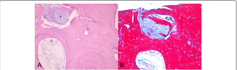
In some cases, small bone segments were acquired from the graft site of the osteotomy with a trephine bur during fixture placement or the graft over cover screws with the tissue punch during uncovering. Decalcified tis-sue sections were then prepared using routine proce-dures and stained with hematoxylin–eosin (H&E) and Masson’s trichrome (MT).
In-vitro assessment of the auto-FDT graft
demineralization. Extracted maxillary premolars (n = 5) were dried and their weight was measured. The teeth were then processed in an ultrasonic chamber as for clinical use, and their dry weight were measured again. EDS (TEAM System, EDAX Inc., NJ, USA) was used to check the rela-tive calcium content (wt%) at 5 points up to a depth of 700 μm from the surface of the dentin. Data were collected in the 2θ range of 8°–90°, with a step size of 0.02° and a counting time of 20 s at each step. The data bank from the International Center for Diffraction Data was used in a search/match program for phase identification.
Scanning electron microscope (SEM) studies were per-formed using the Nanoscope IIIa instrument (Digital Instruments, Tonawanda, NY, USA) after coating samples with a gold–palladium alloy. The processed tooth mate-rials and untreated tooth surfaces were then observed.
Statistical analysis
Wilcoxon signed rank test was used for in-vitro ana-lysis of calcium using SPSS 20 software (IBM SPSS Inc., Chicago, IL, USA) with the significant level of 0.05, and the clinical data are described using descriptive statistics. Figure 1 The demineralized tooth was processed to either blocks or chips. A. Autogenous fresh demineralized tooth (auto-FDT) blocks.
B. Auto-FDT chips.
The defects at the osteotomy site requiring implant placement were restored with graft material prepared using the teeth from the original extraction sites (n = 30) or third molars (n = 8). Horizontal and vertical tations were performed in 31 patients and sinus augmen-tation (lateral approach) in seven. A total of 58 implants were placed, with the mandibular posterior region be-ing the most common site (26 implants). The implants ranged from 3.5 to 4.5 mm in diameter and 7 to 13 mm in length.
The graft materials were covered with a collagen mem-brane in 29 patients and a titanium mesh in nine. Eight pa-tients received implants 3–6 months after bone grafting. All patients exhibited uneventful healing. No significant complications were observed, such as compromised wound healing, uncontrolled dehiscence, local wound infection, graft failure, and dental implant failure. Four patients showed minor wound dehiscence in the early stage of soft tissue healing; however, complete healing by secondary intention was observed within 4 weeks.
Three to 6 months after grafting or final prosthesis placement, increased radiopacity with homogeneity was observed on panoramic radiographs, while CBCT images confirmed good implant support (Figure 2). The mean Implant Stability Quotient (ISQ) at the time of final prosthesis fabrication was 72.7 ± 5.2 (68–81), which was a clinically acceptable range. No case of implant failure was observed during the follow up period.
granulomatous reaction (Figures 3A, B).
In-vitro assessment of the auto-FDT graft
EDS confirmed significant changes in the calcium and phosphorus (mineral components of the tooth) and car-bon, nitrogen, and oxygen (elements of the organic matrix) levels after treatment; 81.1% calcium was removed (from 31.59 ± 1.81 to 5.98 ± 2.47 wt%), while 46.6% dry weight was lost during processing.
On the surface of the auto-FDT graft, SEM observa-tions revealed the absence of enamel and cementum and the presence of distinct dentinal tubules surrounded by a dense collagen matrix. The dentinal tubules on the surface of the graft material were wider than those ob-served in untreated teeth (Figures 4A, B).
Discussion
According to search of the Pubmed database, the first use of demineralized dentin for bone regeneration in humans dates back to 1975; in that case, an allogenic source was used [11], whereas an autogenous source was first used only in 2003 [12]. The present study is likely to be the first human trial of auto-FDT prepared at the chairside and implanted on the day of extraction. Placement of a tooth into an alveolar socket after treatment is called“tooth re-plantation” or“tooth transplantation”. Auto-FDT grafting for alveolar bone regeneration can be referred to as a “tooth osteoplantation”. Demineralization is a crucial step
in tooth grafting. Although a nondemineralized form of tooth powder showed good results [13], demineralization of bone and teeth increases the bioavailability of matrix-associated noncollagenous proteins such as osteocalcin, osteonectin, bone sialoprotein, phosphophoryn, and bone morphogenetic protein; these may enhance new bone formation [14-16]. Considering the higher mineralization and crystallinity of the tooth compared with those of bone, a decrease in mineral components and crystallinity may be more important for tooth-based graft materials [17]. Tooth demineralization is time consuming (usually 2–6 days), thus limiting the use of FDT as a graft material. Never-theless, FDT has shown great potential in alveolar bone re-generation [18,19]. Another drawback of demineralization is that prolonged acid exposure may negatively affect non-collagenous proteins involved in new bone formation [20]. Murata et al. reported a device and method to decrease the demineralization time for tooth graft material, but this method only provides the powder type and uses an organic acid, which is not recommended for process-ing. In addition, these authors did not describe a specific final sterilization method for the graft material [10].
To overcome these limitations, we adopted a modified ultrasonic technology. The preliminary tests revealed that a regular ultrasonic cleaner does not dramatically decrease the processing time because the tooth is comprised of nu-merous microsized dentinal tubules surrounded by peri-tubular dentin, which is more sclerotic than interperi-tubular dentin [21]. Periodic negative pressure can eliminate ultra-sonic pocketing and the implosion loss of cavitation, which decrease the efficacy of ultrasound, and allows deep pene-tration of the cavitation energy and reagents into the den-tinal tubules [22,23]. A thermoelectric cooler prevents the temperature increase caused by strong ultrasonic power and maintains the temperature under 40°C. These ad-vances facilitate quicker demineralization and prevent ther-mal damage to the tooth proteins. The short duration of the entire process enables grafting on the same day of ex-traction and also increases the clinical availability of
auto-FDT, because the block form of the graft can be trimmed or easily converted into chips or powder depending on the condition of the bone defect during surgery.
Implant stability was confirmed by a radio frequency device and histological examination in this study. The ISQ of all implants was satisfactory for final prosthesis placement. Histological examination revealed appos-itional bone growth around the auto-FDT graft, and its resorption implies that the demineralized tooth was osteoconductive with a remodeling characteristic. The interesting histological features observed in this study included the appearance of fusion-like integration be-tween two matries of the graft and new bone at the interface with an undistinguishable border and new bone formation in the auto-FDT graft (Figure 3). This implies that dentinal tubules provide niches for cells involved in osteogenesis. Possible reasons for the good results in this study include the high biocompatibility of autogenous tis-sue, its collagen-based porous structure that is beneficial for cell function, minimal changes in the structure of min-eralized collagen without dehydration (freeze drying), and rehydration procedures and preservation of beneficial non-collagenous proteins involved in mineralization. This study strongly supports the view that auto-FDT grafts provide a useful biological scaffold comprising three-dimensional macro- and microarchitecture with osteopromotive osteo-conduction suitable for new bone formation [24].
Conclusions
The author declares that he has no competing interests.
Acknowledgments
This study was supported by a 2013 grant of Dankook University (No. 113526). The author would like to thank Mr. Jae-Young Lee and Mr. Hyun-Sik Kim for technical support. I also appreciate Dr. Jong-Gyu Paik’s advice on the EDS and SEM studies.
Received: 25 December 2014 Accepted: 5 February 2015
References
1. Kim YK, Kim SG, Byeon JH, Lee HJ, Um IU, Lim SC, Kim SY (2010) Development of a novel bone grafting material using autogenous teeth. Oral Surg Oral Med Oral Pathol Oral Radiol Endod 4:496–503
2. Qian JJ, Bhatnagar RS (1996) Enhanced cell attachment to anorganic bone mineral in the presence of a synthetic peptide related to collagen. J Biomed Mater Res 4:545–554
3. Liu Y, Luo D, Liu S, Fu Y, Kou X, Wang X, Sha Y, Gan Y, Zhou Y (2014) Effect of nanostructure of mineralized collagen scaffolds on their physical properties and osteogenic potential. J Biomed Nanotechnol 6:1049–1060 4. Beniash E (2011) Biominerals–hierarchical nanocomposites: the example of
bone. Wiley Interdiscip Rev Nanomed Nanobiotechnol 1:47–69
5. Pashley DH (1989) Dentin: a dynamic substrate-a review. Scanning Microsc 1:161–174
6. Nanci A (2008) Ten Cate’s oral histology: development, structure and function, 8th edn. Mosby Elsevier, St Louis, Mo, pp 108–192
7. Dirksen TR, Marinetti GV (1970) Lipids of bovine enamel and dentin and human bone. Calcif Tissue Res 1:1–10
8. Yeomans JD, Urist MR (1967) Bone induction by demineralized dentine implanted into oral, osseous and muscle tissues. Arch Oral Biol 12:999–1008 9. Butler WT, Mikulski A, Urist MR, Bridges G, Uyeno S (1977) Noncollagenous
proteins of a rat dentin matrix possessing bone morphogenetic activity. J Dent Res 3:228–232
10. Murata M, Akazawa T, Takahata M, Ito M, Tazaki J, Nakamura K, Iwasaki N, Shibata T, Arisue M (2010) Bone induction of human tooth and bone crushed by newly developed automatic mill. J Ceramic Soc Jpn 1378:434–437 11. Nordenram A, Bang G, Bernhoft CH (1975) A clinical-radiographic study of
allogenic demineralized dentin implants in cystic jaw cavities. Int J Oral Surg 2:61–64
12. Murata M (2003) Autogenous demineralized dentin matrix for maxillary sinus augmentation in humans: the first clinical report. 81th International Association for Dental Research, Gothenburg
13. Atiya BK, Shanmuhasuntharam P, Huat S, Abdulrazzak S, Oon H (2014) Liquid nitrogen-treated autogenous dentin as bone substitute: an experimental study in a rabbit model. Int J Oral Maxillofac Implants 2: e165–e170
14. Rezende ML, Consolaro A, Sant’ana AC, Damante CA, Greghi SL, Passanezi E (2014) Demineralization of the contacting surfaces in autologous onlay bone grafts improves bone formation and bone consolidation. J Periodontol 5:e121–e129
15. Gomes MF (2006) Densitometric analysis of the autogenous demineralized dentin matrix on the dental socket wound healing process in humans. Braz Oral Res 4:324–330
16. Kim YK, Lee JY, Kim SG, Lim SC (2013) Guided bone regeneration using demineralized allogenic bone matrix with calcium sulphate: case series. J Adv Prosthodont 2:167–171
17. Lee EY, Kim ES, Lim KW (2014) Scanning electron microscopy and energy dispersive X-ray spectroscopy studies on processed tooth graft material by vacuum-ultrasonic acceleration. Maxillofac Plast Reconstr Surg 3:103–110
22. Behrend O, Schubert H (2001) Influence of hydrostatic pressure and gas content on continuous ultrasound emulsification. Ultrason Sonochem 8:271–276
23. ASM Handbook Committee (1994) ASM Handbook: Volume 5: Surface Engineering. In: Chap. 6 ultrasonic cleaning 10thed. ASM International, Ohio, USA, pp 44–47
24. Bakhshalian N, Hooshmand S, Campbell SC, Kim JS, Brummel-Smith K, Arjmandi BH (2013) Biocompatibility and microstructural analysis of osteopromotive property of allogenic demineralized dentin matrix. Int J Oral Maxillofac Implants 6:1655–1662
Submit your manuscript to a
journal and benefi t from:
7Convenient online submission 7Rigorous peer review
7Immediate publication on acceptance 7Open access: articles freely available online 7High visibility within the fi eld
7Retaining the copyright to your article