Abstract
Serum response factor (SRF) acts as a multifunctional transcription factor regulated by mutually exclusive
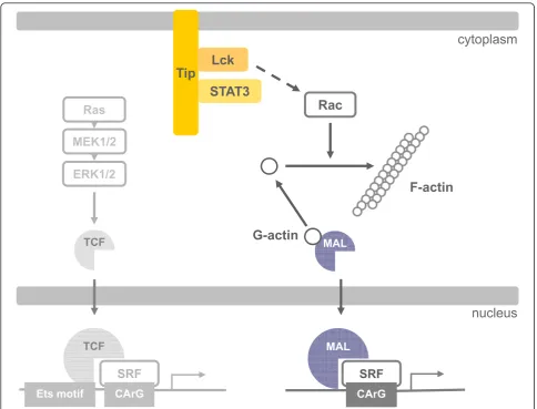
interactions with ternary complex factors (TCFs) or myocardin-related transcription factors (MRTFs). Binding of Rho-and actin-regulated MRTF:SRF complexes to target gene promoters requires an SRF-binding site only, whereas MAPK-regulated TCF:SRF complexes in addition rely on flanking sequences present in the serum response element (SRE). Here, we report on the activation of an SRE luciferase reporter by Tip, the viral oncoprotein essentially contributing to human T-cell transformation by Herpesvirus saimiri. SRE activation in Tip-expressing Jurkat T cells could not be attributed to triggering of the MAPK pathway. Therefore, we further analyzed the contribution of MRTF complexes. Indeed, Tip also activated a reporter construct responsive to MRTF:SRF. Activation of this reporter was abrogated by overexpression of a dominant negative mutant of the MRTF-family member MAL. Moreover, enrichment of monomeric actin suppressed the Tip-induced reporter activity. Further upstream, the Rho-family GTPase Rac, was found to be required for MRTF:SRF reporter activation by Tip. Initiation of this pathway was strictly dependent on Tip’s ability to interact with Lck and on the activity of this Src-family kinase. Independent of Tip, T-cell stimulation orchestrates Src-family kinase, MAPK and actin pathways to induce SRF. These findings establish actin-regulated transcription in human T cells and suggest its role in viral oncogenesis.
Keywords:Actin, Herpesvirus saimiri, Lck, MRTF, Oncoprotein, Serum response factor, T lymphocyte
Background
Serum response factor (SRF) is widely expressed in both invertebrates and vertebrates. SRF plays an essential role in embryogenesis, but is also involved in multiple pro-cesses in developed organisms including neuronal and muscle cell function.
SRF binds as a dimer to a specific DNA sequence known as the CArG box in the promoter of hundreds of target genes. Selective binding is determined by interac-tions with more than 60 different cofactors, which turn SRF into a versatile transcription factor translating cell-and stimulus-specific signaling into selective target gene expression [1,2].
Well-known SRF cofactors are members of the ternary complex factor (TCF) family of Ets domain proteins, like Elk-1, SAP-1 and Net. They are regulated by
phosphorylation via the classical mitogen-activated pro-tein kinase (MAPK) pathway involving the GTPase Ras, which activates the serine-threonine kinases Raf, MEK and ERK. Their recruitment to DNA depends on a defined DNA sequence, called Ets motif (C/A)(C/A) GGA(A/T), next to the SRF-binding CArG box [3,4]. A serum response element (SRE), first described in the c-fos promoter, contains an Ets motif adjacent to the CArG box [5].
Another group of SRF cofactors are the myocardin-related transcription factors (MRTFs). Myocardin, the founding member of this family, is selectively expressed in cardiac and smooth muscle cells and constitutively binds SRF. In contrast, MRTF-A (MAL, MKL1, BSAC) and MRTF-B (MAL16, MKL2) are widely expressed in many cell types [6]. Their cofactor function is controlled by GTPases of the Rho family (RhoGTPases), which are considered as important regulators of the actin cytoske-leton. Activation of the RhoGTPases RhoA, Rac1 and Cdc42 results in the formation of focal adhesion
* Correspondence: [email protected]
1
Institut für Klinische und Molekulare Virologie, Friedrich-Alexander-Universität Erlangen-Nürnberg, Erlangen, Germany
Full list of author information is available at the end of the article
complexes, lamellipodia and filopodia, respectively [7]. These processes involve actin polymerization and thereby reduce the levels of monomeric, globular actin (G-actin). G-actin binds to N-terminal RPEL motifs of MRTF and thereby sequesters and negatively regulates MRTF. RhoGTPase-mediated reduction of G-actin liber-ates MRTF, resulting in its nuclear accumulation and SRF cofactor function. SRF-bound MRTF dimers directly contact DNA near the SRF binding sequence. However, a specific MRTF binding sequence, similar to the Ets motif, has not yet been found [1,6].
Differential regulation of SRF target genes is based on gene-specific cofactor preferences and cofactor competi-tion for a common binding site on SRF [8-11]. In this context, specific SRF functions are defined only for a limited set of cell types and assignment of cofactors is lagging. Conditional knock-out approaches were recently used to elucidate the function of SRF and the role of TCFs and MRTFs in mouse T cells. Elimination of SRF
by a CD4-Cre transgene at the CD4+CD8+ double
posi-tive stage impairs T-cell development and results in the absence of peripheral T cells [12]. An earlier elimination
of SRF by a hCD2-Cre transgene at the CD4-CD8-
dou-ble negative stage severely reduces the numbers of single positive thymocytes, thymic Tregand NK T cells.
Intro-duction of recombinant SRF lacking the ability to bind TCFs or MRTFs fails to restore thymocyte maturation. In contrast, reconstitution was successful upon intro-duction of wild-type SRF or a fusion of the recombinant SRF with Elk [13]. While this study documents an essential role of TCF:SRF complexes in T-cell develop-ment, activation and function of MRTF:SRF complexes in T cells remain to be established.
Herpesvirus saimiri (HVS) is the T-lymphotropic pro-totype ofg2-herpesviruses. In contrast to the apathogenic appearance in its natural host, the squirrel monkey (Sai-miri sciureus), HVS causes severe T-cell lymphoma in experimentally infected non-natural primate hosts [14].
Most notably, in vitro infection of human peripheral
blood mononuclear cells with HVS strain C488 gives rise to continuously proliferating T-cell lines [15]. Deletions of viral genomic sequences coding for the oncoproteins StpC (Saimiri transformation-associated protein of sub-group C) and Tip (Tyrosine kinase interacting protein) obviate human T-cell transformation as well as patho-genicity in non-human primates [16]. Conditional expression of Tip alone in transgenic mice leads to T-cell lymphoma [17]. Tip engages the Src-family kinase (SFK) Lck, a central mediator of proliferation in response to T-cell receptor stimulation [18,19]. Lck interaction and activation relies on two motifs in Tip, a sequence homo-logous to the C-terminus of Src-family kinase domains (CSKH) and a proline-rich Src homology domain 3 bind-ing sequence (SH3B) [18,20,21]. The integrity of both
motifs, CSKH and SH3B, is required for Tip to support human T-cell transformation [22]. However, pro-prolif-erative downstream effectors of Tip:Lck interaction are not defined yet. Pro-oncogenic functions are character-ized for signal transducer and activator of transcription 3 (STAT3) [23]. Indeed, STAT3 is activated by Lck in the presence of Tip and is constitutively phosphorylated in HVS-C488 transformed lymphocytes [21,24-26]. How-ever, mutation of tyrosine residue 114 (Y114) in Tip abrogates constitutive STAT3 phosphorylation, but not viral transformation of human T cells [27,28]. Thus, alternative Tip:Lck effectors must be involved to trigger T-cell proliferation. Given the central role of mitogen-activated protein kinases (MAPK) for growth regulation in general, we previously analyzed MAPK phosphoryla-tion and activaphosphoryla-tion of MAPK-regulated transcripphosphoryla-tion in the presence of the HVS-C488 oncoproteins, StpC and Tip [29]. In Jurkat T cells, neither StpC nor Tip induce the phosphorylation of MEK1/2 and ERK1/2 or the activ-ity of the MAPK-regulated transcription factor AP-1. Nevertheless, Tip specifically triggers SRF activity in this test system [29].
In this work, we now address the mechanism of SRF activation by the viral oncoprotein Tip. We demonstrate an SRF activation in T cells that depends on actin poly-merization and on the cofactor MAL and is abrogated by dominant-negative Rac1. Tip requires Lck interaction and Src kinase activity to induce this pathway, which may also be a target of T-cell receptor stimulation.
Results
Tip induces SRF-regulated transcription independent of MAPK activity
p3D.A luciferase reporter contains a mutated TCF-binding Ets motif within its SRE and is therefore more sensitive to activation by MRTF:SRF complexes. Relative to the SRE reporter, the p3D.A construct displayed a high basal activ-ity in vector-transfected cells. An enhanced activactiv-ity of this reporter was observed for the MEK inhibitor U0126, but not for PD0325901, indicating off-target functions and restricting the validity of U0126 data. Tip induced a 3-fold increase of the basal activity, and this enhancement was not significantly affected by the MEK inhibitor PD0325901. In contrast, PMA stimulation of vector-trans-fected cells enhanced the activity about 7-fold, and this effect was completely abrogated by U0126 and PD0325901. Taken together, the viral oncoprotein Tip induced SRF-responsive luciferase reporters independent of MAPK activity and ERK phosphorylation. Activation of the p3D.A luciferase reporter further points at SRF activa-tion by Tip independent of the MAPK-TCF pathway.
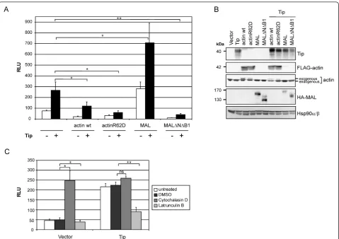
SRF activation involves actin dynamics and the cofactor MAL
To corroborate MAPK and, thus, TCF independence of Tip-mediated SRF activation, we next addressed the actin-MRTF pathway. To this end, we transfected Jurkat T cells with expression plasmids for wild-type actin (actin wt), an actin polymerization mutant (actinR62D), wild-type
full-length MAL (MAL) and a MAL deletion mutant unable to
bind actin and SRF (MALΔNΔB1) alone or in
combina-tion with Tip (Figure 2A). Expression of the transfected constructs was controlled by immunoblot analysis (Figure 2B). Overexpression of actin, presumably resulting in excess globular actin, diminished the basal and Tip-induced reporter activity by 3.5- and 2.2-fold, respectively. This effect became more evident when globular actin was enriched by overexpression of actinR62D, which reduced the Tip-induced signal below basal levels. Upon overex-pression of MAL, the basal reporter activity was 3.7-fold higher compared to vector alone, and this was further enhanced about 2.5-fold by coexpression of Tip. In con-trast, the MAL deletion mutant completely abrogated the signal. To strengthen these observations, we treated trans-fected cells with Latrunculin B, an inhibitor of actin poly-merization and promoter of filamentous actin disassembly. As a positive control we used Cytochalasin D, which binds G-actin irreversibly (Figure 2C). While enrichment of monomeric actin by Latrunculin B inhibited both basal and Tip-induced reporter activity, Cytochalasin D increased the basal activity about 4-fold, but did not further enhance the Tip effect. Thus, actin polymerization and the cofactor MAL indeed play an important role in SRF activation by Tip.
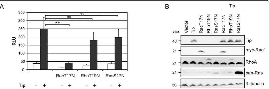
Dominant-negative Rac1 prevents Tip-mediated MAL:SRF activation
The importance of actin dynamics for Tip-induced SRF activation raised the question whether the small GTPases RhoA, Rac1, Cdc42, inducers of actin polymer-ization and actin filament stabilpolymer-ization, play a role in this process. Therefore, we used dominant-negative expression constructs for Rac1 (Rac1-T17N) and RhoA (RhoA-T19N) to further elucidate their role in p3D.A reporter induction. Dominant-negative H-Ras (H-Ras-S17N), a regulator of MAPK and TCFs, was used as a control for interference between the small G proteins (Figure 3A). Expression of the transfected constructs was controlled by immunoblot analysis (Figure 3B). Coexpression of RhoA-T19N and H-Ras-S17N did not significantly reduce Tip-mediated reporter activity. However, overexpression of Rac1-T17N impaired both Tip’s effect on the reporter and background activity in
vector-transfected cells. Effector pull-down assays to detect GTP-loaded Rac1/2/3 and Cdc42 (GST-PAK-CRIB), RhoA (GST-Rhotekin) and H-Ras (GST-Raf-RBD) suggested an activation of Rac and Cdc42, but not RhoA and H-Ras by Tip (data not shown). However, these findings were not constantly reproducible due to high basal levels of activated Rac1/2/3 and Cdc42 in vector-transfected cells. Nevertheless, the luciferase reporter assays demonstrate a major role of the GTPase Rac1, but not of RhoA and H-Ras, in the actin polymer-ization- and MAL-dependent SRF activation by Tip.
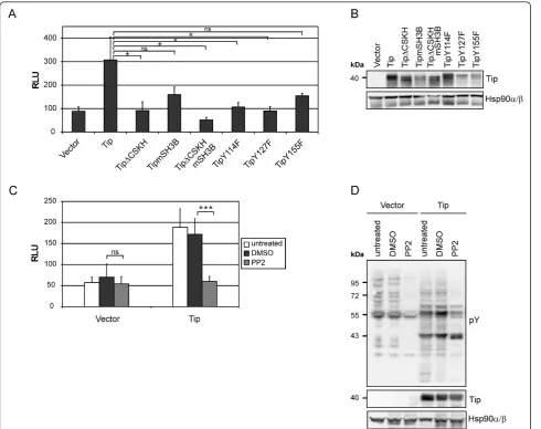
p3D.A reporter activation by Tip depends on Src-family kinase interaction and activity
To test for the properties of Tip required to induce SRF activity, we used mutants of Tip defective in its major effector function, the recruitment and activation of the Src-family kinase (SFK) Lck, or carrying substitutions of Figure 2Effects of actin dynamics and the cofactor MAL on SRF activity.(A)p3D.A-Luc activity of Jurkat T cells transiently transfected with expression plasmids coding for actin wt, actinR62D, MAL and MALΔNΔB1 alone or in combination with a Tip expression construct.(B)
the conserved tyrosine residues Y114, Y127 and Y155, which may be targets of Lck [22,27,30] (Figure 4A). Expression of the transfected constructs was controlled by immunoblot analysis (Figure 4B). Deletion of the CSKH motif (TipΔCSKH and TipΔCSKHmSH3B) or individual point mutations of tyrosine residues 114 (TipY114F) and 127 (TipY127F) significantly reduced SRF reporter activity to vector levels. The repression observed upon mutation of the SH3 binding motif (TipmSH3B) or tyrosine residue 155 (TipY155F) was not significant. Furthermore, interpretation of the data for TipY127F and TipY155F is restricted by their expression levels, which were reproducibly reduced rela-tive to the wild-type protein. The abolishment of Tip-mediated reporter activation by the highly specific SFK inhibitor PP2 verified the requirement of Src-kinase activity (Figure 4C). Immunoblot analysis of protein tyr-osine phosphorylation monitored a modulating function of Tip and the inhibitory efficacy of PP2 (Figure 4D). Hence, Tip relies on both, Lck interaction and SFK activity, to trigger MAL:SRF reporter activity. Further-more, tyrosine residues Y114 and Y127, known to be critical for STAT3 activation [28] and IL-2-independent T-cell transformation [22], respectively, likely contribute to Tip-induced SRF activity.
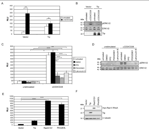
TCR stimulation induces p3D.A reporter activity
The viral oncoprotein Tip activated SRF in T cells via the actin-regulated cofactor MAL (Figure 2), while previous reports demonstrated SRF activation via the MEK-ERK pathway in response to TCR stimulation of Jurkat T cells and in mouse T-cell development [13,31]. This discrepancy prompted us to assess whether TCR stimulation alone can trigger the p3D.A luciferase reporter or further enhance
the Tip effect (Figure 5A). TCR and coreceptor engage-ment via CD3/CD28 antibodies resulted in a 10-fold enhanced reporter activity in vector-transfected Jurkat T cells relative to unstimulated cells. In contrast, CD3/CD28 antibody treatment did not significantly augment the Tip-triggered signal. As ERK phosphorylation was absent in Tip-transfected cells (Figure 5B), this lack of cooperation correlated with an impaired CD3/CD28-induced signaling, which is in accordance with suppression of TCR signaling by Tip [32]. In order to specify the TCR-triggered pathway involved, CD3/CD28-stimulated and unstimulated vector-transfected cells were treated with inhibitors of SFK (PP2), MEK (PD0325901), and actin polymerization (Latrunculin B) (Figure 5C). TCR-induced reporter activity was signifi-cantly reduced in all treated samples. All three inhibitors were similarly effective, with low but significant residual activities relative to unstimulated cells. Unexpectedly, the residual activities in PD0325901- and Latrunculin B-treated cells did not add up to the activity of solvent-treated cells (DMSO). This finding may be related to the partial reduc-tion of ERK phosphorylareduc-tion by Latrunculin B (Figure 5D). The impact of actin polymerization on SRF activation in T cells was further addressed by the expression of constitu-tively active Rac1 (Rac1-G12V) and RhoA (RhoA-Q63L) in the Jurkat system (Figure 5E). Rac1-G12V and RhoA-Q63L (Figure 5E) were equally effective and even more potent than CD3/CD28 stimulation (Figure 5A, C) in inducing 3D.A reporter activity. In conclusion, TCR stimulation relied on both, MAPK signaling and actin polymerization, to activate SRF.
Discussion
(SRF) in T cells. This activation mainly depends on actin-mediated MRTF coactivation, with minor contri-butions of MEK-mediated TCF coactivation. Discrimina-tion of coactivator involvement was assessed using two SRF-dependent luciferase reporter constructs, based on thec-fos SRE, considered to be specific for TCF coacti-vation, and on a mutated SRE (3D.A), considered to respond preferentially to MRTF coactivation. However, largely MEK-independent SRE activation by Tip and MEK-sensitive 3D.A activation by PMA revealed a restricted specificity of the reporters in the Jurkat T cells used throughout this study. Hence, we included chemical inhibitors and overexpression of mutant
signaling intermediates to assign Tip-induced SRF acti-vation to the actin-dependent MRTF coactiacti-vation path-way. Targeting of this pathway by a viral T-cell oncoprotein was unexpected, as SRF function in T cells had previously been linked mainly to the TCF pathway [13].
accordance with early results on SRE-dependent tran-scription in Jurkat T cells [31]. Thus, Lck-dependent MRTF coactivation, which we suggest for Tip, may as well apply to T-cell stimulation. However, while Tip trig-gers SRF largely independent of MAPK activity, stimula-tion-induced SRF activation substantially involves MAPK signaling and likely integrates different intracellular sig-naling routes. The interference of Tip with
receptor-mediated SRF activation most likey occurs further upstream. Dependent on its localization in lipid rafts, Tip induces the internalization of TCR complexes [36-38]. Independent of its lipid raft association, Tip blocks TCR-mediated intracellular signaling most likely through sequestration of Lck [32,38]. Consequently, Tip-expres-sing cells are refractory to receptor ligation by stimulat-ing antibodies.
The dependence of Tip-induced SRF activation on Lck interaction, Src-family kinase (SFK) activity and the potential Lck phosphorylation sites in Tip, Y114 and Y127, draws the attention to the Tip:Lck effectors involved in this pathway. So far, only STATs, especially STAT3, are described as direct targets of Tip-activated Lck [21,24-27]. Tip-induced STAT3 activation depends on residue Y114, which is not required for human T-cell transformationin vitro[28]. However, the potential of STAT3 to promote invasion in various cancers [23] may well relate to the massive tissue invasion by HVS-lymphoma cells [26,39], which is not reflected in the cell culture system. Therefore, while effectors of Tip essen-tial for viral T-cell transformation are still not identified, we suggest that Tip Y114 contributes to viral oncogen-esis through STAT3-regulated lymphocyte invasion. In this context, STAT3 would be expected as an upstream regulator of RhoGTPases. However, an emerging model positions STAT3 downstream of Rac1 and Cdc42 in the regulation of cell proliferation and migration [40]. Alter-natively, transcriptional regulation of genes involved in MRTF:SRF activation by Tip-induced STAT3 appears conceivable. Such an indirect mechanism might also be elicited by STAT5, a recently identified target of Tip [41] likely related to the strict IL-2 dependence of viral transformation in the presence of TipY127F [22]. In any case, a functional link between STAT3 or STAT5 and MRTF:SRF, to the best of our knowledge, has not been reported. Hence, Tip-activated Lck may trigger SRF acti-vation through alternative, yet unknown effectors like the various RhoGTPase guanine nucleotide exchange factors (GEFs) expressed in T cells [42]. Altogether, mechanisms of MRTF:SRF activation proximal to the Tip:Lck complex remain to be established.
The RhoGTPases RhoA, Rac1 and Cdc42 directly reg-ulate actin cytoskeleton organization [7] and therefore share the potential to modulate cellular G-actin pools, which in turn determine MRTF coactivator availability [1]. We expressed constitutively active Rac1 and RhoA and thereby proved the inducibility of MRTF:SRF by both GTPases in T cells independent of Tip. Dominant-negative versions of Rac1, RhoA and Ras were used to test for the involvement of these GTPases in Tip-mediated SRF activation. The missing influence of domi-nant-negative Ras corroborated the TCF independence of Tip-induced SRF activation. Suppression of the Tip effect by inhibitory Rac1 and not RhoA is in contrast to the initial report on SRF activation by MAL in NIH3T3 fibroblasts [8], but in accordance with MAL signaling in epithelial cells [43]. We assumed that Tip induces SRF via Rac1, but not RhoA. Accordingly, active (GTP-bound) RhoA and H-Ras were not detected in Tip-expressing cells, whereas cellular levels of basally active Rac1 and Cdc42 were enhanced by Tip in some, but not
all effector pull-down assays performed. We further
used the Rac1/Cdc42-glucosylating C. difficiletoxins
that have been shown to inhibit SRF activation induced by Ca2+-dependent dissociation of epithelial integrity [43]. Unexpectedly, the C. difficile toxins failed to sup-press Tip-induced reporter activity in our Jurkat system (data not shown). This observation is apparently incon-sistant with our observation that Rac1-T17N strongly reduces Tip-induced SRF activation. In general, either pronounced Rac1/Cdc42 activation or pronounced Rac1/Cdc42 phosphorylation by Akt1 protects Rac1/ Cdc42 from toxin-catalyzed glucosylation and inactiva-tion [44,45]. In particular, protective phosphorylainactiva-tion of Rac1/Cdc42 has to be taken into account, as Jurkat T cells are deficient in expression of PTEN, a major nega-tive regulator of PI3K/Akt signaling [46]. Based on the data available, we would exclude RhoA and Ras and suggest Rac1 and Cdc42 activation in response to Tip expression as the crucial step in SRF induction. The mechanism of the Tip-mediated activation of Rac1/ Cdc42, however, remains to be clarified.
Besides the critical role of Rac1 in Tip-induced SRF activation, our results substantiate an essential role of actin and actin-regulated MRTF in SRF activation by Tip in T cells. The syngergism between ectopic MAL and the viral oncoprotein, which is in contrast to the effects obtained with the cellular oncoprotein OTT-MAL [47], points at limiting OTT-MAL expression levels and clearly positions Tip upstream in the activation cascade. However, although we used wild-type and mutant MAL expression constructs, our assays are not suited to dis-criminate the contribution of the individual MRTF-family proteins, MAL/MRTF-A and MRTF-B, which may add another layer of complexity to SRF regulation.
MRTF:SRF functions in T cells are not characterized yet, and T cell-specific target genes of this transcription factor complex are not known. However, transcription of cytoskeletal regulators like MYH9 and MYL9 is ele-vated in different non-lymphoid cancer cell lines, which depend on MRTFs and SRF for cell spreading, adhesion, and motility [48]. Thus, MRTF:SRF activation by Tip, a viral oncoprotein essential for the development of fulmi-nant T-cell lymphoma characterized by infiltration of multiple organs [16,26], may well contribute to viral oncogenesis and tissue invasion of tumor cells.
Conclusion
reveal the detailed mechanisms and target genes of the pathway triggered by Tip as well as its applicability to T cells in general. This approach is anticipated to resolve the functional relevance of MRTF:SRF activity in T-cell regulation and in viral oncogenesis.
Methods
Cell culture
Jurkat T cells (E6.1, ATCC TIB-152) were cultured in RPMI 1640 medium supplemented with 10% fetal calf
serum (FCS), glutamine (350 μg/ml) and gentamicin
(100μg/ml) at a maximum concentration of 0.5-1 ×10⁶
cells/ml.
Transient transfection of Jurkat T cells
Transfection of 5-10 ×106 cells/ml Jurkat T cells was
carried out by electroporation in medium without anti-biotics at 250 V, 1,500μF using a Gene pulser × cell™
Electroporation System (Bio-Rad). For each sample, a
total of 50μg plasmid DNA was used and appropriate
empty vector was included to equalize plasmid DNA amounts. Transfected cells, cultured in complete med-ium without antibiotics, were harvested after 48 h, washed with phosphate-buffered saline (PBS) and pro-cessed for luciferase reporter gene assays or immunoblot analysis.
Expression plasmids
Jurkat T cells were transfected with 20μg of expression constructs coding for wild type and mutants of the viral oncoprotein Tip derived from HVS-C488: pEF1-Tip, pEF1-TipΔCSKH, pEF1-TipmSH3B, pEF1-TipΔCSKHm SH3B, pEF1-TipY114F, pEF1-TipY127F, pEF1-TipY155F [49]. All Tip constructs are N-terminally myc-tagged. The expression plasmids actin wt; pEF-FLAG-actinR62D, coding for a FLAG-tagged polymerization
TCF
MAL
ERK1/2
MAL
SRF
F-actin
G-actin
CArG
nucleus
SRF
CArG Ets motif
MALMAL TCF
mutant of actin; pEF-MAL-HA (f.l.), encoding HA-tagged full-length murine MAL; pEF-MALΔNΔB1-HA, coding for a MAL deletion mutant unable to bind to actin and SRF, were described previously [8,50]. Sequences coding for dominant-negative Rac1 (RacT17N) and RhoA (RhoT19N) and constitutively active Rac1 (RacG12V) and RhoA (RhoQ63L) were amplified by PCR with oligonu-cleotide primers introducing terminal BamHI and EcoRI restriction sites and a N-terminal myc-tag (myc-RacT17N, myc-RacG12V, myc-RhoQ63L) (primers available upon request) were cloned into pEF1 to yield the expression constructs myc-RacT17N, RhoT19N, pEF1-myc-RacG12V and pEF1-myc-RhoQ63L. Dominant-nega-tive Ras was expressed using the plasmid pcDNA3-RasS17N (kindly provided by A. Wittinghofer, Dortmund, Germany). Integrity of the coding sequences was con-firmed by automated DNA sequencing (ABI 3130, Applied Biosystems).
Immunoblot analysis
Jurkat T cells were lysed in RIPA buffer and processed as previously described to generate whole cell lysates [29]. Protein extracts of 0.5-1 ×10⁶ Jurkat T cells were loaded on SDS-polyacrylamide gels and transferred to polyvinylidene difluoride membranes (GE Healthcare). After blocking with 5% milk powder in 0.1% Tween20-PBS or NET-gelatine (150 mM NaCl; 5 mM EDTA; 50 mM Tris-HCl pH7.5; 0.05% TritonX-100; 2.5 g/ml gela-tine), the membranes were probed with antibodies direc-ted against: phosphotyrosine (4 G10, Millipore), pERK1/
2 (pY204 in ERK1), Hsp90a/b (Santa Cruz), ERK1/2,
RhoA, Rac1/2/3 (Cell Signaling Technology), Pan-Ras (Calbiochem), Tip [17], Myc-epitope (9E10; ATCC CRL-1729), FLAG-epitope (M2, HRP-coupled; Sigma), HA-epitope (Convance),b-tubulin (GE Healthcare). Binding of primary antibodies was detected using horseradish peroxidase-coupled secondary antibodies directed against mouse or rabbit immunoglobulins (Dako). Pri-mary and secondary antibodies were diluted in blocking buffer. Immunodetection was performed by chemilumi-nescence and documented with a Kodak Image Station 4000 MM PRO camera.
Luciferase reporter gene assay
Jurkat T cells were transfected with 20μg of the indivi-dual effector plasmids and 10μg of the reporter plasmid pSRE-luc containing five SRE of the c-fos promoter (Stratagene) or p3D.A-Luc [51] comprising three SRE with a mutated Ets motif. Cells were harvested 48 h post transfection and divided equally for luciferase activ-ity quantification and immunoblots. For luciferase reporter gene assay, cells were lysed and luminescence intensity was measured as described [52]. Raw data were normalized to the protein content of each sample as
determined by a BCA assay (Uptima) and indicated as relative light units (RLU). Data were statistically evalu-ated with two-tailed t-tests for correlevalu-ated (Figure 1A) or independent (all other figures) samples using the online-tools provided by the VassarStats Website for Statistical Computation [http://faculty.vassar.edu/lowry/Vassar-Stats.html]. Results were assigned to the categories p > 0.05 (ns, not significant), p < 0.05 (*), p < 0.01 (**), p < 0.001 (***).
Inhibitor treatment and CD3/CD28 ligation
For inhibitor treatment, transfected Jurkat T cells were seeded in a 12-well plate at a density of approximately
0.5 ×10⁶ cells/ml. The SFK inhibitor PP2 (Sigma; 10
μM) and the MAPK inhibitors U0126 (Biomol Germany;
25 μM) and PD0325901 (1μM) were added 8 h post
transfection and remained in the cultures until harvest-ing of the cells. 12-O-tetradecanoylphorbol-13-acetate (PMA; Sigma; 20 ng/ml), combined with MAPK inhibi-tors if applicable, was added for 15 h. To modulate actin polymerization, cells were treated with Latrunculin
B (Calbiochem; 1 μM), Cytochalasin D (Applichem; 1
μM) for 24 h. Under these conditions all inhibitors were not toxic to Jurkat T cells as measured by propidiumio-dide staining and flow cytometry. T cell-receptor stimu-lation of transfected Jurkat T cells was carried out for 14 h in a 6-well plate at a density of approximately 1 × 10⁶ cells/ml previously coated with antibodies against
CD3 (OKT3; Janssen-Cilag; 10μg/ml) and CD28 (a gift
from R. Kroczek, Robert Koch-Institut, Berlin; 5μg/ml).
Abbreviations
CSKH: Sequence homologous to the C-terminus of Src-family kinase domains; ERK: Extracellular signal-regulated kinase; G-actin: Globular actin; GEF: Guanine nucleotide exchange factor; GTP: Guanosine triphosphate; HVS: Herpesvirus saimiri; MAPK: Mitogen-activated protein kinase; MRTFs: Myocardin-related transcription factors; MYH9: Myosin heavy chain 9: non muscle; MYL9: Myosin light chain 9: regulatory; PMA: 12-O
-tetradecanoylphorbol-13-acetate; RhoGTPases: Guanosine triphosphatases of the Rho family; SFK: Src-family kinase; SH3: Src homology domain 3 binding sequence; SRE: Serum response element; SRF: Serum response factor; STAT: Signal transducer and activator of transcription; StpC: Saimiri transformation-associated protein of subgroup C; TCF: Ternary complex factor; Tip: Tyrosine interacting protein.
Acknowledgements
We thank Richard Kroczek (Berlin) and Alfred Wittinghofer (Dortmund) for kindly providing antibodies and plasmids. This work was supported by grants GRK1071/C3 and Bi465/5-1 from the German Research Foundation (DFG) and by the BIGSS program (State of Bavaria).
Author details
1
Institut für Klinische und Molekulare Virologie, Friedrich-Alexander-Universität Erlangen-Nürnberg, Erlangen, Germany.2Institut für Toxikologie,
Medizinische Hochschule Hannover, Hannover, Germany.3Department
Molecular Biology, Max-Planck-Institut für Biochemie, Munich, Germany.
Authors’contributions
inducibility of the human c-fos promoter.Cell1989,56:563-572. 4. Treisman R:Ternary complex factors: growth factor regulated
transcriptional activators.Curr Opin Genet Dev1994,4:96-101. 5. Treisman R:Journey to the surface of the cell: fos regulation and the
SRE.EMBO J1995,14:4905-4913.
6. Olson EN, Nordheim A:Linking actin dynamics and gene transcription to drive cellular motile functions.Nat Rev Mol Cell Biol2010,11:353-365. 7. Spiering D, Hodgson L:Dynamics of the Rho-family small GTPases in
actin regulation and motility.Cell Adh Migr2011,5:170-180.
8. Miralles F, Posern G, Zaromytidou AI, Treisman R:Actin dynamics control SRF activity by regulation of its coactivator MAL.Cell2003,113:329-342. 9. Wang Z, Wang DZ, Hockemeyer D, McAnally J, Nordheim A, Olson EN:
Myocardin and ternary complex factors compete for SRF to control smooth muscle gene expression.Nature2004,428:185-189.
10. Zaromytidou AI, Miralles F, Treisman R:MAL and ternary complex factor use different mechanisms to contact a common surface on the serum response factor DNA-binding domain.Mol Cell Biol2006,26:4134-4148. 11. Gineitis D, Treisman R:Differential usage of signal transduction pathways
defines two types of serum response factor target gene.J Biol Chem
2001,276:24531-24539.
12. Fleige A, Alberti S, Grobe L, Frischmann U, Geffers R, Muller W, Nordheim A, Schippers A:Serum response factor contributes selectively to
lymphocyte development.J Biol Chem2007,282:24320-24328.
13. Mylona A, Nicolas R, Maurice D, Sargent M, Tuil D, Daegelen D, Treisman R, Costello P:The essential function for serum response factor in T-cell development reflects its specific coupling to extracellular signal-regulated kinase signaling.Mol Cell Biol2011,31:267-276. 14. Ensser A, Fleckenstein B:T-cell transformation and oncogenesis by
gamma2-herpesviruses.Adv Cancer Res2005,93:91-128.
15. Biesinger B, Müller-Fleckenstein I, Simmer B, Lang G, Wittmann S, Platzer E, Desrosiers RC, Fleckenstein B:Stable growth transformation of human T lymphocytes by herpesvirus saimiri.Proc Natl Acad Sci USA1992,
89:3116-3119.
16. Duboise SM, Guo J, Czajak S, Desrosiers RC, Jung JU:STP and Tip are essential for herpesvirus saimiri oncogenicity.J Virol1998,72:1308-1313. 17. Wehner LE, Schröder N, Kamino K, Friedrich U, Biesinger B, Rüther U:
Herpesvirus saimiri Tip gene causes T-cell lymphomas in transgenic mice.DNA Cell Biol2001,20:81-88.
18. Biesinger B, Tsygankov AY, Fickenscher H, Emmrich F, Fleckenstein B, Bolen JB, Bröker BM:The product of the Herpesvirus saimiri open reading frame 1 (tip) interacts with T cell-specific kinase p56lckin transformed cells.J Biol Chem1995,270:4729-4734.
19. Smith-Garvin JE, Koretzky GA, Jordan MS:T cell activation.Annu Rev Immunol2009,27:591-619.
20. Jung JU, Lang SM, Friedrich U, Jun T, Roberts TM, Desrosiers RC, Biesinger B:
Identification of Lck-binding elements in tip of herpesvirus saimiri.J Biol Chem1995,270:20660-20667.
21. Lund TC, Prator PC, Medveczky MM, Medveczky PG:The Lck binding domain of herpesvirus saimiri tip-484 constitutively activates Lck and STAT3 in T cells.J Virol1999,73:1689-1694.
22. Heck E, Friedrich U, Gack MU, Lengenfelder D, Schmidt M, Müller-Fleckenstein I, Müller-Fleckenstein B, Ensser A, Biesinger B:Growth transformation of human T cells by herpesvirus saimiri requires multiple Tip-Lck interaction motifs.J Virol2006,80:9934-9942.
is independent of STAT3 activation.J Virol2005,79:5713-5720. 29. Glanz A, Albrecht JC, Heinemann S, Fleckenstein B, Isakov N, Biesinger B:
NF-kappaB activation by the viral oncoprotein StpC enhances IFN-gamma production in T cells.Immunol Cell Biol2008,86:622-630. 30. Ensser A, Thurau M, Wittmann S, Fickenscher H:The genome of
herpesvirus saimiri C488 which is capable of transforming human T cells.Virology2003,314:471-487.
31. Charvet C, Auberger P, Tartare-Deckert S, Bernard A, Deckert M:Vav1 couples T cell receptor to serum response factor-dependent transcription via a MEK-dependent pathway.J Biol Chem2002,
277:15376-15384.
32. Cho NH, Feng P, Lee SH, Lee BS, Liang X, Chang H, Jung JU:Inhibition of T cell receptor signal transduction by tyrosine kinase-interacting protein of Herpesvirus saimiri.J Exp Med2004,200:681-687.
33. Wiese N, Tsygankov AY, Klauenberg U, Bolen JB, Fleischer B, Bröker BM:
Selective activation of T cell kinase p56lck by herpesvirus saimiri protein tip.J Biol Chem1996,271:847-852.
34. Lund T, Medveczky MM, Medveczky PG:Herpesvirus saimiri Tip-484 membrane protein markedly increases p56lck activity in T cells.J Virol
1997,71:378-382.
35. Hartley DA, Hurley TR, Hardwick JS, Lund TC, Medveczky PG, Sefton BM:
Activation of the Lck Tyrosine-protein Kinase by the binding of the tip protein of herpesvirus Saimiri in the absence of regulatory tyrosine phosphorylation.J Biol Chem1999,274:20056-20059.
36. Park J, Lee BS, Choi JK, Means RE, Choe J, Jung JU:Herpesviral protein targets a cellular WD repeat endosomal protein to downregulate T lymphocyte receptor expression.Immunity2002,17:221-233.
37. Park J, Cho NH, Choi JK, Feng P, Choe J, Jung JU:Distinct roles of cellular Lck and p80 proteins in herpesvirus saimiri Tip function on lipid rafts.J Virol2003,77:9041-9051.
38. Cho NH, Kingston D, Chang H, Kwon EK, Kim JM, Lee JH, Chu H, Choi MS, Kim IS, Jung JU:Association of herpesvirus saimiri tip with lipid raft is essential for downregulation of T-cell receptor and CD4 coreceptor.J Virol2006,80:108-118.
39. Fleckenstein B, Desrosiers RC:Herpesvirus saimiri and Herpesvirus ateles.
InThe Herpesviruses.Edited by: Roizman B. New York: Plenum Publishing Corporation; 1982:253-332.
40. Raptis L, Arulanandam R, Geletu M, Turkson J:The R(h)oads to Stat3: Stat3 activation by the Rho GTPases.Exp Cell Res2011,317:1787-1795. 41. Katsch K, de Jong SJ, Schmidt M, Müller-Fleckenstein I, Fleckenstein B,
Albrecht JC, Biesinger B:Species restriction ofHerpesvirus saimiriand
Herpesvirus ateles: human lymphocyte transformation correlates with distinct signaling propertiesof viral oncoproteins.Virus Research2012. 42. Tybulewicz VL, Henderson RB:Rho family GTPases and their regulators in
lymphocytes.Nat Rev Immunol2009,9:630-644.
43. Busche S, Descot A, Julien S, Genth H, Posern G:Epithelial cell-cell contacts regulate SRF-mediated transcription via Rac-actin-MAL signalling.J Cell Sci2008,121:1025-1035.
44. Brandes V, Schelle I, Brinkmann S, Schulz F, Schwarz J, Gerhard R, Genth H:
45. Halabi-Cabezon I, Huelsenbeck J, May M, Ladwein M, Rottner K, Just I, Genth H:Prevention of the cytopathic effect induced by Clostridium difficile Toxin B by active Rac1.FEBS Lett2008,582:3751-3756. 46. Shan X, Czar MJ, Bunnell SC, Liu P, Liu Y, Schwartzberg PL, Wange RL:
Deficiency of PTEN in Jurkat T cells causes constitutive localization of Itk to the plasma membrane and hyperresponsiveness to CD3 stimulation.
Mol Cell Biol2000,20:6945-6957.
47. Descot A, Rex-Haffner M, Courtois G, Bluteau D, Menssen A, Mercher T, Bernard OA, Treisman R, Posern G:OTT-MAL is a deregulated activator of serum response factor-dependent gene expression.Mol Cell Biol2008,
28:6171-6181.
48. Medjkane S, Perez-Sanchez C, Gaggioli C, Sahai E, Treisman R: Myocardin-related transcription factors and SRF are required for cytoskeletal dynamics and experimental metastasis.Nat Cell Biol2009,11:257-268. 49. Richter S:The influence of Herpesvirus saimiri oncoprotein Tip on
apoptosis signaling pathways in T cells.PhD Thesis Friedrich-Alexander-Universität Erlangen-Nürnberg, Naturwissenschaftliche Fakultät; 2010. 50. Posern G, Sotiropoulos A, Treisman R:Mutant actins demonstrate a role
for unpolymerized actin in control of transcription by serum response factor.Mol Biol Cell2002,13:4167-4178.
51. Geneste O, Copeland JW, Treisman R:LIM kinase and Diaphanous cooperate to regulate serum response factor and actin dynamics.J Cell Biol2002,157:831-838.
52. de Jong SJ, Albrecht JC, Schmidt M, Müller-Fleckenstein I, Biesinger B:
Activation of noncanonical NF-kappaB signaling by the oncoprotein Tio.
J Biol Chem2010,285:16495-16503. doi:10.1186/1478-811X-10-5
Cite this article as:Katschet al.:Actin-dependent activation of serum response factor in T cells by the viral oncoprotein tip.Cell
Communication and Signaling201210:5.
Submit your next manuscript to BioMed Central and take full advantage of:
• Convenient online submission
• Thorough peer review
• No space constraints or color figure charges
• Immediate publication on acceptance
• Inclusion in PubMed, CAS, Scopus and Google Scholar
• Research which is freely available for redistribution