Turkish Journal of Fisheries and Aquatic Sciences 16: 865-871 (2016)
www.trjfas.org ISSN 1303-2712 DOI: 10.4194/1303-2712-v16_4_13
RESEARCH PAPER
© Published by Central Fisheries Research Institute (CFRI) Trabzon, Turkey in cooperation with Japan International Cooperation Agency (JICA), Japan
Structural Analysis of the Erythrocytes of
Channa punctatus
(Bloch)
exposed to Mercuric Chloride using Scanning Electron Microscopy
Introduction
Water pollution is of major concern to environmentalists as well as toxicologists as it is a potential threat to aquatic organisms. Aquatic ecosystem gets contaminated with toxic chemicals released from industrial wastes and agricultural run-off. Due to bioaccumulation, heavy metal pollution has always been a global concern. Heavy metals in water bodies induce structural and functional alterations in the organisms and pose serious threat to non-target species (Mwachiro and Druve, 1997; Fazio et al., 2014). Mercury is discharged into the environment by effluents of various industries like paper and pulp, electroplating, thermal power stations, metal industries, electronic wastes etc. It is also used as fungicide in agriculture, in chemistry for production of various mercury compounds, in manufacturing of paints and dental amalgams and is used in medicines as tropical antiseptic.
Various reports have documented the toxicity of mercuric chloride and its impact on fish species. Hedayati and Ghaffar (2013) reported significant changes in hematological, biochemical and immunological parameters in silver carp, Hypophthalmichthys molitrix exposed to mercuric chloride. Behavioral manifestation and alterations in
biochemical content in mercury treated fish Danio rerio was reported by Vutukuru and Basani (2013). Further, light microscopic studies were previously performed on liver, gills and testis in fish, Gymnotus carapo (Vergilio et al., 2012) and electron microscopic studies on gills in Channa punctatus (Gupta and Dua, 2002). Saloch et al. (2014) observed hematological changes in fish Clarias gariepinus exposed to mercuric chloride for a period of 60 days.
Fish lives in direct contact with water so they are vulnerable to physico-chemical change occurring in them (Faggio et al., 2014; Fazio et al., 2013a; Fazio et al., 2013b). They are highly sensitive to environmental changes and occupy different trophic levels in the food chains. Therefore, fish are considered as reliable indicators of heavy metal pollution. Fish serve as a good protein source for human consumption. Hence, unhealthy fishes become health hazard for human population.
Hematology is a valuable tool to evaluate pathological condition of the fish. Hesser (1960) recommended the use of hematological parameters as indicators of fish health. Erythrocytes are important subject for toxicological studies, as it has no internal compartments. Thus morphological alteration in erythrocytes in response to water pollution can be studied through ultrastructural analysis. According to
Shweta Maheshwari
1,*, Anish Dua
11
Aquatic Biology Laboratory, Department of Zoology, Guru Nanak Dev University, Amritsar 143005, Punjab, India.
* Corresponding Author: Tel.: 91.183 245601-614; Fax: +91.183 2258819; E-mail: maheshwari_shweta03@yahoo.com
Received 23 April 2016 Accepted 08 June 2016
Abstract
Fish are susceptible to harmful effects of heavy metals and other effluents which influence the hematological profile. Hematological parameters are considered as pathophysiological indicators and are important in diagnosing the structural and functional status of fish exposed to toxicants. Ultrastructural analyses can be used to study the alterations in erythrocytes due to environmental stress. The aim of the present study was to evaluate the toxicity of mercuric chloride on erythrocytes of fish,
Channa punctatus. The experiments were conducted in accordance with the standard methods given in APHA (2005) and EPA (1996). Erythrocytes of the fish are nucleated and elliptical in shape. Scanning electron micrographs revealed the presence of spherocytes, crenation of membrane, membrane internalization and development of lobopodial projections in the erythrocytes of exposed fish. The study substantiates the hypothesis that assessment of morphological damage to erythrocytes through scanning electron microscopy can be effectively used as a toxicological tool.
Juneja and Mahajan (1983) pollutants as well as other environmental factors can induce morphological changes in the blood parameters.
Scanning electron microscopy (SEM) is considered as a powerful tool for analyzing the effects of toxicants and pollutants on the organism. SEM provides information about structural variations in the cells of organism exposed to toxicants. In view of such considerations, in this study the morphological alterations in the erythrocytes of Channa punctatus exposed to sublethal concentrations of mercuric chloride were evaluated by means of scanning electron microscopy.
Materials and Methods
Test Organism
Healthy specimens of fish, Channa punctatus (Bloch) were procured from local market, Amritsar and carefully brought to the laboratory. Fish were acclimated in plastic tanks (60x35x60cm) containing tap water under normal laboratory conditions (Photoperiod- natural 12-h light:dark cycle, Temp- 17.4±0.1°C, pH- 7.19±0.01, dissolved oxygen- 6.65±0.02 mg/L, Salinity- 0.29±0.01 mg/L and Electrical Conductivity- 601.67±0.6μS/cm) for 15 days. Each tank was stocked with fish with stocking density 0.8g/L as recommended by EPA (1996) and APHA (2005). These were static bioassay studies. The species was selected for experimental study because it is available throughout the year, easy to maintain in laboratory and sensitive to pollutants.
Chemicals
Mercuric chloride (AR) manufactured by SD fine chemicals limited, Mumbai, India having a molecular weight of 271.50 was used during the present study. Mercuric chloride solutions having concentration i.e. 0.020, 0.027 and 0.040 mg/L were prepared using double distilled water.
Chronic Toxicity Bioassay
Specimens of freshwater fish, Channa punctatus having weight = 16.74±0.6 mg, total length= 11.56±0.03 cm and fork length= 9.53±0.14 cm were used for bioassay. 96h LC50 value of mercuric chloride for Channa punctatus was calculated using Probit analysis. Static non-renewal chronic toxicity bioassays were conducted according to standard methods of the U.S. Environmental Protection Agency (U.S. EPA, 2002) and APHA (2005).
Fish were transferred to experimental tanks (n = 7 per treatment and duration; total fish = 84) containing sub lethal concentrations of mercuric chloride (1/40, 1/30, 1/20 of LC50) i.e. 0.020, 0.027, 0.040 mg/L. Simultaneously control tank containing tap water was run for the same exposure durations containing fish (n = 7 per treatment and duration; total = 28). All the tests were carried out in triplicates. Fish were exposed to mercuric chloride for a period of 15, 30, 60 days and 30 days of recovery. During
recovery, fish from three exposed groups were transferred from experimental tanks to tanks containing normal tap water. Water in all the four tanks was changed after every two days and individuals were fed with boiled egg protein daily during recovery period of 30 days.
Sample Collection and Processing
Blood samples were collected from the heart region by cardiac puncture using syringes having needle size 0.55x25mm/24Gx1” (Hindustan syringes & medical devices ltd, Faridabad, India) from both control and exposed groups on completion of 15, 30, 60 days of exposure as well as 30 days of recovery. Few drops of blood were immediately fixed in 2.5% glutaraldehyde prepared in 0.1M phosphate buffer (pH 7.4) for 2-3h in cliklok micro centrifuge tubes (Tarsons product pvt ltd, Kolkata, India) and the sample was centrifuged at 1500 rpm for 5 min. Supernatant was discarded and the erythrocyte pellet was washed 2-3 times with phosphate buffer. The pellet was then suspended in small volume of distilled water and dehydrated with increasing concentrations of ethanol. A small drop of erythrocyte suspension was applied to a coverslip (circular, 10mm). Air-dried samples were sputter coated with gold and examined under scanning electron microscope (Zeiss EVO LS10, Carl Zeiss, Germany) at an accelerating voltage of 15-20 kV.
Results
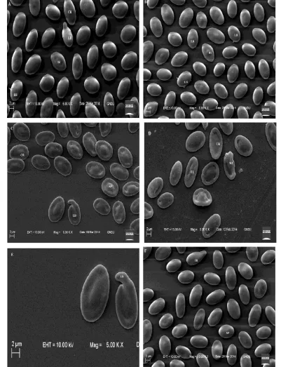
Scanning electron micrograph of erythrocyte in the control group showed elliptical shape (Figure 1A). On exposure to mercuric chloride, alterations in the morphology of erythrocytes were observed.
Change in the shape of erythrocytes was observed from elliptical to spherical (spherocytes), when the fish was exposed to 0.020 mg/L for 15 days (Figure 1B). An increase in the frequency of spherocytes was noticed with increasing exposure duration i.e., 30 days (Figure 1F) and 60 days (Figure 2B). Increasing concentration of the toxicant for different exposure period also enumerated an increase in the number of such cells. Number of elongated cells also increased with exposure concentration (Figure 1C-D).
concentration. The recovery experiments after 30 days showed a little improvement but still alterations were observed (Figure 2F, 3A-B).
Discussion
The results of the present study explicitly exhibit substantial changes in the morphology of erythrocytes of fish Channa punctatus exposed to sublethal concentrations of mercuric chloride. Marked increase in the alterations was observed in a duration and dose dependent manner. Stress condition such as presence of heavy metals in water lead to physiopathological
changes, which can be studied through hematological profile of the organism (Nussey et al., 1995). Changes in the morphology of the erythrocytes of fish exposed to mercuric chloride indicate poor health status of the fish. Round and swollen RBCs are named as spherocytes (Sawhney and Johal, 2000). According to Dey et al., 1999; Sawhney and Johal, 2000; Massar et al., 2012, alterations in the morphology of erythrocyte in the blood of fish are associated with pesticide toxicity as well as heavy metal pollution.
Mercury has high affinity for sulfhydryl groups (SH). According to Ribarov et al. (1983), binding of mercury to sulfhydryl groups leads to their
Figure 1. Scanning electron micrograph of erythrocytes of Channa punctatus: (A) control group – elliptical shape; (B)
inactivation, which further decreases antioxidant activity of the membrane. Calcium homeostasis is also affected by inorganic mercury. There is a rapid increase in the intracellular calcium in the cells exposed to mercury due to influx from the extracellular medium. ATP depleted cells as well as cells having increased Ca2+ produce echinocytes but
restoration of appropriate ATP and calcium levels result in the return of normal shape of RBC (Nelson et al., 1983). Due to interlacing of the spicules of the crenated erythrocytes, blood flow as well as viscosity is affected (Reinhart and Chien, 1986).
The transformation of normal red blood cells of human to crenated cells can be attributed to extrinsic
Figure 2. Scanning electron micrographs of erythrocytes from treated fish showing: (A) crenated cell (CR), irregular
factors or intrinsic factors, such as ATP depletion, which is associated with the ageing of red cells as testified by Brecher and Bessis (1972). The amount of ATP acts as determinant factor for the genesis of echinocytes (Birchmeier and Singer, 1977; Feo and Mohandas, 1977; Reinhart and Chien, 1987). According to Nikinmaa and Huestis (1984), adequate amount of ATP is required for the maintenance of structural integrity of nucleated RBC.
The present observations suggest that mercuric chloride have echinocytogenic properties, as crenation is the early stage of echinocyte formation. Crenation of RBC membrane has been reported in fish, Cyprinus carpio, inhabiting a polluted lake in North East India by Massar et al. (2012). Echinocyte formation results in the expansion of plasma membrane, which leads to swelling of RBC before lysing was also observed by Isomaa et al. (1986). Therefore, various alterations induced by mercuric chloride in the current study can be correlated with altered surface membrane area to volume ratio of the cell as reported by Naskar et al. (2006) in Clarias batrachus due to aluminium toxicity. Hemolysis of erythrocytes may be witnessed at the higher concentrations of mercuric chloride or other echinocytogenic agents. Spherocytes observed in the current study were also reported by Agarwal et al. (1990) on giving single as well as repeated exposures of methyl isocynate to rat. According to Tanaka and Nakai (1977), the normal biconcave shape of rat erythrocyte changes to spherocyte on exposure to mercuric chloride.
The formation of lobopodia and contraction of erythrocyte from one side as observed in the present study has also been observed in the fish, Channa punctatus exposed to malathion (Sawhney and Johal, 2000). Similar results have also been reported by Massar et al. (2012). Abnormally shaped red blood cells, codocytes observed in the current study has been reported by Naskar et al., 2006.
Zeni et al. (2002) in a study on fish Ictalurus melas observed occurrence of echinocytosis when exposed to sublethal concentrations of an anionic
detergent. The ultrastructural changes on exposure to azo dye, basic violet- 1(Cl: 42535) in erythrocyte of Labeo rohita were irregular margins, echinocytosis, lobopodial projections, spherocytosis and tear- drop shaped cells as reviewed by Kaur and Kaur (2015). The frequency of such alterations increased with increasing concentration and time duration.
According to Barnhart et al. (1983), alterations in the morphology of RBC symbolize the pathologic state. On exposure to the toxicant, fish counteract reduced oxygen level by increasing the blood components that are responsible for uptake of oxygen (Gill et al., 1991). Therefore alteration in the red cell morphology can be correlated to the fulfillment of oxygen requirement of the animal. Escudero et al. (1998) suggested that the change in the morphology of surface membrane could be attributed to the deviation in the normal membrane lipid composition.
In conclusion, the scanning electron microscopic observations revealed morphological damage to the erythrocytes of Channa punctatus. The degree of damage is directly proportional to the exposure duration and concentration of toxicant and is developed to help fish to encounter hypoxic and toxic ambient conditions. The monitoring of morphological alterations in fish blood cells is a highly sensitive way to assess the effects of toxicants.
Acknowledgements
The authors would like to thank Department of Zoology, Guru Nanak Dev University, Amritsar for providing necessary laboratory facilities, SEM facility of the university for carrying out the work and Department of Science and Technology, New Delhi for providing financial assistance.
Declaration of Conflicting Interests
The author(s) declared no potential conflicts of interest with respect to the research, authorship, and/or publication of this article.
Figure 3. Scanning electron micrographs of erythrocytes exposed to recovery: (A) 0.027 mg/L for 30d; (B) 0.040 mg/L,
References
Agarwal D, Gupta GSD, Shukla JS, Dutta KK, Ray PK. 1990. Effect of methyl isocynate (MIC) on rate erythrocytes. Archives of Toxicology. 64(4):332-335. doi:10.1007/BF01972995
2APHA/AWWA/WEF. 2005. Standard methods for the examination of water and wasterwater. Vol 21. American Public Health Association, American Water Works Association, Washington, U.S.A.
Barnhart MI, Wallace MA, Lusher LM. 1983. Red blood cells. In: Biomedical Research Applications of Scanning Electron Microscopy (eds. Hodges, GM and Carr KE). Academic Press New York. 3:171-243. Birchmeier W, Singer SJ. 1977. On the mechamism of
ATP-induced shape changes in human erythrocyte membranes. II: The role of ATP. Journal of Cell Biology. 73(3):647-659.
Brecher G, Bessis M. 1972. Present status of spiculed red cells and their relationship to the discocyte-echinocyte transformation: A critical review. Blood. 40:333-344. Dey S, Arjun J, Das M. 1999. Erythrocyte membrane
dynamics in albino mice offspring born to females with lead-induced toxicity during pregnancy: A scanning electron microscopic study. Biomedical Letters. 59(231):55-66.
EPA. 1996. Fish acute toxicity test, freshwater and marine. Environmental Protection Agency, United States. pp:1-11.
EPA. 2002. Short-term methods for estimating the chronic toxicity of effluents and receiving waters to freshwater organisms. 4th edn. Environmental Protection Agency, United States. pp:1-45.
Escudero A, Montilla JC, Garcia JM, Sanchez- Quevedo MC, Periago JL, Hortelano P, Suarez MD. 1998. Effect of dietary (n-9), (n-6) and (n-3) fatty acid on membrane composition and morphology of rat erythrocytes.
Biochimica et Biophysica Acta. 1394(1):65-73. doi:10.1016/S0005-2760(98)00095-2
Faggio C, Piccione G, Marafioti S, Arfuso F, Fortino G, Fazio F. 2014. Metabolic response to monthly variations of Sparus aurata reared in mediterranean off-shore tanks. Turkish Journal of Fisheries and Aquatic Sciences.14:567-574. doi:10.4194/1303-2712-v14_2_28
Fazio F, Marafioti S, Arfuso F, Piccione G, Faggio C. 2013a. Influence of different salinity on haematological and biochemical parameters of the widely cultured mullet, Mugil cephalus. Marine and Freshwater Behaviour and Physiology. 46(4):211-218.
Fazio F, Faggio C, Marafioti S, Torre A, Sanfilippo M, Piccione G. 2013b. Effect of water quality on hematological and biochemical parameters of Gobius niger caught in Faro lake (Sicily). Iranian Journal of Fisheries Sciences. 12(1):219-231.
Fazio F, Piccione G, Tribulato K, Ferrantelli V, Giangrosso G, Arfuso F, Faggio C. 2014. Bioaccumulation of heavy metals in blood and tissue of striped mullet in two Italian Lakes. Journal of aquatic animal health.
26(4):278-284.
Feo C, Mohandas N. 1977. Clarification of role of ATP in red-cell morphology and function. Nature. 265:166-168.
Gill TS, Pande J, Tewari H. 1991. Hemopathological changes associated with experimental aldicarb poisoning in fish (Puntius conchonius Hamilton).
Bulletin of Environmental Contamination and
Toxicology. 47:628-633.
Gupta N, Dua A. 2002. Mercury induced architectural alterations in gill surface of a fresh water fish, Channa punctatus. Journal of Environmental Biology.23(4):383-386.
Hedayati A, Ghaffari Z. 2013. Effect of mercuric chloride on some hematological, biochemical parameters in silver carp (Hypophthalmichthys molitrix).
International Journal of Veterinary Medicine:
Research & Reports. pp:1-11. doi:
10.5171/2013.183410
Hesser EF. 1960. Methods for routine on fish hematology. The Progressive Fish-Culturist. 22(4):164-171. doi: 10.1577/1548-8659(1960)22[164:MFRFH]2.0.CO;2 Isomaa B, Hagerstrand H, Paatero G, Engblom AC. 1986.
Permeability alterations and antihemolysis induced by amphiphiles in human erythrocytes. Biochimica et Biophysica Acta. 860(3):510-524. doi:10.1016/0005-2736(86)90548-1
Juneja CJ, Mahajan CL. 1983. Hematological and haemopoietic changes in fish Channa punctatus due to mercury pollution in water. Indian Journal of Animal Research. 17(2):63-71.
Kaur K, Kaur A. 2015. Fish erythrocytes as biomarkers for the toxicity of sublethal doses of an azo dye, basic violet-1 (CI: 42535). Microscopy and Microanalysis. 21(1):264-273.
Massar B, Dey S, Barua R, Dutta, K. 2012. Microscopy and microanalysis of hematological parameters in common carp, Cyprinus carpio, inhabiting a polluted lake in north east India. Microscopy and Microanalysis. 18(5):1077-1087. doi: 10.1017/S1431927612001432 Mwachiro EC, Druve VS. 1997. Heavy metal status of the
reservoir Bari near Udaipur (Rajasthan) and the accumulation of metals in fish organs. Pollution Research.162:67-74.
Naskar R, Sen NS, Ahmad MF. 2006. Aluminium toxicity induced poikilocytosis in an air breathing teleost,
Clarias batrachus (Linn.). Indian Journal of Experimental Biology. 44(1):83-85.
Nelson GA, Andrews ML, Karnovsky MJ. 1983. Control of erythrocyte shape by calmodulin. Journal of Cell Biology. 96(3):730-735.
Nikinmaa M, Huestis WH. 1984. Shape changes in goose erythrocytes. Biochimica et Biophysica Acta. 773(2):317-320. doi:10.1016/0005-2736(84)90096-8 Nussey G, Vuren JHJ Van, Preez HH du. 1995. Effect of
copper on the haematology and osmoregulation of the Mozambique tilapia, Oreochromis mossambicus
(Cichlidae). Comparative Biochemistry and Physiology
Part C: Pharmacology, Toxicology and
Endocrinology.111(3):369-380. doi:10.1016/0742-8413(95)00063-1
Reinhart WH, Chien S. 1986. Red cell rheology in stomatocyte-echinocyte transformation: roles of cell geometry and cell shape. Blood. 67(4):1110-1118. Reinhart WH, Chien S. 1987. Echinocyte-stomatocyte
transformation and shape control of human red blood cells: morphological aspects. American Jounal of Haematology. 24(1):1-14.
Ribarov SR, Benov LC, Benchev IC. 1983. On the mechanism of mercury-induced hemolysis. General Physiology and Biophysics. 2:81-84.
Sawhney AK, Johal MS. 2000. Erythrocyte alterations induced by malathion in Channa punctatus (Bloch).
Bulletin of Environmental Contamination and
Toxicology. 64(3):398-405.
doi:10.1007/s001280000014.
Tanaka R, Nakai K. 1977. Hemolysis and morphological changes in rat erythrocytes with mercurial. Japanese Journal of Pharmacology. 27(3):413-419.
Vergilio CS, Carvalho CEV, Melo EJT. 2012. Accumulation and Histopathological Effects of Mercury Chloride after Acute Exposure in Tropical
Fish Gymnotus carapo. Journal of Chemical Health Risks. 2(4):01-08.
Vutukuru SS, Basani K. 2013. Acute effects of mercuric chloride on glycogen and protein content of Zebra fish, Danio rerio. Journal of Environmental Biology. 34(2): 277.