Lidia Usnarska-Zubkiewicz
1, A, C–D, Marta Strutyńska-Karpińska
2, A–B, F,
Agnieszka Zubkiewicz-Kucharska
3, B–C, Paweł Zarębski
2, C–D,
Krzysztof Grabowski
2, ESoluble Urokinase-Type Plasminogen Activator
Receptor and Ferritin Concentration in Patients
with Advanced Alimentary Tract Carcinoma.
Relationship to Localization, Surgical Treatment
and the Stage of the Disease – Preliminary Report
1 Department of Haematology, Blood Neoplasms, and Bone Marrow Transplantation,Wroclaw Medical University, Poland
2 Department of Gastrointestinal and General Surgery, Wroclaw Medical University, Poland
3 Department of Endocrinology and Diabetology for Children and Adolescents, Wroclaw Medical University, Poland
A – research concept and design; B – collection and/or assembly of data; C – data analysis and interpretation;
D – writing the article; E – critical revision of the article; F – final approval of article; G – other
Abstract
Background. The urokinase plasminogen activation system is associated with metastatic potential of cancer in several tumors. Its specific membrane receptor (uPAR) is released from cancer cells and can be detected as the soluble fraction of the plasminogen activator receptor (suPAR). Ferritin (FRT) is a poor prognostic factor in vari-ous neoplasms.
Objectives. We analyzed the serum concentrations of suPAR and FRT in patients with gastrointestinal cancer. Tumor localization, stage of the disease, possibility of surgery and histopathological diagnosis were considered.
Material and Methods. The analysis involved 48 patients (8 females/40 males) treated in the Department of Gastrointestinal and General Surgery, Wroclaw Medical University. Thirty two patients had esophageal, 7–gastric, 9-colorectal cancer. Fifteen patients underwent resection surgery, 33 palliative therapy. The control comprised 10 healthy donors. The serum concentration of suPAR was determined by enzyme-linked immunosorbent assay (ELISA), expressed in pg/mL. Concentration of the serum FRT was detected using immunonephelometry method, expressed in µg/L.
Results. Serum concentration of suPAR ranged from 1789 –7320, x = 3676.2, SD = 1042 and was significantly higher (p = 0.0002) than in the control group. In patients who underwent palliative therapy, the concentration of suPAR was significantly higher (p = 0.05) than in those after resection, also in patients with esophageal cancer com-pared to those with colorectal one (p = 0.02). Serum concentration of FRT in patients with gastrointestinal cancer was significantly higher than in control group. Serum FRT concentration was higher in patients with esophageal cancer compared to patients with gastric cancer (p = 0.05), in persons with IV compared to patients with I–III stage of the disease, patients who underwent palliative compared to surgical therapy.
Conclusions. In patients with gastrointestinal cancer the level of suPAR is high, with highest values in advances disease with remote metastases. The FRT concentration is sensitive indicator of the disease process: its level is high-est in pts with IV stage who underwent palliative therapy (Adv Clin Exp Med 2014, 23, 6, 959–967).
Key words: colorectal cancer, esophageal cancer, gastric cancer, suPAR concentration, FRT concentration. Adv Clin Exp Med 2014, 23, 6, 959–967
ISSN 1899–5276
ORIGINAL PAPERS
© Copyright by Wroclaw Medical University
The stage of the tumor is one of the most im-portant prognostic factors in alimentary tract can-cers, and the evaluation of the clinical stage of the
The invasion of cancer cells, both, in situ, as well as leading to metastasis, is affected by the uro-kinase-type plasminogen activator (uPA) system which consists of the urokinase-type plasminogen activator (uPA), tissue-type plasminogen activator (tPA), the uPA receptor (the urokinase-type plas-monogen activator receptor: uPAR or CD87), its form deprived of the glycolipid anchor, the soluble form of uPAR: suPAR, and 2 major inhibitors: the plasmionogen activator inhibitor-1 (PAI-1) and plasminogen activator inhibitor-2 (PAI-2). The tPA is mainly involved in intravascular thrombol-ysis, while uPA is involved in pericellular prote-olysis, and plays the main role in the process of plasmin formation from plasminogen. Plasmin hydrolyses fibrin bonds, fibronectin and lamin and activates metalloproteinases, which digest col-lagen and other matrix proteins [1, 2]. This tissue remodeling together with matrix desorganization stimulate the development of endothelium and the formation of new blood vessels which supply the tumor tissue and enable the tumor cells to pass the anatomical barrier separating the tumor from the host’s healthy tissues [3–5]. Recent studies have demonstrated that neovascularisation is an essen-tial metastasis risk factor in patients suffering from colorectal cancer [6–8]. It was shown that blocking the activity of uPA and/or uPAR, e.g. by means of endostatin, may inhibit angiogenesis and the inva-sion of the tumor [9]. Przybyłowska demonstrat-ed a correlation between the level of uPAR and the density of blood vessels in cancer tissue of patients with colorectal cancer [10]. Thus uPAR is func-tionally engaged in cancer invasion and its high levels affect the survival time in patients with gas-tric, endometrial and breast cancers [11–13]. As a result of enzymatic exfoliation of uPAR from the surface of tumor cells and/or matrix cells, the plas-ma level of suPAR in patients with cancer of lung, breast, ovarian and colorectal was significantly higher than in healthy subjects [14–17]. Stephens, who examined 591 patients before surgical treat-ment, was first to prove that high level of suPAR is a poor prognostic factor in cancers in the lower part of alimentary tract [18]. Similar observations were reported by Fernebro and Riisbro [17, 19].
Association between the level of stored iron and cancers risk has been investigated by research-ers for many years. As the data shows, a high lev-el of iron may be observed in cancer of the pan-creas, liver, esophagus, stomach, colon, breast, as well as multiple myeloma and other hematological malignancies [20–25].
FRT is the major Fe storage protein in the cells and at the same time an acute phase protein. One FRT molecule is able to store about 4500 Fe3 +. FRT is present in the liver, spleen, bone marrow cells
and serum. An estimation of serum concentrations of FRT is a good indicator of resources of Fe and is the most common clinical method of assessing the concentration of Fe in patients without conditions that cause acute phase responses such as inflamma-tion, infection. The main reason for the increased concentration of FRT are states of Fe overload, or block of the availability of Fe.
In Fe overload conditions, unbound Fe gener-ates oxygen free radicals which are mutagens and cause damage to cellular and sub-cellular mem-branes, cellular organelles memmem-branes, leading to failure of cells, tissues and organs [26]. Stevens first reported that there is a link between the satu-ration of the transferin (iron binding transporter), and the risk of colorectal cancer [27]. In 33 stud-ies analyzed by Nelson in 2001 it has been shown that there is a positive association between iron in diet and colon cancer risk [21]. Nelson stated that the development of precancerous lesions in colon, colonic adenomas and polyps is correlated with body iron storage [20]. In 2003 Elmberg described an increased risk of extrahepatic cancers and he-patocellular cancer in patients suffering from hae-mochromatosis [28]. In addition, a risk of cancers among blood donors is lower, and it correlates with the frequency of donation and in the same time iron loss [29]. Data may indicate the partici-pation of Fe in the process of carcinogenesis.
The study was undertaken to evaluate the se-rum concentration of suPAR and ferritin in pa-tients with alimentary tract cancer in relation to tumor localization, stage of the disease, possibility of surgery and histopathological diagnosis.
Material and Methods
Blood samples (5 mL) for analysis were tak-en at diagnosis; the serum conctak-entration of su-PAR was determined by enzyme-linked immuno-sorbent assay (ELISA) and expressed in pg/mL. In all patients serum FRT concentration was detected using immunonefelometry method and expressed in µg/L. Patients with iron deficiency anemia were excluded, all were free of infectious complication at the time of study.
The control group comprised 10 (5/5 F/M) healthy donors. Written informed consent was ob-tained from all patients and donors.
Statistical evaluation was performed using the Student’s t, c2 and Fisher’s exact tests. All the re-sults were considered statistically significant for p< 0.05. All statistical analyses were performed in Statistica software (StatSoft, Inc. (2011). STATIS-TICA (data analysis software system), version 10. www.statsoft.com.).
Results
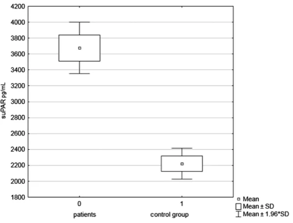
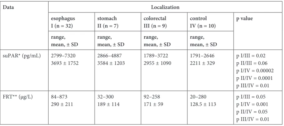
The serum concentration of suPAR in patients with gastrointestinal cancer ranged from 1789 to 7320, x = 3676.2, SD = 1042 and was significant-ly higher (p = 0.0002) than in the control group: 1791–2646, x = 2211, SD = 329 (Fig. 1). The serum concentration of suPAR in patients with esopha-geal and gastric cancer as well as colorectal cancer was significantly higher than in the control group p = 0.00002, p = 0.0001, p = 0.01 respectively. In patients with esophageal cancer, suPAR concentra-tion was significantly higher than in patients with colorectal cancer (p = 0.02) and ranged respec-tively 2799–7320, x = 3693, SD = 1752 and 1789– –3722, x = 2955, SD = 1090. The suPAR concen-tration did not differ between patients with gastric and colorectal cancer; however, patients with gas-tric cancer revealed a tendency to have a higher su-PAR concentration (p = 0.06) (Table 2).
Table. 1. Basic clinical data of patients with advanced alimentary tract carcinoma at diagnosis
Number of patients (F/M) 48 (8/40)
Age (years), range, mean 39–82 (mean: 61.2)
Localization (F/M) esophagus 32 (3/29)
stomach 7 (2/5)
colon 9 (1/8)
Clinical stage esophagus II//III/IV 1/3/28
stomach I/III/IV 1/3/3
colon III/IV 4/5
Treatment palliative 26
resection (esophagus/stomach/colon) 15 (5/4/6)
The serum concentration of FRT in patients with esophageal and gastric cancer as well as colorec-tal cancer was significantly higher than in the con-trol group p = 0.001, p = 0.05, p = 0.01 respectively. The serum FRT concentration was the highest in patients with esophageal cancer compared with pa-tients with colorectal cancer (p = 0.05) and ranged respectively 84–873 (mean = 290, SD = 211) and 92–258 (mean = 171, SD = 59).
Patients in the clinical stages I, II and III (sub-group A) had a lower expression of suPAR than patients in stage IV (subgroup B), but the differ-ence was not statistically significant (Table 3). The serum FRT concentration displayed a similar trend with range of 32–271 (mean = 126, SD = 156) and 65–873 (mean = 293, SD = 210) respective-ly, and the difference was statistically significance (p = 0.01).
Fifteen patients who underwent total resec-tion of the neoplastic lesion were markedly young-er (p = 0.02) than those with advanced stage who were subjected for palliative therapy; it ranged 58 ± 6.2 years and 65 ± 8.4 years respectively. The suPAR concentration in patients who underwent resection was significantly lower (p = 0.05) than in patients who underwent palliative therapy, it being 1789–3745, mean = 3078, SD = 922 and 2573–7320, mean = 3898, SD = 1155 respectively (Table 4). A similar trend was observed for FRT concentra-tion, which was lower in the surgical group com-pared with the palliative one (p = 0.01).
Patients with squamous cell esophageal carci-noma had the same concentration of suPAR as the patients with gastric or colorectal adenocarcino-ma (Table 5). However, the serum concentration of FRT was higher in patients with squamous cell
Table 2. The serum concentration of suPAR and FRT in patients with alimentary tract carcinoma according to localization
Data Localization
esophagus
I (n = 32) stomachII (n = 7) colorectalIII (n = 9) controlIV (n = 10) p value
range,
mean, ± SD range, mean, ± SD range, mean, ± SD range, mean, ± SD
suPAR* (pg/mL) 2799–7320
3693 ± 1752 2866–48873584 ± 1203 1789–37222955 ± 1090 1791–26462211 ± 329 p I/III = 0.02p II/III = 0.06 pI/IV = 0.00002 p II/IV = 0.0001 p III/IV = 0.01 FRT** (µg/L) 84–873
290 ± 211 32–300189 ± 114 92–258 171 ± 59 20–280128.5 ± 113 p I/III = 0.05p I/IV = 0.001 p II/IV = 0.05 pIII/IV = 0.01 *suPAR – soluble urokinase-type plasminogen activator receptor.
** FRT – ferritin.
Table 3. The serum concentration of suPAR and FRT in patients with alimentary tract carcinoma according to stage of the disease
Stage of the disease
I/II/III IV p value
Data A (n = 12) B (n = 36)
range, mean, ± SD range, mean, ± SD
suPAR* (pg/mL) 1789–4203
3178 ± 972 2751–73203842 ± 1159 p A/B = ns.p A/C = 0.005 p B/C = 0.00004
FRT** (µg/L) 32–271
126 ± 156 65–873293 ± 210 p A/B = 0.01
carcinoma compared to adenocarcinoma (p = 0.04) and ranged 84–873 (mean = 292, SD = 211) and 32–300, (mean = 148, SD = 118) respectively.
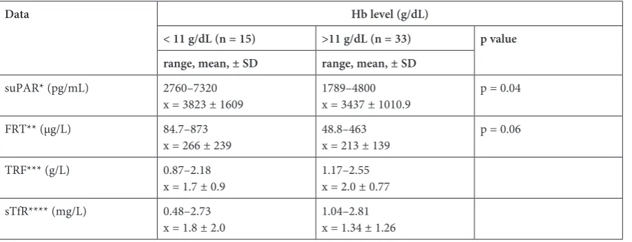
Hemoglobin levels ranged from 7.6 to 15.8 g/dL. (mean = 12.8, SD = 1.8 g/dL). Fifteen patients (10/32 with esophageal, 2/7 with gastric and 3/9 with colorectal cancer) were diagnosed with anaemia (Hb 7.6–11.0 g/dL). All the patients had normocytic anaemia; they did not demon-strate bleeding from the digestive tract (negative benzidine test, normal iron level) or haemolysis (normal or decreased reticulocyte number, normal serum bilirubin level). In patients with anaemia (subgroup A) suPAR concentration was signifi-cantly higher (p = 0.04) than in the patients with normal Hb levels (subgroup B), it being x = 3823.3, SD = 1609 and x = 3437, SD = 1010.9 respectively. A similar trend was observed for FRT concentra-tion (Table 6). The serum concentraconcentra-tion of FRT in subgroups A and B was significantly higher than in the control group, p = 0.05. The sTfR (serum transferrin receptor)/log ferritin ratio (sTfR/FRT index) in both group was below one, 0.248–0.938, mean = 0.742 and 0.615–1.25 mean = 0.97.
Discussion
According to the world data of 2003 on the in-cidence of malignant tumors, every third patient from 10 million new cancer cases in the world was diagnosed with digestive cancer [30]. Second place in this group is occupied by colorectal can-cer, with the same incidence rate for males and fe-males. On the other hand, as far as mortality rate is concerned, stomach cancer occupies 2nd place as the reason of death in men and 4th in women, while oesophageal carcinoma, which has the poor-est prognosis among digpoor-estive cancers, with high prevalence among men, occupies 8th place among malignant tumors [30–33].
Surgical treatment remains the central ther-apeutic modality. The selection of an adequate therapeutic modality is strictly correlated with the severity of the tumor [34–36]. In patients with esophageal carcinoma, despite extensive surgical intervention, 5-year survival after surgery alone is observed in only about 20% of patients. Almost half of the patients with locally advanced tumors will die following cancer recurrence within the first 2 years after surgical resection [32, 36]. This means that at the time of diagnosis and surgical
Table 4. The serum concentration of suPAR and FRT in patients with alimentary tract carcinoma qualified for surgery or palliative therapy
Data Treatment
surgical (n = 15) palliative (n = 33) p value
range, mean, ±SD range, mean, ± SD
suPAR* (pg/mL) 1789–3745
3078, ± 922 2573–73203898, ± 1155 p = 0.05
FRT** (µg/L) 32–408
84–873 96.8 ± 56295.7 ± 214 p = 0.01
* suPAR – soluble urokinase-type plasminogen activator receptor. ** FRT – ferritin.
Table 5. The serum concentration of suPAR and FRT in patients with alimentary tract carcinoma according to histopatho-logical diagnosis
Data Histopathological diagnosis
squamous cell carcinoma
(n = 32) adenocarcinoma (n = 16) p value
range, mean, ± SD range, mean, ± SD
suPAR* (pg/mL) 2799–7320
3693 ± 1752 1789–48873269 ± 1213 p = ns.
FRT** (µg/L) 84–873
290 ± 211 96.8 ± 56295.7 ± 214 p = 0.01
intervention the cancer has already spread far be-yond its primary focus in the gastrointestinal tract. Thus, the search for parameters which would pre-dict systemic changes in the course of oesophageal carcinoma has become an urgent need.
Numerous changes in the activity of uPA, uPAR and PAI-1 in matrix cells, among others, uPA expression on fibroblast-like cells, uPAR on macrophages and cancer cells as well as PAI-1 ex-pression on myofibroblasts were demonstrated in digestive cancers. The changes evidence a direct participation of fibrinolysis in remodeling and dis-semination of the tumor [37–39]. Seetoo demon-strated that the overexpression of uPA, uPAR and PAI 1 was significantly associated with metasta-sis to the liver in patients suffering from colorec-tal cancer [40]. Shiomi examined 56 patients after esophagectomy and did not observe the presence of uPA, uPAR and PAI-2 in healthy oesophageal tissue, while uPA was found on cancer cells on the tumor margins. Patients with uPA-positive cells more often displayed tumor infiltration beyond muscularis propria and metastases to the lymph nodes [41].
Moreover, other authors reported a neg-ative correlation between uPAR, uPA and/or PAI-1 expression in cancer cells and the survival time and stage of tumor in gastric and colorectal cancer [42, 43].
In our material the serum concentration of su-PAR was significantly higher than in the control group. The highest levels were observed in patients with oesophageal carcinoma and they were signif-icantly higher than in colorectal cancer. Oesopha-geal carcinoma develops insidiously and for a long time does not produce characteristic symptoms.
Difficulties with swallowing, as the first and prima-ry symptom of cancer, as a rule occur in advanced stages of the disease, when the neoplastic infiltra-tion has already involved all layers of the oesoph-ageal wall and extended beyond them. The detec-tion of a potentially early form of carcinoma in the asymptomatic stage is associated with implemen-tation of a screening programme, which unfor-tunately has not been considered justified yet. In the studied group of 32 patients with oesophageal carcinoma, only 4 patients (12.5%) were in a stage lower than IV, and resection surgery was possible only in 5 persons (15.6%). Among patients with stomach cancer and colorectal cancer, 50% were staged lower than IV and 62.5% were qualified for resection surgery. The level of suPAR in the sub-group of patients in stage I–III of the disease was lower than in patients in stage IV, this finding was similar to data reported by Stephens and Riisbro; moreover, in patients undergoing resection it was significantly lower in comparison to patients who did not qualify for resection surgery [17, 18]. In the study by Stephens, high preoperative level of su-PAR increased significantly the risk of death [18]. Our studies may also correspond to the findings by other authors who analyzed the levels of uPA and uPAR as well as uPA and uPAR mRNA in tumour tissue. Chen investigated 67 patients with stomach cancer and demonstrated that the level of uPA and uPAR was significantly increased in stomach can-cer; moreover, patients with uPA and uPAR ex-pression had significantly lower survival time in comparison to patients who were free from such an expression [11].
In the studies by Zhanga performed on 105 gastric tumor tissue specimens, cells with uPA and
Table 6. The serum concentration of suPAR, and FRT, TRF and sTfR in patients with alimentary tract carcinoma in relation to the level of hemoglobin
Data Hb level (g/dL)
< 11 g/dL (n = 15) >11 g/dL (n = 33) p value
range, mean, ± SD range, mean, ± SD
suPAR* (pg/mL) 2760–7320
x = 3823 ± 1609 1789–4800x = 3437 ± 1010.9 p = 0.04
FRT** (µg/L) 84.7–873
x = 266 ± 239 48.8–463x = 213 ± 139 p = 0.06
TRF*** (g/L) 0.87–2.18
x = 1.7 ± 0.9 1.17–2.55x = 2.0 ± 0.77
sTfR**** (mg/L) 0.48–2.73
x = 1.8 ± 2.0 1.04–2.81x = 1.34 ± 1.26 * suPAR – soluble urokinase-type plasminogen activator receptor.
** FRT – ferritin. *** TRF – transferrin.
uPAR expression were found to infiltrate the mus-cular coat, the peritoneum and the greater omen-tum [44]. Hogdall analyzed 567 patients suffering from colorectal cancer and demonstrated that in-creased levels of suPAR in association with low levels of PAI-1 were a poor prognostic factor and indicated the presence of remote metastases [45]. As soon as in 1995, Heiss proved that the expres-sion of uPAR in solid tumors may be an indica-tor of tumor cells dissemination in the bone mar-row [46]. Prior examinations revealed the presence of microdeposits of epithelial cells in bone marrow of patients with oesophageal carcinoma [46].
Already in 1983, Ludwig and Linkesch showed that the concentration of FRT in myeloma patients correlated with tumor mass and the concentration of beta-2-microglobulin [48]. Ludwig research published in 2014 confirmed the importance of fer-ritin as an independent prognostic factor for PFS and OS in myeloma patients undergoing autolo-gous transplantation [22]. Italian research group showed that ferritin subunit L increases angiogen-esis and cells proliferation rate in an iron-indepen-dent manner [49]. In cases of inhibition of ferri-tin expression, the growth of cells arrested in G1/S phase was resumed. An antiapoptotic effect of FRT not related with its iron-biding capacity was shown [50]. FRT is inducted by proinflamatory cy-tokines, on the other hand, FRT inhibits apopto-sis inducted by TNF-alpha by suppressing reactive oxygen species. [51]. The correlation between NF- -kappa B and ferritin has been shown for inflam-matory cells and fibroblasts, which play a great role in the microenvironment of tumor in cancer.
The serum concentration of FRT in patients with esophageal and gastric cancer as well as colorectal cancer was significantly higher than in the control group, in particular it was high in pa-tients with esophageal cancer compared to papa-tients with colorectal cancer (p = 0.05), in persons with IV stage compared to patients with I, II and III stage of the disease, patients who underwent palliative therapy, compared to patients who underwent to
surgical therapy. Consistent with our observations are Alkhateeb et al. studies in breast cancer, which indicated that the concentration of FRT correlates with tumor stage and histological grade. Moreover, the authors demonstrated that ferritin may be pro-duced by tumor infiltrating macrophages.
The level of FRT in squamous cell carcinoma was significantly higher than in adenocarcinomas. Squamous cell carcinomas involved patients with oesophageal carcinoma in most advanced stages. However we cannot exclude that the difference may have resulted from different metastatic poten-tials of both histological types of the tumor. Signif-icantly elevated level of FRT in comparison to the control group was also observed among patients with anaemia. In this group iron deficiency anemia was excluded by calculation of sTfR/log ferritin ra-tio (sTfR/FRT index), which was 0.742. Interest-ingly, both the level of suPAR as well as FRT con-centration were highest in oesophageal carcinoma. Just as in myeloma patients, FRT can be produced by stromal cells of the tumor and stimulate tumor cell proliferation. This requires further research and observation on a larger group of patients.
Concluding, suPAR as well as the serum lev-el of FRT in patients suffering from oesophageal, stomach and colorectal cancers, who did not de-velop bleeding from the digestive tract, is a sensi-tive marker of the activity of the pathological pro-cess and may be a significant prognostic marker while making therapeutic decisions.
According to Dass, the investigation of the uPA system may be a useful tool in the treatment of cancer patients, the evaluation of the efficacy of prophylaxis and sensitivity of neoplasms to new chemotherapeutic agents [52]. Already in 1994, Fazioli presented a concept of therapy for patients suffering, among others, from colorectal cancer, which consisted in the administration of recom-bined uPA receptor or receptor-blocking anti-bodies in order to inhibit the activity of uPA sys-tem [53]. Studies on gene therapy used to inhibit uPAR expression are currently undergoing [54].
References
[1] Dano K, Andreasen PA, Grondahl-Hansen J, Kristensen P, Nielsen LS, Skriver L: Plasminogen activators, tissue degradation and cancer. Adv Cancer Res 1985, 44, 139–266.
[2] Egeblad M, Werb Z: New function for the matrix metalloproteinases in cancer progression. Nat Rev Cancer 2002, 2, 163–176.
[3] Yu HR, Schultz RM: Relationship between secreted urokinase plasminogen activator activity and metastatic potential in murine B16 cells transfected with human urokinase sense and antisense genes. Cancer Res 1990, 50, 7623–7633.
[4] Shapiro RL, Duquette JG, Roses DF, Nunes I, Harris MN, Kamino H, Wilson EL, Rifkin DB: Induction of primary cutaneous melanocytic neoplasms in urokinase-type plasminogen activator (uPA)-deficient type mice: cellular blue nevi invade but do not progress to malignant melanoma in uPA-deficient animals. Cancer Res 1996, 56, 3597–3604.
[6] Shpitz B, Gochberg S, Neufeld D, Grankin M, Buklan G, Klein E, Bernheim J: Angiogenic switch in earliest stages of human colonic tumorigenesis. Anticancer Res 2003, 23, 5153–5157.
[7] Pang RW, Poon RT: Clinical implications of angiogenesis in cancers. Vasc Health Risk Manag 2006, 2, 97–108.
[8] Rajaganeshan R, Prasad R, Guillou PJ, Chlmers CR, Scott N, Sarkar R, Poston G, Jayne DG: The influence of invasive growth pattern and microvessel density on prognosis in colorectal cancer and colorectal liver metastases. Br J Cancer 2007, 96, 1112–1117.
[9] Dkhissi F, Lu H, Soria C, Opolon P, Griscell F, Liu H, Khattar P, Mishal Z, Pericaudet M, Li H: Endostatin exhibits a direct antitumor effect in addition to its antiagiogenic activity in colon cancer cells. Hum Gene Ther 2003, 14, 997–1008.
[10] Przybyłowska K, Szemraj J. Kulig A, Dziki A, Ulanska J, Blasiak J: Antigen levels of urokinaze type plasminogen activator receptor and its gen polymorphism relate to microvassel density in colorectal cancer. Acta Bioch Pol 2008, 2, 357–363.
[11] Ji F, Chen YL, Jin RY, Wang WL, Yang ZL, Li YM: Relationship between matrix metalloproteinase-2 mRNA expression and clinicopathological and urokinase-type plasminogen activator system parameters and prognosis in human gastric cancer. World J Gastroenterol 2005, 11, 3222–3226.
[12] Memarzadeh S, Kozak KR, Chang L, Natorajan S, Shintaku P, Reddy ST, Farias-Eisner R: Urokinase plasmino-gen activator receptor: Prognostic biomarker for endometrial cancer. Proc Natl Acad Sci USA 2002, 99, 10647– –10652.
[13] Stillfried GE, Saunders DN, Ranson M: Plasminogen binding and activation at the breast cancer cell surface: the integral role of urokinase activity. Breast Cancer Res 2007, 9.
[14] Pappot H, Høyer-Hansen G, Rønne E, Hansen HH, Brünner N, Danø K, Grøndahl-Hansen J: Elevated plasma lev-els of urokinase plasminogen activator receptor in non-small lung cancer patients. Eur J Cancer 1997, 33, 867–872.
[15] Riisbro R, Christensen IJ, Piiroren T, Greenall M, Larsen B, Stephens RW, Han C, Heyer-Hansen G, Smith K, Brünner N, Harris AL: Prognostic significance of soluble urokinase plasminogen activator receptor in serum and cytosol of tumor tissue from patients with primary breast cancer. Clin Cancer Res 2002, 8, 1132–1141.
[16] Sier CF, Stephens R, Bizik J, Mariani A, Bassan M, Pedersen N, Frigerio L, Ferrari A, Dano K, Brünner N, Blasi F: The level of urokinase – type plasminogen activator receptor is increased in serum of ovarian cancer patients. Cancer Res 1998, 58, 1843–1849.
[17] Riisbro R, Christensen IJ, Nielsen HJ, Brünner N, Nilbert M, Fernebro E: Preoperative plasma soluble uro-kinase plasminogen activator receptor as prognostic marker in rectal cancer patients. An EORTC-Receptor and Biomarker Group collaboration. Int J Biol Markers 2005, 20, 93–102.
[18] Stephens RW, Nielsen HJ, Christensen IJ, Thorlacius-Ussing O, Sorensen S, Dano K, Brünner N: Plasma uroki-nase receptor levels in patients with colorectal cancer: Relationship to prognosis. J Nat Cancer 1999, 10, 869–874.
[19] Fernebro E, Madsen RR, Fernö M, Brünner N, Bendahl P, Christensen IJ, Johnson A, Nilbert M: Prognostic importance of the soluble plasminogen activator receptor, suPAR, in plasma from rectal cancer patients. Eur J Cancer 2001, 37, 486–491.
[20] Nelson RL, Davis FG, Sutter E, Sobin LH, Kikkendal JW, Bowen P: Body iron stores and risk of colonic neopla-sia. J Natl Cancer Inst 1994, 86, 455–460.
[21] Nelson RL: Iron and colorectal cancer risk: Human studies: Nutr Rev 2001, 59, 140–148.
[22] Strasser-Weippl K, Ludwig H: Ferritin as prognostic marker in multiple myeloma in patient undergoing autolo-gous transplantation. Leukemia Lymphoma 2014, in press.
[23] Kalousová M, Krechler T, Jáchymová M, Kuběna AA, Zák A, Zima T: Ferritin as an independent mortality pre-dictor in patients with pancreas cancer. Results of a pilot study. Tumor Biol 2012, 33, 1685–1700, 1695–177.
[24] Alkhateeb AA, Han B, Connor JR: Ferritin stimulates breast cancer cells through an iron independent mechanism and is localized within tumor-associated macrophages. Breast Cancer Res Treat 2013, 137, 733–744.
[25] Kikuchi S, Kobune M, Iyama S, Sato T, Murase K, Kawano Y, Takada K, Ono K, Hayashi T, Miyanishi K, Sato Y, Takimoto R, Kato J: Prognostic significance of serum ferritin level at diagnosis in myelodysplastic syn-drome. Int J Hematol 2012, 95, 527–534.
[26] Dizdaroglu M, Jaruga P: Mechanisms of free radical-induced demage to DNA. Free Radic Res 2012, 26, 382–419.
[27] Stevens RG, Jones DY, Miccozzi MS, Taylor PR: Body iron stores and the risk of cancer. N Engl J Med 1988, 319, 1047–1052.
[28] Elmberg M, Hultcrantz R, Ekbom A, Brandt L, Olsson S, Olsson R, Lindgren S, Lööf L, Stål P, Wallerstedt S, Almer S, Sandberg-Gertzén H, Askling J: Cancer risk in patients with hereditary hemochromatosis and in their first-degree relatives. Gastroenterology 2003, 125, 1733–1741.
[29] Edgren G, Reilly M, Hjalgrim H, Tran TN, Rostgaard K, Adami J, Titlestad K, Shanwell A, Melbye M, Nyrén O:
Donation frequency, iron loss, and risk of cancer among blood donors. J Natl Cancer Inst 2008, 100, 572–579.
[30] Lambert R: Overview of the epidemiology and prevention of digestive cancer. World Gastroenterology News 2003, 82, 21–25.
[31] Brenner H, Rothenbacher D, Arndt V: Epidemiology of stomach cancer. Methods Mol Biol 2009, 472, 467–477.
[32] Crew KD, Neugut AI: Epidemiology of gastric cancer. World J Gastroenterol 2006, 12, 354–362.
[33] Eslick GD: Epidemiology of esophageal cancer. Gastroenterol Clin North Am 2009, 38, 17–25.
[35] Khosravi Shahi P, Diaz Munoz de la Espada VM, Garcia Alfonso P, Encina Garcia S, Izarzugaza Peron Y, Arranz Cozar JL, Hernandez Maria B, Perez Manga G: Management of gastric adenocarcinoma. Clin Transl Oncol 2007, 9, 438–442.
[36] Wilkinson N, Scott-Conner CE: Surgical therapy for colorectal adenocarcinoma. Gastroenterol Clin North Am 2008, 37, 253–267.
[37] Romer J, Nielsen BS, Ploug M: The urokinase receptor as a potential target in cancer therapy. Curr Pharm Des 2004, 10, 2359–2376.
[38] Illemann M, Hansen U, Nielsen HJ, Andreasen PA, Høyer-Hansen G, Lund LR, Danø K, Nielsen BS: Leading-edge myelofibroblast in human colon cancer express plasminogen activator inhibitor 1. Am J Clin Pathol 2004, 122, 256–265.
[39] Beyer BC, Heiss MM, Simon EH, Gruetzner KU, Babic R, Jauch KW, Schildberg FW, Allgayer M: Urokinase System expression in gastric carcinoma. Prognostic impact in an independent patient series and first evidence of predictive value in preoperative biopsy and intestinal metaplasia specimes. Cancer 2006, 106, 1026–1035.
[40] Seetoo DQ, Crowe PJ, Russell PJ, Yang JL: Quantitative expression of protein markers of plasminogen activation system in prognosis in colorectal cancer. J Surg Oncol 2003, 82, 184–193.
[41] Shiomi H, Eguchi Y, Tani T, Kodama M, Hattori T: Cellular distribution and clinical value of urokinase-type plasminogen activator, its receptor and plasminogen activator inhibitor-2 in esophageal squamous cell carcinoma. Am J Pathol 2000, 156, 567–575.
[42] Heiss MM, Allgayer H, Gruetzner KU, Babic R, Jauch KW, Schildberg FW: Clinical value of an extended bio-logical staging by bone marrow micrometastases and tumor associated proteases in gastric cancer. Ann Surg 1997, 226, 726–745, 736–744, discussion 744–745.
[43] Mulcahy HE, Duffy MJ, Gibbons D, McCarthy P, Parfrey NA, O’Donoghue DP, Sheahan K: Urokinase type plasminogen activator and outcome in Dukes’B colorectal cancer. Lancet 1994, 344, 583–684, 583–584.
[44] Zhang L, Zhao ZS, Ru GQ, Ma J: Correlative studies on uPA mRNA, uPAR mRNA expression with vascular endothelial growth factor, microvessel density, progression and survival time of patients with gastric cancer. World J Gastroenterol 2006, 12, 3970–3976.
[45] Hogdall CK, Christensen IJ, Stephens RW, Sorensen S, Norgaard-Petersen B, Nielsen HJ: Serum tetranectin is an independent prognostic marker in colorectal cancer and weakly correlated with plasma suPAR, plasma PAI-1 and serum CEA. APMIS 2002, 110, 630–638.
[46] Heiss MM, Allgayer H, Gruetzner KU, Funke I, Babic R, Jauch KW, Schildberg FW: Individual development and uPA receptor expression of disseminated tumour cells in bone marrow: a reference to early systemic disease in solid cancer. Nat Med 1995, 1, 1035–1039.
[47] Usnarska-Zubkiewicz L, Strutyńska-Karpińska M, Podolak-Dawidziak M, Nienartowicz M, Grabowski K, Prajs I, Kuliczkowski K: Epithelial bone marrow cells in patients with advanced esophageal squamous cell carci-noma. Neoplasma 2009, 56, 245–251.
[48] Linkesch W, Ludwig H: Serum ferritin and beta-2-microglobulin in patients with multiple myeloma. Cancer Detect Prev 1983, 6, 297–301.
[49] Cozzi A, Corsi B, Levi S, Santambrogio P, Biasiotto G, Arosio P: Analysis of the biologic functions of H and L ferritin in HeLa cells by transfection with siRNAs and cDNAs: evidence for a proliferative role of L-ferritin. Blood 2004, 103, 2377–2383.
[50] Cozzi A, Levi S, Corsi B, Santambrogio P, Campanella A, Gerardi G, Arosio P: Role of iron and ferritin in TNF alpha induced apoptosis in HeLa cells. FEBS lett 2003, 537, 187–192.
[51] Pham CG, Bubici C, Zazzeroni F, Papa S, Jones J, Alvarez K, Jayawardena S, De Smaele E, Cong R, Beaumont C, Torti FM, Torti SV, Franzoso G: Ferritin heavy chain upregulation by NF-kappa B inhibits TNF alpha-induced apoptosis by suppressing reactive oxygen species. Cell 2004, 119, 529–542.
[52] Dass K, Ahmed A, Azmi AS, Sarkar SH, Sarkar FH: Evolving role of uPA/uPAR system in human cancers. Cancer Treat Rev 2008, 34, 122–136.
[53] Fazioli F, Blasi F: Urokinase-type plasminogen activator and its receptor: new targets for anti-metastatic therapy? Trends Pharmac Sci 1994, 15, 25–29.
[54] Pillay V, Dass CR, Choong PF: The urokinase plasminogen activator receptor as a gene therapy target for cancer. Trends Biotechnol 2007, 25, 33–39.
Address for correspondence:
Lidia Usnarska-Zubkiewicz
Department of Haematology, Blood Neoplasms, and Bone Marrow Transplantation Wroclaw Medical University
Pasteura 4 50-376 Wrocław Poland
Tel.: +48 71 784 25 96
E-mail: [email protected]
Conflict of interest: None declared