R E V I E W
Open Access
Aspirin and multiple sclerosis
Sheila Tsau
1, Mitchell R. Emerson
2, Sharon G. Lynch
3and Steven M. LeVine
1*Abstract
Aspirin is widely used to lessen the risks of cardiovascular events. Some studies suggest that patients with multiple sclerosis have an increased risk for some cardiovascular events, for example, venous thromboembolism and perhaps ischemic strokes, raising the possibility that aspirin could lessen these increased risks in this population or subgroups (patients with limited mobility and/or antiphospholipid antibodies). However, aspirin causes a small increased risk of hemorrhagic stroke, which is a concern as it could potentially worsen a compromised blood-brain barrier. Aspirin has the potential to ameliorate the disease process in multiple sclerosis (for example, by limiting some components of inflammation), but aspirin also has the potential to inhibit mitochondrial complex I activity, which is already reduced in multiple sclerosis. In an experimental setting of a cerebral ischemic lesion, aspirin promoted the proliferation and/or differentiation of oligodendrocyte precursors, raising the possibility that aspirin could facilitate remyelination efforts in multiple sclerosis. Other actions by aspirin may lead to small improvements of some symptoms (for example, lessening fatigue). Here we consider potential benefits and risks of aspirin usage by patients with multiple sclerosis.
Keywords:Antiphospholipid antibodies, Aspirin, Experimental autoimmune encephalomyelitis, Fatigue, Multiple sclerosis, Salicylate, Stroke, Thrombosis
Introduction
Multiple sclerosis (MS) is a debilitating chronic disease characterized by inflammation, demyelination, axonal transection, and neurodegeneration in the central ner-vous system (CNS), leading to motor, sensory, and cog-nitive difficulties [1, 2]. Although MS is thought to have an autoimmune component, other mechanisms contrib-ute to disease progression, for example, mitochondrial dysfunction, activated microglia, and intracerebral vascu-lar changes [3–6]. Vascuvascu-lar changes include blood-brain barrier (BBB) leakage [7, 8], areas of decreased or increased cerebral perfusion [9–13], and vessel occlusion [14–16]. In line with these vascular changes, some reports have sug-gested that patients with MS have a greater risk of ischemic stroke and venous thrombosis [17–23]. These patients also have a higher incidence of antiphospholipid antibodies (APLAs), the main feature of antiphospholipid syndrome (APS); and patients with APS have an elevated risk of ische-mic stroke and thrombosis.
Aspirin (acetylsalicylic acid, ASA) is a popular and readily available drug that has a variety of effects including alleviat-ing pain and reducalleviat-ing fever, and it is often used for the sec-ondary prevention of cardiovascular events in patients at risk [24–26]. Given the elevated risks for stroke and venous thrombosis in MS, ASA may help counter the development of these conditions [27–29]. ASA may also positively im-pact other facets of MS disease activity (for example, it may reduce inflammation, lessen fatigue, and promote remyeli-nation). However, ASA usage increases the risk for hemorrhagic stroke [30–32], indicating the possibility that ASA could worsen BBB disruption in MS. ASA also has the potential to interfere with mitochondrial complex I ac-tivity [33], which has reduced acac-tivity in MS [34, 35]. Thus, the risk-to-benefit ratio for ASA usage by MS patients is unclear. This paper will review the potential benefits (Table 1) and risks (Table 2) associated with ASA usage in patients with MS.
Cardiovascular risks in MS
Patients with MS may have an increased risk for cardiovascular disease
Multiple studies have examined the risks for stroke in patients with MS, but questions remain about the * Correspondence:[email protected]
1
Department of Molecular and Integrative Physiology, University of Kansas Medical Center, Kansas City, KS, USA
Full list of author information is available at the end of the article
findings. MS patients had a greater likelihood of being hospitalized for ischemic stroke (odds ratio (OR): 1.66, 95 % CI = 1.33–2.09) when compared to non-MS con-trols [36], but there were no differences or elevated risk for hemorrhagic stroke [36, 37]. A study on 32 different immune-mediated diseases (including MS) found that the risk for ischemic stroke was significantly increased (standardized incidence ratio = 3.05) in the year follow-ing a hospitalization for MS [37]. A study of 13,963 pa-tients with MS compared to 66,407 non-MS controls from the Danish National Registry of Patients [17] also
found a heightened risk of stroke shortly after MS diag-nosis (for example, within a year) with the elevated risk being most pronounced in younger MS patients and ab-sent in older MS subjects (≥56 years) [17]. However, the opposite trend for the effect of age on the risk of strokes in patients with MS was observed in a Swedish study [18]; complicating the interpretation, it is unclear if the same types of strokes were evaluated in these studies. When considering these studies, however, it is important to recognize that patients who are having an MS relapse are often misdiagnosed as having a stroke, particularly Table 1Potential benefits of ASA usage in MS
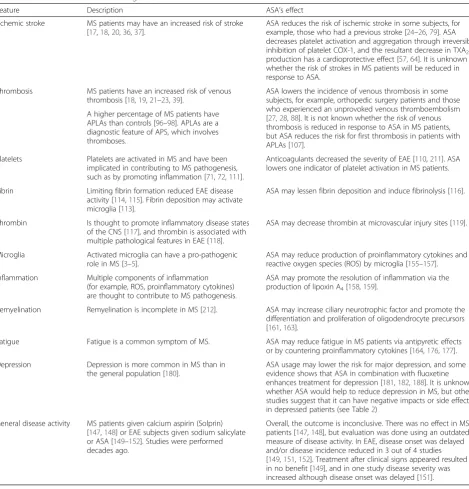
Feature Description ASA’s effect
Ischemic stroke MS patients may have an increased risk of stroke [17,18,20,36,37].
ASA reduces the risk of ischemic stroke in some subjects, for example, those who had a previous stroke [24–26,79]. ASA decreases platelet activation and aggregation through irreversible inhibition of platelet COX-1, and the resultant decrease in TXA2 production has a cardioprotective effect [57,64]. It is unknown whether the risk of strokes in MS patients will be reduced in response to ASA.
Thrombosis MS patients have an increased risk of venous thrombosis [18,19,21–23,39].
ASA lowers the incidence of venous thrombosis in some subjects, for example, orthopedic surgery patients and those who experienced an unprovoked venous thromboembolism [27,28,88]. It is not known whether the risk of venous thrombosis is reduced in response to ASA in MS patients, but ASA reduces the risk for first thrombosis in patients with APLAs [107].
A higher percentage of MS patients have APLAs than controls [96–98]. APLAs are a diagnostic feature of APS, which involves thromboses.
Platelets Platelets are activated in MS and have been implicated in contributing to MS pathogenesis, such as by promoting inflammation [71,72,111].
Anticoagulants decreased the severity of EAE [110,211]. ASA lowers one indicator of platelet activation in MS patients.
Fibrin Limiting fibrin formation reduced EAE disease activity [114,115]. Fibrin deposition may activate microglia [113].
ASA may lessen fibrin deposition and induce fibrinolysis [116].
Thrombin Is thought to promote inflammatory disease states of the CNS [117], and thrombin is associated with multiple pathological features in EAE [118].
ASA may decrease thrombin at microvascular injury sites [119].
Microglia Activated microglia can have a pro-pathogenic role in MS [3–5].
ASA may reduce production of proinflammatory cytokines and reactive oxygen species (ROS) by microglia [155–157]. Inflammation Multiple components of inflammation
(for example, ROS, proinflammatory cytokines) are thought to contribute to MS pathogenesis.
ASA may promote the resolution of inflammation via the production of lipoxin A4[158,159].
Remyelination Remyelination is incomplete in MS [212]. ASA may increase ciliary neurotrophic factor and promote the differentiation and proliferation of oligodendrocyte precursors [161,163].
Fatigue Fatigue is a common symptom of MS. ASA may reduce fatigue in MS patients via antipyretic effects or by countering proinflammatory cytokines [164,176,177].
Depression Depression is more common in MS than in the general population [180].
ASA usage may lower the risk for major depression, and some evidence shows that ASA in combination with fluoxetine enhances treatment for depression [181,182,188]. It is unknown whether ASA would help to reduce depression in MS, but other studies suggest that it can have negative impacts or side effects in depressed patients (see Table2)
General disease activity MS patients given calcium aspirin (Solprin) [147,148] or EAE subjects given sodium salicylate or ASA [149–152]. Studies were performed decades ago.
early in the course of their disease. Thus, some studies showing elevated risks for stroke in patients with MS may be inaccurate and reflect misdiagnoses or an in-crease in the surveillance (for example, MRI and more frequent physician visits) of this patient population iden-tifying asymptomatic lesions suggestive of strokes [38].
Venous thromboembolism (VTE) appeared to be ele-vated in patients with MS compared to controls [18, 19, 21–23]. Ocak and colleagues [39] found an increased risk of venous thrombosis in MS patients: the OR for venous thrombosis in MS was 2.4 (95 % CI 1.3–4.3), and the OR increased to 12.5 (95 % CI 1.5-107.9) in patients with both MS and increased factor VIII levels, which in-dicates the additional risk factor of thrombophilia. Also, APLAs, which are a diagnostic feature of APS that re-sults in thromboses, occur in a greater percentage of MS patients compared to controls (discussed later in the subsection“ASA and antiphospholipid antibodies”).
Individuals who have an occupation that involves prolonged sedentary behavior or patients experiencing immobilization have an increased risk of venous throm-bosis compared to more mobile people [39, 40]. A
sedentary lifestyle is also associated with an increased risk of stroke [41]. The activity level of patients with MS is less than that of healthy control subjects [42]. Physical inactivity, particularly in patients needing a wheelchair or who are bedridden, may contribute to the higher prevalence of thrombosis and ischemic strokes in pa-tients with MS [43, 44].
Whether patients with MS have an increased risk for myocardial infarction is unclear [20]. In one study, MS patients were found to have decreased risks of being hospitalized for myocardial infarction and ischemic heart disease [36]; however, a recent study conducted with an initial cohort of 8,281 MS patients in Sweden deter-mined that the risks for myocardial infarction, heart fail-ure, and stroke were increased in individuals with MS compared to matched controls, and that these risks were more pronounced in women than men [18]. Shared pathological factors (for example, inflammation, oxida-tive stress, thrombogenic factors) between MS and car-diovascular diseases may explain the association between these conditions [18]. Of note, MS patients with≥1 car-diovascular risks had increased MRI indications of Table 2Potential risks of aspirin usage in MS
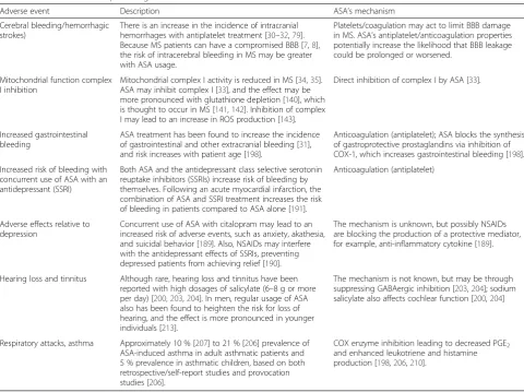
Adverse event Description ASA’s mechanism
Cerebral bleeding/hemorrhagic strokes)
There is an increase in the incidence of intracranial hemorrhages with antiplatelet treatment [30–32,79]. Because MS patients can have a compromised BBB [7,8], the risk of intracerebral bleeding in MS may be greater with ASA usage.
Platelets/coagulation may act to limit BBB damage in MS. ASA’s antiplatelet/anticoagulation properties potentially increase the likelihood that BBB leakage could be prolonged or worsened.
Mitochondrial function complex I inhibition
Mitochondrial complex I activity is reduced in MS [34,35]. ASA may inhibit complex I [33], and the effect may be more pronounced with glutathione depletion [140], which is thought to occur in MS [141,142]. Inhibition of complex I may lead to an increase in ROS production [143].
Direct inhibition of complex I by ASA [33].
Increased gastrointestinal bleeding
ASA treatment has been found to increase the incidence of gastrointestinal and other extracranial bleeding [31], and risk increases with patient age [198].
Anticoagulation (antiplatelet); ASA blocks the synthesis of gastroprotective prostaglandins via inhibition of COX-1, which increases gastrointestinal bleeding [198].
Increased risk of bleeding with concurrent use of ASA with an antidepressant (SSRI)
Both ASA and the antidepressant class selective serotonin reuptake inhibitors (SSRIs) increase risk of bleeding by themselves. Following an acute myocardial infarction, the combination of ASA and SSRI treatment increases the risk of bleeding in patients compared to ASA alone [191].
Anticoagulation (antiplatelet)
Adverse effects relative to depression
Concurrent use of ASA with citalopram may lead to an increased risk of adverse events, such as anxiety, akathesia, and suicidal behavior [189]. Also, NSAIDs may interfere with the antidepressant effects of SSRIs, preventing depressed patients from achieving relief [190].
The mechanism is unknown, but possibly NSAIDs are blocking the production of a protective mediator, for example, anti-inflammatory cytokine [189].
Hearing loss and tinnitus Although rare, hearing loss and tinnitus have been reported with high dosages of salicylate (6–8 g or more per day) [200,203,204]. In men, regular usage of ASA also has been found to heighten the risk for loss of hearing, and the effect is more pronounced in younger individuals [213].
The mechanism is not known, but may be through suppressing GABAergic inhibition [203,204]; sodium salicylate also affects cochlear function [200,204]
Respiratory attacks, asthma Approximately 10 % [207] to 21 % [206] prevalence of ASA-induced asthma in adult asthmatic patients and 5 % prevalence in asthmatic children, based on both retrospective/self-report studies and provocation studies [206].
disease activity, that is, more brain atrophy and an in-crease in lesion burden [45].
Treatments for MS may also increase the risk for car-diovascular diseases [20]. For example, systemic gluco-corticoids have been reported to increase risks of stroke, myocardial infarction, and atrial fibrillation [20]. Add-itionally, a positive association was found for cardiovas-cular risk factors and the use of disease modifying therapies (DMTs) such as interferon-β and glatiramer acetate [46]. For example, about 20 % of MS patients on DMTs versus about 5 % of MS patients naïve to DMTs had diastolic blood pressure above 90 mmHg, and about 25 % of MS patients on DMTs versus about 14 % MS patients naïve to DMTs had glucose > 100 mg/dL [46]. The association was more pronounced in chronic pro-gressive MS patients compared to relapsing remitting MS (RRMS) patients, but the correlation to disease ac-tivity (for example, rate of clinical relapse) was weak [46]. Although similar studies have not been conducted for more recent DMTs, cardiac side effects can be asso-ciated with these agents. For example, an increase in hypertension has been noted for teriflunomide (4 % treated versus 2 % placebo [47]). A reduction of heart rate can occur within six hours of fingolimod initiation [48], and there can be an increase in blood pressure in a small percentage of patients over the long term [49].
Increased risk of dying from cardiovascular disease in patients with MS
Besides a possibly increased risk of cardiovascular dis-ease (CVD), studies generally suggest that MS patients have a heightened risk of dying from CVD [43, 50]. As reviewed in Christiansen [20], patients with MS have a 6–34 % greater risk of death by CVD or stroke than for the overall population, and greater than 10 % of MS pa-tients may have stroke as the cause of death. An analysis of 9,881 MS patients in the Danish Multiple Sclerosis Registry revealed that the mortality rate from CVD was significantly higher in MS patients compared to matched controls [50], and an analysis of over 6,000 patient deaths in the Danish MS Registry revealed that 17.6 % of deaths were caused by vascular or cardiac diseases, which was the most frequently listed cause of death out-side of MS itself [43]. The standard mortality rate (the number of subjects who died from the specific cause di-vided by the number of deaths expected from population mortality statistics) of cardiac or vascular diseases in this same patient population was 1.34, indicating about a 34 % greater chance of death by CVD in MS patients when compared to general population mortality statistics [43]. In contrast, a study in South Wales revealed that while CVD caused 16 % of deaths in the MS population sur-veyed, this rate did not differ from the expected death rates in the general population [51].
While it appears that patients with MS have a greater likelihood of dying from cardiovascular-related issues, the underlying causative reason is not clear. These pa-tients may lead a more sedentary lifestyle than healthy individuals due to motor symptoms and fatigue, for ex-ample [42], particularly as the disease progresses, and a less active lifestyle has been associated with cardiovascu-lar risk factors such as impaired glucose tolerance and the development of metabolic syndrome in the general population [52, 53]. Furthermore, altered metabolic re-sponses in patients with MS, such as increased adipose lipolytic activity, could be a factor in lower physical per-formance [54]. Several studies (reviewed in [55]) suggest that patients with MS are more likely to develop vascu-lar disease and comorbidities related to metabolic syn-drome (such as obesity, impaired glucose tolerance, and dyslipidemia), but these factors may develop or worsen as a result of progressively debilitating MS symptoms. Because many of these studies are retrospective, correl-ational designs, the exact cause and effect relationship between MS and CVD risk and mortality cannot be eas-ily discerned. It is unclear whether CVD risk factors contribute to MS disease pathogenesis, whether MS symptoms promote the development of CVD and mor-tality, or whether a shared underlying disturbance, such as in glucose metabolism [56], underlies both disease processes.
Treatment potential of ASA for cardiovascular disease in patients with MS
ASA mechanisms of action
Aspirin (ASA) is a traditional nonsteroidal anti-inflammatory drug (tNSAID) used to treat inflammation, pain, and fever, and to inhibit platelet activation and aggre-gation. This latter effect inhibits thrombus formation, thus providing cardioprotection, and is the basis for the use of ASA in the prevention of myocardial infarction [31, 57, 58]. The many therapeutic uses of ASA are due to its inhibition of cyclooxygenases (COXs), enzymes that catalyze a step in the production of prostanoids (Fig. 1). Prostanoids include prostaglandins, prostacyclin, and thromboxanes, molecules with pleiotropic effects on a large number of physiologic systems [59].
A unique quality of ASA that differentiates it from other tNSAIDs is its ability to covalently acetylate COXs [60], whereas other tNSAIDS are competitive, reversible inhibitors [59, 61]. The irreversible linkage by ASA inhibits the enzyme’s ability to convert arachidonic acid to pros-taglandin H2 (PGH2), a committed step in prostanoid
found in monocytes, endothelial cells, and fibroblasts, al-though it is also constitutively expressed in some cells within the brain, testes, and kidney [62]. Therefore, COX-1 is thought to be the dominant source of prostanoids for housekeeping functions and COX-2 is considered to be a main source in inflammation, although platelet-derived prostanoids generated through COX-1 are linked to pro-motion of an inflammatory state [59, 63–65].
Once formed, PGH2may be converted to prostaglandin
E2(PGE2) by prostaglandin-E synthase; prostacyclin (PGI2)
by prostaglandin-I synthase; or thromboxane A2(TXA2) by
thromboxane synthase [66, 67]. These mediators interact with specific G-protein coupled receptors that utilize either cAMP or IP3/DAG/Ca2+as second messengers ultimately
to elicit physiologic responses that often are in opposition to each other. In terms of vascular smooth muscle tone and platelet reactivity, PGI2and TXA2act conversely, with
PGI2 decreasing and TXA2 increasing these parameters
[64]. Also, PGI2inhibits vascular smooth muscle cell
prolifer-ation and TXA2promotes it [64]. Increases or decreases in
vascular tone and platelet reactivity can be elicited by PGE2,
depending on which of its five receptors are activated [64].
Platelets participate in the thrombus formation process, and as such, hyperactivity built upon underlying athero-sclerosis can contribute to the development of pathological cardiovascular events that may result in decreased blood flow, acute coronary syndrome, and stroke [68, 69]. Through irreversible inhibition of platelet COX-1, and the resultant decrease in TXA2 production, ASA decreases
limit production of proinflammatory eicosanoids and at-tenuate the inflammatory state.
Since the COX inhibition by ASA is irreversible, the antiplatelet effect is dependent on the synthesis of new platelets. Therefore, lower doses of ASA (50–100 mg/ day) are effective due to action within platelets, thus making it useful in the treatment of coronary artery disease [31, 57, 58, 64, 74]. Distinct from this platelet ef-fect, higher doses are used for analgesic, antipyretic, and anti-inflammatory effects and are usually taken only as needed as opposed to daily [59].
ASA use in preventing cardiovascular disease
In order to assess the potential impact of ASA to coun-ter cardiovascular events in MS, it is helpful to address its role in reducing CVD in other segments of the popu-lation. In the general population, regular ASA use for prevention of cardiovascular events ranges between 18– 41 %, and usage can be more prevalent in subpopula-tions, for example, diabetics and older individuals who are at higher risk for CVD [75–78]. Long-term adminis-tration of ASA at low doses helps prevent strokes, heart attacks, and blood clot formation in people at high risk of these events [24–26]. In an analysis of antiplatelet treatment following acute ischemic stroke, Sandercock et al. [79] found a significant decrease in recurrent ische-mic stroke and death in patients who began ASA treat-ment no more than 14 days following a presumed ischemic stroke occurrence (OR 0.95, 95 % CI 0.91– 0.99). It has also been noted that although there is a slight increase in the incidence of intracranial hemor-rhages with antiplatelet treatment, the benefits of pre-venting repeat ischemic strokes and other cardiovascular events such as pulmonary embolism (PE) outweigh the risks of intracranial bleeding [30, 79]. ASA monotherapy (50–325 mg/day), clopidogrel, or extended-release dipyr-idamole (ER-DP) combined with ASA are therapies rec-ommended by the American Heart Association and American Stroke Association, as well as the Eighth American College of Chest Physicians (ACCP), for the secondary prevention of ischemic strokes in individuals with a history of ischemic events including stroke [25, 80, 81]. The combination of ASA and ER-DP is prefera-ble to ASA alone, while the combination of ASA with clopidogrel has a heightened risk of bleeding events, and thus should be avoided [25, 26, 80–83].
Although ASA has been established as effective for preventing secondary cerebrovascular events [26], its prophylactic efficacy is less clear. A significant reduction in the risks for a first myocardial infarction was found with ASA usage, but results for ASA’s efficacy in pre-venting a first stroke and CVD were inconclusive [31, 84]. A review of 27 studies on the effectiveness of ASA for the primary prevention of cardiovascular events (for
example, in patients not at risk for CVD) revealed only modest/minor benefits that did not outweigh the risks of increased bleeding and hemorrhagic strokes [32].
Platelets were traditionally considered to play a greater role in arterial rather than venous thrombosis, but platelets have been shown to have a role in venous thromboembol-ism (VTE) by inducing the formation of neutrophil extra-cellular traps, releasing proinflammatory mediators and microparticles, and aggregating as a component of thrombi themselves [27]. While some studies such as the Longitu-dinal Investigation on Thromboembolism Etiology did not find a reduction in VTE by ASA users [85], other trials re-port that ASA lowers the risk of deep vein thrombosis (DVT) [29]. The INSPIRE study combined data from two previous studies where patients were given ASA after a first unprovoked VTE to determine its effects on VTE recur-rence. The INSPIRE analysis found that ASA reduces the overall risk of recurrent VTE by 42 % (P= 0.005) with only minor bleeding concerns, supporting the use of ASA for secondary prophylaxis of VTE [28]. In the large Pulmonary Embolism Prevention (PEP) trial, which included over 13,000 patients undergoing surgery for hip fracture or elect-ive arthroplasty, PE or DVT was experienced in 105 out of 6,679 (1.5 %) patients receiving 160 mg ASA compared to 165 of 6,677 (2.5 %) patients receiving a placebo, which equals a proportional reduction of 36 % PE/DVT in those treated with ASA (P= 0.0003) [86]. ASA was also found to be effective for the prevention of PE in high-risk patients by the Antithrombotic Trialists’Collaboration in a 2002 meta-analysis: patients taking an antiplatelet (for example, ASA) had a 25 % reduced risk of acute PE compared with pa-tients on placebo (P< 0.01) [87]. Based on results such as these and the low cost and relatively low risk of bleeding of ASA when compared to warfarin and newer anticoagulants, the American Academy of Orthopedic Surgeons in 2009 and the ACCP in 2012 included ASA as a method in their guidelines to prevent VTE in high-risk orthopedic surgery patients [27, 88].
Considerations regarding ASA use for cardiovascular disease in patients with MS
of patients with MS [92] could be performing a protect-ive role by limiting BBB leakage, and if ASA disrupted their deposition, then this could slow the resolution of the leakage. However, ASA can act to limit vascular leak-age: ASA reduced arachidonate-induced vascular leakage in the peritoneum [93] and induced lipoxin A4, which
was found to reduce vascular leakage following acute ear inflammation [94]. ASA also lowered the permeability of the BBB in non-stroke patients [95].
It might be important to identify patients with MS who have a heightened risk of a cardiovascular event, as they would be expected to have the most favorable risk-to-benefit ratio when using ASA. Besides the traditional risk factors for cardiovascular events, there are features of MS that may potentially lead to increased risk of CVD within the MS population. For example, a more sedentary or immobile state in MS patients [42, 44] or taking an MS DMT [46–49] might be associated with an increased risk of cardiovascular events.
ASA and antiphospholipid antibodies
Multiple studies have reported that APLAs are more prevalent in patients with MS than in the general popu-lation [96–98]. APLAs are the main diagnostic feature of APS, an autoimmune disease characterized by throm-bosis and/or pregnancy morbidity and elevated levels of anticardiolipin antibodies, lupus anticoagulant, and/orβ2 glycoprotein I [99]. MS and APS share various features, and one disorder can sometimes be misdiagnosed for the other [96, 99]. Although the role that APLAs may have in MS is presently unclear, recent studies indicate a relationship between APLAs and a more severe MS disease course [97, 98, 100–102]. In a prospective three-year study following interferon-β(INF-β)-treated MS pa-tients with or without APLAs, Zivanidov et al. found that the APLA-positive patients showed a greater disease progression as measured by higher MRI lesion volumes, increased tissue damage, and loss of brain volume, as well as more clinical relapses [102]. The presence of anti-INF-βbinding antibodies may decrease the efficacy of INF-βtreatment, and Garg et al. [103] found a signifi-cant co-occurrence of high APLAs in MS patients with anti-INF-β binding antibodies. A higher frequency of APLAs has been reported in secondary progressive MS (SPMS) compared to RRMS, which is consistent with the idea that the presence of APLAs is related to a more chronic, advanced stage of the disease [97, 102, 104].
Given that thromboses are a main symptom in APS, anticoagulation, antiaggregation, and ASA are all used as treatment for this syndrome [96, 99, 102]. Although many physicians prescribe daily low-dose ASA for asymptomatic APLA-positive patients to prevent a first thrombotic event, results from studies on the benefit of ASA prophylaxis in this population are mixed [105]. The
Antiphospholipid Antibody Acetylsalicylic Acid study found that 81 mg of ASA daily for APLA-positive but asymptomatic patients was not more effective than pla-cebo in protecting against a thrombotic event [106]. In contrast, in an individual patient-level meta-analysis from five international cohort studies, Arnaud and col-leagues [107] found that prophylactic low-dose ASA in patients with APLAs significantly reduced the risk of a first thrombotic event. Although the role of APLAs in the pathogenesis of MS is not clear, given that MS is as-sociated with an increased risk of thrombosis and car-diovascular events (see, for example, [18, 20, 21]), MS patients with APLAs may benefit from anticoagulation or ASA treatment.
Potential effects of ASA on vascular pathology in MS
In addition to affecting thrombosis and stroke in MS pa-tients, ASA could affect other components of vascular pathology in MS. Pathological changes to the vascular tissue in the CNS have been observed since the earliest descriptions of MS (see, for example, [89, 108]), and they include platelet and fibrin deposits associated with ves-sels in active lesions, vessel occlusion, vascular thicken-ing, enhanced deposition of perivascular collagen, BBB disruption, and perivascular inflammation [7, 8, 14–16, 109]. Proteomic analysis revealed the deposition of an array of coagulation proteins in chronic active plaques [110], and anticoagulants decreased the severity of ex-perimental autoimmune encephalomyelitis (EAE) [110], an animal model of MS, indicating that vascular changes have a detrimental role in the disease.
Besides platelets acting to promote inflammation in EAE [73], fibrin deposition has been credited with acti-vation of microglia, which have been associated with tis-sue damage [113]. Depending on the type, microglia can have different influences on disease activity in MS; that is, they can induce tissue damage or promote repair [5]. Blockage of fibrin formation lessened disease activity in EAE [114, 115]. ASA may promote fibrinolysis or inter-fere with fibrin deposition [116].
In addition to fibrin, thrombin activity has been asso-ciated with worsening of inflammatory CNS disease states such as MS [117], and thrombin activity in the spinal cord of mice with EAE is associated with multiple pathological features [118]. ASA may decrease thrombin levels at sites of microvascular injury [119]. In experi-mental APS, which models a condition that results in clots in deep veins and in organs such as the brain, ASA also reduced tumor necrosis factor alpha (TNFα) and prostaglandin E synthesis and increased thrombin inhibi-tors [120].
Vascular changes may contribute to the decreased per-fusion observed in MS [9–12, 92, 121]. Decreased perfu-sion has been observed in the cortex [9, 10, 12], deep gray matter structures (for example, thalamus, caudate) [10–12], and normal-appearing white matter (NAWM) [92] of patients with MS. In addition, white matter le-sions from patients with RRMS had altered perfusion (some with decreased perfusion and others with an in-creased perfusion compared to that from white matter in control subjects) [10, 122]. The extent of white matter lesions was correlated with decreased cortical blood flow [123], and decreased cerebral blood flow has also been observed in EAE [124, 125].
Decreased perfusion could impair tissue oxygenation. Decreased tissue oxygenation in MS patients has been detected in white matter and cortex gray matter by positron emission tomography [126], and decreased utilization of oxygen in MS was revealed in periventricu-lar veins by susceptibility-weighted imaging (SWI) [127] and by T2-relaxation-under-spin-tagging of venous sinus blood [128]. Additionally, lesions observed by SWI in the spinal cord of EAE mice were thought to be detected largely due to deoxyhemoglobin, whose presence was likely a result of hypoxia [129]. At the molecular level, upregulation of hypoxia-inducible factor-1α (HIF-1α) has been observed in active MS plaques by immunohis-tochemistry [130, 131], and the upregulation of genes associated with ischemic preconditioning, including HIF-1α, has been observed in NAWM in patients with MS [132]. Additionally, endoplasmic reticulum stress proteins, which have been associated with ischemic in-jury, are overexpressed in active MS lesions [133, 134] and in gray matter MS lesions [135]. Although altered perfusion may reduce oxygen delivery and impair energy
production [136], vascular changes, that is, microvascu-lar thrombosis, as the cause of hypoxia-like changes in MS have been questioned, since hypoxia-like changes have been observed in MS patients in the absence of vascular pathology [137].
Mitochondrial dysfunction has been observed in mul-tiple sites in the CNS of MS patients, including NAWM, lesions, and cortex, and altered mitochondria function may help create a hypoxic state [35, 137–139]. ASA may act to inhibit complex I of the respiratory chain [33]. Complex I is decreased in chronic active white matter lesions and in the motor cortex of patients with MS [34, 35]. Thus, ASA has the potential to further lower com-plex I activity in MS, and ASA’s inhibitory effect may be more pronounced with depletion of glutathione [140], which is thought to occur in patients with MS [141, 142]. Furthermore, inhibition of complex I activity can lead to an increased production of reactive oxygen spe-cies (ROS) [143], which could amplify cellular damage.
Cerebrovascular reactivity, the ability of the cerebral vasculature to increase local blood flow via arteriole dila-tion in response to neural activity, is impaired in MS, perhaps as a result of vascular desensitization from chronic high levels of nitric oxide stemming from in-flammation [144]. Interestingly, ASA has been found to increase endothelial nitric oxide synthase activity [145, 146], which would favor an increase in blood flow, but not if vascular desensitization has developed [144].
ASA effects on MS symptoms
ASA in MS, EAE, and related studies
including those examining the vasculature and blood flow, and other more current measures of MS disease activity could provide greater insights regarding the ef-fect of ASA on disease progression.
Studies examining ASA in animal models of MS also have been relatively sparse. One study published in 1949 examined the prophylactic and therapeutic effects of so-dium salicylate (the main metabolite of ASA) and para-aminobenzoic acid, either alone or in combination, in a guinea pig EAE model [149]. Although neither com-pound showed effects against disease onset and progres-sion at moderate dosage levels, the combination of sodium salicylate and para-aminobenzoic acid, as well as larger doses of sodium salicylate by itself, seemed to delay onset, lessen incidence, and inhibit disease severity if administration was begun prior to or shortly after (5 days or less) EAE induction [149]. Treatment with so-dium salicylate or the combined drugs after the animals became sick had no effect on the disease [149]. When ASA was tested in the guinea pig EAE model by another group, there was no beneficial effect [150]. In the Lewis rat model of EAE, ASA delayed the onset of disease but increased the severity of disease [151], while so-dium salicylate postponed disease onset and reduced clinical signs [152].
More recent studies have looked at the role of COX-1 and COX-2 in EAE. Naproxen, a COX-1 and COX-2 in-hibitor, was shown to delay EAE onset and reduce the severity of the disease when treatment was started on the day of EAE immunization [153]. In another study, celecoxib, a new generation COX-2 inhibitor, reduced EAE incidence and/or severity when animals were treated beginning on the day of EAE induction or 8 days post-induction [154]. But celecoxib also reduced disease severity in COX-2-deficient mice; and nimesulide, an-other COX-2 inhibitor, did not affect disease develop-ment or severity, which indicates that the mechanism of action of celecoxib on EAE is not via the COX-2 path-way [154].
ASA was found to limit the production of ROS and proinflammatory cytokines (for example, TNFα and IL-1β) by a microglial cell line treated with the activator lipopolysaccharide [155, 156], and ASA limited proin-flammatory cytokine production and microglial activa-tion following middle cerebral arterial occlusion in the rat [157]. Activated microglia can mediate tissue damage in MS [3–5], and lessening their production of inflam-matory mediators by ASA could have possible benefits. Also, it may be that ASA could promote resolution of inflammation in MS by inducing lipoxin A4[158], which
is an anti-inflammatory mediator [159].
In salt-loaded, stroke-prone, spontaneously hyperten-sive rats, ASA suppressed BBB damage and reduced several markers of inflammation (for example, matrix
metalloproteinase-9 activity, superoxide production, and macrophage accumulation) [160], raising the pos-sibility that ASA could limit similar pathological pro-cesses in MS.
Recent studies found that ASA upregulated the pro-duction of the ciliary neurotrophic factor [161], which augments myelin formation [162]. ASA also induced the proliferation and differentiation of oligodendrocyte pre-cursors and limited demyelination following a cerebral ischemic lesion [163]. If ASA acted similarly in response to MS lesions, then it could promote remyelination ef-forts [161].
ASA and fatigue
Fatigue is a pervasive and debilitating symptom associ-ated with a marked decrease in the quality of life for pa-tients with MS. The cause of fatigue is not understood. Some possible theories concerning the causes of fatigue have included: elevated body temperature (in RRMS pa-tients) [164], sleep disturbances and depression [165, 166], proinflammatory cytokines [167], reduced metabol-ism and degeneration of cerebral and deep gray matter structures [168, 169], reduced connectivity [170], and re-duced perfusion of deep gray matter [11]. There are a limited number of approaches used to counter fatigue in MS, for example, amantadine, modafinil, vitamin D ana-log, treatment for sleep disorder, and exercise [171–175]. Overall, studies have been conflicting as to the benefits of these modalities, and better management of fatigue is sorely needed.
A subsequent randomized double-blind crossover clin-ical trial for 52 patients with MS was conducted over a ten-week period to study the effectiveness of ASA and amantadine for alleviating fatigue in MS. Half of the pa-tients were randomly assigned to receive 500 mg ASA orally once daily for the first four weeks, and following a two-week washout period were switched to 100 mg amantadine orally twice a day for the final four weeks; the other half of the patients received the treatments in reverse order [177]. A significant decrease in self-reported fatigue levels measured using the FSS with both ASA and amantadine was found following a baseline measurement [177]. After the first round of treatment, mean FSS scores decreased by 1.1 (from a maximum of 7) for ASA and by 0.8 for amantadine [177]. During the two-week washout period, the mean FSS scores in-creased back to baseline levels [177]. In the second phase of the study, where patients received crossover treatments, self-reported fatigue scores were once again reduced significantly for each treatment regimen: mean FSS decreased by 0.7 for ASA and by 1.6 for amantadine [177]. The authors noted that both ASA and amantadine were well-tolerated by patients with few and mild side effects, none of which led to participant dropouts. Given the promising results shown by both treatments in these studies, they suggest that ASA and amantadine deserve further consideration as potential treatments to combat fatigue in MS [177].
A placebo-controlled, double-blind, multicenter study was performed comparing placebo, 162 mg/d and 1,300 mg/d of ASA [178]. Although the study was not com-pleted, an intermediate analysis of the placebo and high dose groups revealed a difference of 4.6 points on the MFIS, that is, adjusted mean scores of 42.7 versus 38.1 in the respective groups. However, the study was under-powered and it did not reveal a statistically significant effect [178]. The authors indicated that it is unlikely that ASA provides a clinically relevant benefit for MS pa-tients [178]. It is possible that confounding factors, such as an undiagnosed sleep disorder [166], could interfere with ASA effect on fatigue.
The mechanism of action by which ASA might lessen fatigue, even minimally, is unclear. ASA’s reduction of fatigue symptoms may be through the drug’s antipyretic effects [176, 177], which is supported by a recent study that found a correlation between fatigue, as measured by FSS and MFIS, and elevated body temperature in RRMS patients [164]. In addition, ASA could have affected other systems (for example, autonomic or neuroendo-crine) involved in the perception of fatigue [176, 177]. ASA also could have countered proinflammatory cyto-kines [176, 177], which may contribute to fatigue [167, 179]. ASA may also act on fatigue associated with some form of interferon-β therapy, which was taken by 5/30
patients in the Wingerchuck et al. (2005) study [176] and by 52/52 patients in the Shaygannejad et al. (2012) study [177].
ASA and depression
Patients with MS are more likely to experience depres-sion than the general population [180]. Although depression has traditionally been thought of as a neurotransmitter-related/driven disease, evidence sug-gests that inflammation may play a role in the disorder [181]. As such, drugs that reduce inflammation may be beneficial in depression [181].
The use of ASA may lower the risk of major depres-sion [182]. A study on depresdepres-sion and anxiety in patients with myocardial infarction found that those taking ASA reported fewer depression and anxiety symptoms (P < 0.01) as measured by the Hamilton Depression and Hamilton Anxiety Rating Scales, respectively [183]; and an analysis of 174 male coronary angiography pa-tients (99 on ASA) found fewer depressive symptoms in those taking ASA regularly (range from 80 mg every other day to 325 mg daily), both by self-report (P = 0.016) and reported perceptions from a significant other (P= 0.048) [184].
In an established rodent model of depression, ASA lessened immobility in a forced swim test in rats and concurrently attenuated cytokine levels (IL-6 and
Potential risks associated with ASA use in MS Due to the known pleiotropic effects of the prostaglan-dins and thromboxanes on multiple physiological sys-tems, it is not unexpected that inhibition of their production by ASA could potentially cause various ad-verse effects. Many of the major adad-verse effects related to ASA use are discussed below and summarized in Table 2.
While the incidence of cerebral hemorrhage during ASA treatment is low in most studies, the antiplatelet ef-fect of ASA can contribute to the development of in-creased cerebral bleeding [30]. The Antithrombotic Trialists’Collaboration performed a meta-analysis (6 pri-mary prevention trials and 16 secondary prevention tri-als) of serious vascular events, including stroke and major bleeds comparing long-term ASA versus control [31]. Their conclusions included that ASA increased incidences of hemorrhagic stroke in both primary (P = 0.05) and secondary (P = 0.07) prevention trials and when analyzed in combination (P = 0.01), while ASA showed a protective effect concerning ischemic stroke (P = 0.005) [31]. Sutcliffe et al. [32] reviewed data from ran-domized trials assessing ASA in the primary prevention of CVD and cancer and concluded that the benefits of ASA for primary prevention of CVD are modest, and are much less than those for secondary prevention. Further-more, while benefits and harms were low based on person-years, they estimated an increased risk of hemorrhagic stroke ranging from 32–38 % [32].
Although the increases in incidents of hemorrhagic stroke noted are slight, it is possible that in disease states like MS, where the BBB is disrupted or compromised [7, 8], the risk of intracerebral bleeding may be greater with ASA usage. Additionally, there is an increased risk of immune thrombocytopenia in patients with MS [196], which would be a counter indication of ASA usage due to the enhanced risk of bleeding with ASA in this pa-tient population [197].
An adverse effect that is well associated with ASA use is upper gastrointestinal (GI) tract injury and bleeding. Because ASA inhibits the production of prostaglandins by GI-located COX-1, these gastroprotective substances are unavailable and damage may occur. The risk of com-plications is increased with aging, concomitant use of anticoagulants, history of NSAID-associated bleeding, and comorbidities [198]. The Antithrombotic Trialists’ Collaboration study noted above determined that ASA usage increased major GI and extracranial bleeds to 0.10 % versus 0.07 % per year in controls (RR = 1.54 [1.30–1.82],
P< 0.0001) [31].
ASA and salicylate, the active metabolite of ASA, are known to cause hearing loss and tinnitus at high doses (for example, 6–8 g/day) [199–203]. The mechanisms behind these effects are unclear, but salicylate seems to
have effects centrally on GABAergic neurotransmission [203, 204], as well as more peripherally by affecting cochlear function [200, 204]. Moreover, a strong linear relationship exists between unbound salicylate plasma concentration and a resultant decrease in auditory sensi-tivity [205], and the salicylate toxicity model is used by auditory scientists to investigate mechanisms underlying tinnitus [204].
Certain asthmatic patients have a sensitivity to ASA that manifests itself as a respiratory/asthma-type attack. Jenkins et al. performed a systematic review and found that the pooled incidence of ASA-induced asthma was 21 % in adults and 5 % in children [206]. This is higher than the value of approximately 10 % that has been pub-lished elsewhere [207, 208]. The mechanism of this reac-tion in ASA sensitive-individuals is thought to occur due to COX inhibition resulting in decreased PGE2and thus
unabated activation of the 5-lipoxygenase pathway. This, in turn, increases production of leukotrienes and mast cell release of histamine, leading to airway hyperreactiv-ity [209, 210].
Prostaglandin-mediated vasodilation is necessary for proper renal plasma flow, especially in individuals with underlying renal disease, congestive heart failure, or cir-rhosis. Through inhibition of renal COX-2, ASA and other NSAIDs can cause volume-dependent renal failure and that resulting from interstitial nephritis and neph-ritic syndrome [198].
Conclusion
Although ASA use is relatively common in the general population, ASA usage by MS patients has the potential for positive and/or negative influences on different facets of the disease, that is, symptoms, disease mechanisms, and associated disease risks. Understanding the impact of ASA use on these features would help to further es-tablish the risk-to-benefit ratio of ASA usage in this pa-tient population. Venous thrombosis and possibly stroke have an elevated likelihood in MS [17–23, 37], and given that ASA can lessen the risks of these cerebrovascular diseases [25, 26, 29], it is likely that ASA confers a simi-lar benefit of lower risk for MS patients. Fatigue is a relatively common symptom in MS, and ASA may ameliorate fatigue in MS patients [176, 177], although the effect size might be small [178]. ASA could also im-pact pathogenic processes. For example, ASA could act to reduce inflammation by limiting the production of proinflammatory mediators from activated microglia [155–157] or by inducing the production of lipoxin A4,
119]. Despite these potential benefits, ASA usage can have negative side effects such as being associated with an elevated risk of hemorrhagic stroke [30–32]. ASA may also worsen some specific components of MS path-ology, for example, enhancing leakage of the BBB and possibly inhibiting mitochondrial complex I activity [33], which is already reduced in MS [34, 35]. It is possible that subgroups of MS patients may find particular bene-fit from ASA, for example, immobile patients with in-creased risk for DVTs or patients with APLAs, which could counterbalance the risks associated with ASA usage. It is also possible that ASA usage has a small beneficial impact on overall disease progression. Thus, further studies are needed to determine the benefits and risks of ASA in patients with MS in order to establish proper guidance for ASA use by this patient population. Given the widespread usage of aspirin and the likelihood that many effects could be small, traditional placebo controlled trials would be unlikely to yield meaningful results. Carefully crafted population-based studies, while not definitive, may help guide our understanding of this complex issue.
Abbreviations
ACCP:American College of Chest Physicians; APLA: antiphospholipid antibody; APS: antiphospholipid syndrome; ASA: aspirin; BBB: blood-brain barrier; CNS: central nervous system; COX-1: cyclooxygenase-1; COX-2: cyclooxygenase-2; COXs: cyclooxygenases; CVD: cardiovascular disease; DMT: disease modifying therapy; DVT: deep vein thrombosis; EAE: experimental autoimmune encephalomyelitis; ER-DP: extended-release dipyridamole; GI: gastrointestinal; HIF-1α: hypoxia-inducible factor-1α; MFIS: Modified Fatigue Impact Scale; MS: multiple sclerosis; NAWM: normal-appearing white matter; OR: odds ratio; PE: pulmonary embolism; PEP: Pulmonary Embolism Prevention (trial); PF4: platelet factor 4; PGE synthase: prostaglandin-E synthase; PGE2: prostaglandin E2; PGH2: prostaglandin H2; PGI2: prostacyclin; ROS: reactive oxygen species; RRMS: relapsing remitting MS; SSRI: selective serotonin reuptake inhibitor; SWI: susceptibility-weighted imaging; tNSAID: traditional nonsteroidal anti-inflammatory drug; TXA2: thromboxane A2; VTE: venous thromboembolism;β-TG:β-thromboglobulin.
Competing interests
SML has received past and current grant support from ApoPharma, Inc. SGL has participated in multi-center clinical trials sponsored by Biogen, Bayer, Teva, Acorda, Medimmune, Alexion, Chugai, Novartis, Genzyme, Roche, Vaccinex, Opexa, Actelion, NMSS, and NIH. Publication costs were also partially funded by intramural funds from the Midwestern University College of Pharmacy-Glendale (MRE) and the University of Kansas Medical Center (SML).
Author’s contributions
Wrote manuscript: ST, MRE, SGL, SML. All authors read and approved the final manuscript.
Acknowledgements
This work was supported by the National Multiple Sclerosis Society (NMSS) and an NICHD Center Grant (HD 02528). Publication costs were also partially funded by intramural funds from the Midwestern University College of Pharmacy-Glendale (MRE) and the University of Kansas Medical Center (SML).
The contents are solely the responsibility of the authors and do not necessarily represent the official views of any of the funding agencies listed above.
Author details
1Department of Molecular and Integrative Physiology, University of Kansas
Medical Center, Kansas City, KS, USA.2Department of Pharmaceutical Sciences, College of Pharmacy-Glendale, Midwestern University, Glendale, AZ,
USA.3Department of Neurology, University of Kansas Medical Center, Kansas City, KS, USA.
Received: 13 February 2015 Accepted: 4 June 2015
References
1. Karussis D. The diagnosis of multiple sclerosis and the various related demyelinating syndromes: a critical review. J Autoimmun. 2014;48–49:134–42. 2. Kutzelnigg A, Lassmann H. Pathology of multiple sclerosis and related
inflammatory demyelinating diseases. Handb Clin Neurol. 2014;122:15–58. 3. Neumann H. Molecular mechanisms of axonal damage in inflammatory
central nervous system diseases. Curr Opin Neurol. 2003;16:267–73. 4. Lassmann H. Axonal and neuronal pathology in multiple sclerosis: what
have we learnt from animal models. Exp Neurol. 2010;225:2–8. 5. Correale J. The role of microglial activation in disease progression. Mult
Scler. 2014;20:1288–95.
6. Witte ME, Mahad DJ, Lassmann H, van Horssen J. Mitochondrial dysfunction contributes to neurodegeneration in multiple sclerosis. Trends Mol Med. 2014;20:179–87.
7. de Vries HE, Kooij G, Frenkel D, Georgopoulos S, Monsonego A, Janigro D. Inflammatory events at blood-brain barrier in neuroinflammatory and neu-rodegenerative disorders: implications for clinical disease. Epilepsia. 2012;53(s6):45–52.
8. Cramer SP, Simonsen H, Frederiksen JL, Rostrup E, Larsson HB. Abnormal blood-brain barrier permeability in normal appearing white matter in multiple sclerosis investigated by MRI. Neuroimage Clin. 2013;4:182–9. 9. Lycke J, Wikkelsö C, Bergh AC, Jacobsson L, Andersen O. Regional cerebral
blood flow in multiple sclerosis measured by single photon emission tomography with technetium-99m hexamethylpropyleneamine oxime. Eur Neurol. 1993;33:163–7.
10. Rashid W, Parkes LM, Ingle GT, Chard DT, Toosy AT, Altmann DR, et al. Abnormalities of cerebral perfusion in multiple sclerosis. J Neurol Neurosurg Psychiatry. 2004;75:1288–93.
11. Inglese M, Park SJ, Johnson G, Babb JS, Miles L, Jaggi H, et al. Deep gray matter perfusion in multiple sclerosis: dynamic susceptibility contrast perfusion magnetic resonance imaging at 3 T. Arch Neurol. 2007;64:196–202. 12. Debernard L, Melzer TR, Van Stockum S, Graham C, Wheeler-Kingshott CA,
Dalrymple-Alford JC, et al. Reduced grey matter perfusion without volume loss in early relapsing-remitting multiple sclerosis. J Neurol Neurosurg Psychiatry. 2014;85:544–51.
13. Wuerfel J, Paul F, Zipp F. Cerebral blood perfusion changes in multiple sclerosis. J Neurol Sci. 2007;259:16–20.
14. Wakefield AJ, More LJ, Difford J, McLaughlin JE. Immunohistochemical study of vascular injury in acute multiple sclerosis. J Clin Pathol. 1994;47:129–33. 15. Adams CW. A color atlas of multiple sclerosis and other myelin disorders.
Dobbs Ferry, NY: Sheridan House Inc.; 1989.
16. Mohan H, Krumbholz M, Sharma R, Eisele S, Junker A, Sixt M, et al. Extracellular matrix in multiple sclerosis lesions: fibrillar collagens, biglycan and decorin are upregulated and associated with infiltrating immune cells. Brain Pathol. 2010;20:966–75.
17. Christiansen CF, Christensen S, Farkas DK, Miret M, Sorensen HT, Pedersen L. Risk of arterial cardiovascular diseases in patients with multiple sclerosis: a population-based cohort study. Neuroepidemiology. 2010;35:267–74. 18. Jadidi E, Mohammadi M, Moradi T. High risk of cardiovascular diseases after
diagnosis of multiple sclerosis. Mult Scler. 2013;19:1336–40.
19. Ramagopalan SV, Wotton CJ, Handel AE, Yeates D, Goldacre MJ. Risk of venous thromboembolism in people admitted to hospital with selected immune-mediated diseases: record-linkage study. BMC Med. 2011;9:1. 20. Christiansen CF. Risk of vascular disease in patients with multiple sclerosis: a
review. Neurol Res. 2012;34:746–53.
21. Christensen S, Farkas DK, Pedersen L, Miret M, Christiansen CF, Sorensen HT. Multiple sclerosis and risk of venous thromboembolism: a population-based cohort study. Neuroepidemiology. 2012;38:76–83.
22. Zöller B, Li X, Sundquist J, Sundquist K. Risk of pulmonary embolism in patients with autoimmune disorders: a nationwide follow-up study from Sweden. Lancet. 2012;379:244–9.
24. Lewis Jr HD, Davis JW, Archibald DG, Steinke WE, Smitherman TC, Doherty 3rd JE, et al. Protective effects of aspirin against acute myocardial infarction and death in men with unstable angina. Results of a Veterans
Administration Cooperative Study. N Engl J Med. 1983;309:396–403. 25. Guthrie R. Review and management of side effects associated with
antiplatelet therapy for prevention of recurrent cerebrovascular events. Adv Ther. 2011;28:473–82.
26. Kirshner HS. Prevention of secondary stroke and transient ischaemic attack with antiplatelet therapy: the role of the primary care physician [corrected]. Int J Clin Pract. 2007;61:1739–48.
27. Becattini C, Agnelli G. Aspirin for prevention and treatment of venous thromboembolism. Blood Rev. 2014;28:103–8.
28. Simes J, Becattini C, Agnelli G. Aspirin for the prevention of recurrent venous thromboembolism: the INSPIRE collaboration. J Vasc Surg. 2014;60:1711. 29. Undas A, Brummel-Ziedins K, Mann KG. Why does aspirin decrease the risk
of venous thromboembolism? On old and novel antithrombotic effects of acetyl salicylic acid. J Thromb Haemost. 2014;12:1776–87.
30. Cattaneo M. Haemorrhagic stroke during anti-platelet therapy. Eur J Anaesthesiol Suppl. 2008;42:12–5.
31. Antithrombotic Trialists’(ATT) Collaboration, Baigent C, Blackwell L, Collins R, Emberson J, Godwin J, et al. Aspirin in the primary and secondary prevention of vascular disease: collaborative meta-analysis of individual par-ticipant data from randomised trials. Lancet. 2009;373:1849–60.
32. Sutcliffe P, Connock M, Gurung T, Freeman K, Johnson S, Ngianga-Bakwin K, et al. Aspirin in primary prevention of cardiovascular disease and cancer: a systematic review of the balance of evidence from reviews of randomized trials. PLoS One. 2013;8:e81970.
33. Sandoval-Acuña C, Lopez-Alarcón C, Aliaga ME, Speisky H. Inhibition of mitochondrial complex I by various non-steroidal anti-inflammatory drugs and its protection by quercetin via a coenzyme Q-like action. Chem Biol Interact. 2012;199:18–28.
34. Lu F, Selak M, O’Connor J, Croul S, Lorenzana C, Butunoi C, et al. Oxidative damage to mitochondrial DNA and activity of mitochondrial enzymes in chronic active lesions of multiple sclerosis. J Neurol Sci. 2000;177:95–103. 35. Dutta R, McDonough J, Yin X, Peterson J, Chang A, Torres T, et al.
Mitochondrial dysfunction as a cause of axonal degeneration in multiple sclerosis patients. Ann Neurol. 2006;59:478–89.
36. Allen NB, Lichtman JH, Cohen HW, Fang J, Brass LM, Alderman MH. Vascular disease among hospitalized multiple sclerosis patients. Neuroepidemiology. 2008;30:234–8.
37. Zöller B, Li X, Sundquist J, Sundquist K. Risk of subsequent ischemic and hemorrhagic stroke in patients hospitalized for immune-mediated diseases: a nationwide follow-up study from Sweden. BMC Neurol. 2012;12:41. 38. Roshanisefat H, Bahmanyar S, Hillert J, Olsson T, Montgomery S. Multiple
sclerosis clinical course and cardiovascular disease risk - Swedish cohort study. Eur J Neurol. 2014;21:1353–e88.
39. Ocak G, Vossen CY, Verduijn M, Dekker FW, Rosendaal FR, Cannegieter SC, et al. Risk of venous thrombosis in patients with major illnesses: results from the MEGA study. J Thromb Haemost. 2013;11:116–23.
40. Healy B, Levin E, Perrin K, Weatherall M, Beasley R. Prolonged work- and computer-related seated immobility and risk of venous thromboembolism. J R Soc Med. 2010;103:447–54.
41. Shah SM, Shah SM, Khan S, Rehman SU, Khan ZA, Ahmed W, et al. “Addressing the impact of stroke risk factors in a case control study in tertiary care hospitals”: a case control study in Tertiary Care Hospitals of Peshawar, Khyber Phukhtoonkhwa (KPK) Pakistan. BMC Res Notes. 2013;6:268.
42. Stuifbergen AK. Physical activity and perceived health status in persons with multiple sclerosis. J Neurosci Nurs. 1997;29:238–43.
43. Koch-Henriksen N, Brønnum-Hansen H, Stenager E. Underlying cause of death in Danish patients with multiple sclerosis: results from the Danish Multiple Sclerosis Registry. J Neurol Neurosurg Psychiatry. 1998;65:56–9. 44. Arpaia G, Bavera PM, Caputo D, Mendozzi L, Cavarretta R, Agus GB, et al.
Risk of deep venous thrombosis (DVT) in bedridden or wheelchair-bound multiple sclerosis patients: a prospective study. Thromb Res. 2010;125:315–7. 45. Kappus N, Weinstock-Guttman B, Hagemeier J, Kennedy C, Melia R, Carl E,
et al. Cardiovascular risk factors are associated with increased lesion burden and brain atrophy in multiple sclerosis. J Neurol Neurosurg Psychiatry. 2015. doi: 10.1136/jnnp-2014-310051. [Epub ahead of print].
46. Sternberg Z, Leung C, Sternberg D, Yu J, Hojnacki D. Disease modifying therapies modulate cardiovascular risk factors in patients with multiple sclerosis. Cardiovasc Ther. 2014;32:33–9.
47. Canadian Agency for Drugs and Technologies in Health. CADTH Common Drug Review. CDR Clinical Review Report for Aubagio [Internet]. Ottawa: The Agency; 2014. Oct [cited 2015 May 15]. https://www.cadth.ca/media/ cdr/clinical/SR0350_Aubagio_CL_Report_e.pdf.
48. Gold R, Comi G, Palace J, Siever A, Gottschalk R, Bijarnia M, et al. Assessment of cardiac safety during fingolimod treatment initiation in a real-world relapsing multiple sclerosis population: a phase 3b, open-label study. J Neurol. 2014;261:267–76.
49. Paolicelli D, Manni A, Direnzo V, D’Onghia M, Tortorella C, Zoccolella S, Trojano M. Long term cardiac safety and tolerability of fingolimod in multiple sclerosis: a post-marketing study. J Clin Pharmacol 2015. doi:10.1002/jcph.519. [Epub ahead of print].
50. Brønnum-Hansen H, Koch-Henriksen N, Stenager E. Trends in survival and cause of death in Danish patients with multiple sclerosis. Brain. 2004;127:844–50.
51. Hirst C, Swingler R, Compston DA, Ben-Shlomo Y, Robertson NP. Survival and cause of death in multiple sclerosis: a prospective population-based study. J Neurol Neurosurg Psychiatry. 2008;79:1016–21.
52. Grundy SM, Hansen B, Smith Jr SC, Cleeman JI, Kahn RA, American Heart Association; National Heart, Lung, and Blood Institute; American Diabetes Association. Clinical management of metabolic syndrome: report of the American Heart Association/National Heart, Lung, and Blood Institute/ American Diabetes Association conference on scientific issues related to management. Arterioscler Thromb Vasc Biol. 2004;24:e19–24.
53. Wens I, Dalgas U, Deckx N, Cools N, Eijnde B. Does multiple sclerosis affect glucose tolerance? Mult Scler. 2013;20:1273–6.
54. Mähler A, Steiniger J, Bock M, Brandt AU, Haas V, Boschmann M, et al. Is metabolic flexibility altered in multiple sclerosis patients? PLoS One. 2012;7:e43675.
55. Wens I, Dalgas U, Stenager E, Eijnde BO. Risk factors related to cardiovascular diseases and the metabolic syndrome in multiple sclerosis - a systematic review. Mult Scler. 2013;19:1556–64.
56. Mathur D, López-Rodas G, Casanova B, Marti MB. Perturbed glucose metabolism: insights into multiple sclerosis pathogenesis. Front Neurol. 2014;5:250.
57. Patrono C, García Rodríguez LA, Landolfi R, Baigent C. Low-dose aspirin for the prevention of atherothrombosis. N Engl J Med. 2005;353:2373–83. 58. Patrono C, Baigent C. Low-dose aspirin, coxibs, and other NSAIDS: a clinical
mosaic emerges. Mol Interv. 2009;9:31–9.
59. Grosser T, Smyth E, FitzGerald G. Anti-inflammatory, antipyretic, and analgesic agents; pharmacotherapy of gout. In: Brunton L, Chabner B, Knollmann B, editors. Goodman and Gilman’s pharmacological basis of therapeutics, 12E. New York, NY: The McGraw-Hill Companies; 2011. 60. Roth GJ, Stanford N, Majerus PW. Acetylation of prostaglandin synthase by
aspirin. Proc Natl Acad Sci U S A. 1975;72:3073–6.
61. Stanford N, Roth GJ, Shen TY, Majerus PW. Lack of covalent modification of prostaglandin synthetase (cyclo-oxygenase) by indomethacin.
Prostaglandins. 1977;13:669–75.
62. Smith W, Garavito R, DeWitt D. Prostaglandin endoperoxide H synthases (cyclooxygenases)-1 and -2. J Biol Chem. 1996;271:33157–60.
63. McAdam B, Mardini I, Habib A, Burke A, Lawson J, Kapoor S, et al. Effect of regulated expression of human cyclooxygenase isoforms on eicosanoid and isoeicosanoid production in inflammation. J Clin Invest. 2000;105:1473–82. 64. Warner TD, Nylander S, Whatling C. Anti-platelet therapy: cyclo-oxygenase inhibition and the use of aspirin with particular regard to dual anti-platelet therapy. Br J Clin Pharmacol. 2011;72:619–33.
65. Langer HF, Chavakis T. Platelets and neurovascular inflammation. Thromb Haemost. 2013;110:888–93.
66. Dubois RN, Abramson SB, Crofford L, Gupta RA, Simon LS, Van De Putte LB, et al. Cyclooxygenase in biology and disease. FASEB J. 1998;12:1063–73. 67. Vane J, Bakhle Y, Botting R. Cyclooxygenases 1 and 2. Ann Rev Pharmacol
Toxicol. 1998;38:97–120.
68. Angiolillo D, Ueno M, Goto S. Basic principles of platelet biology and clinical implications. Circ J. 2010;74:597–607.
69. Gleim S, Stitham J, Tang WH, Martin KA, Hwa J. An eicosanoid-centric view of atherothrombotic risk factors. Cell Mol Life Sci. 2012;69:3361–80. 70. Stokes KY, Granger DN. Platelets: a critical link between inflammation and
microvascular dysfunction. J Physiol. 2012;590:1023–34.
72. Sheremata W, Jy W, Horstman LL, Ahn YS, Alexander JS, Minagar A. Evidence of platelet activation in multiple sclerosis. J Neuroinflammation. 2008;5:27.
73. Langer HF, Choi EY, Zhou H, Schleicher R, Chung KJ, Tang Z, et al. Platelets contribute to the pathogenesis of experimental autoimmune
encephalomyelitis. Circ Res. 2012;110:1202–10.
74. Guyatt G, Akl E, Crowther M, Gutterman D, Schünemann H. Antithrombotic therapy and prevention of thrombosis, 9thed: American College of Chest Physicians evidence-based clinical practice guidelines. Chest. 2012;141:7S–47. 75. Ajani UA, Ford ES, Greenland KJ, Giles WH, Mokdad AH. Aspirin use among
U.S. adults: Behavioral Risk Factor Surveillance System. Am J Prev Med. 2006;30:74–7.
76. Pignone M, Anderson GK, Binns K, Tilson HH, Weisman SM. Aspirin use among adults aged 40 and older in the United States: results of a national survey. Am J Prev Med. 2007;32:403–7.
77. VanWormer JJ, Greenlee RT, McBride PE, Peppard PE, Malecki KC, Che J, et al. Aspirin for primary prevention of CVD: are the right people using it? J Fam Pract. 2012;61:525–32.
78. Zhou Y, Boudreau DM, Freedman AN. Trends in the use of aspirin and nonsteroidal anti-inflammatory drugs in the general U.S. population. Pharmacoepidemiol Drug Saf. 2014;23:43–50.
79. Sandercock PA, Counsell C, Tseng MC, Cecconi E. Oral antiplatelet therapy for acute ischaemic stroke. Cochrane Database Syst Rev. 2014;3:CD000029. 80. Vande Griend JP, Saseen JJ. Combination antiplatelet agents for secondary
prevention of ischemic stroke. Pharmacotherapy. 2008;28:1233–42. 81. Simmons BB, Gadegbeku AB, Cirignano B. Transient ischemic attack: Part II.
Risk factor modification and treatment. Am Fam Physician. 2012;86:527–32. 82. Jamieson DG, Parekh A, Ezekowitz MD. Review of antiplatelet therapy in
secondary prevention of cerebrovascular events: a need for direct comparisons between antiplatelet agents. J Cardiovasc Pharmacol Ther. 2005;10:153–61.
83. Chaturvedi S. Acetylsalicylic acid + extended-release dipyridamole combination therapy for secondary stroke prevention. Clin Ther. 2008;30:1196–205. 84. Hennekens CH. Aspirin in the treatment and prevention of cardiovascular
disease: current perspectives and future directions. Curr Atheroscler Rep. 2007;9:409–16.
85. Tsai AW, Cushman M, Rosamond WD, Heckbert SR, Polak JF, Folsom AR. Cardiovascular risk factors and venous thromboembolism incidence: the Longitudinal Investigation of Thromboembolism Etiology. Arch Intern Med. 2002;162:1182–9.
86. PEP Trial Collaborative Group. Prevention of pulmonary embolism and deep vein thrombosis with low dose aspirin: Pulmonary Embolism Prevention (PEP) trial. Lancet. 2000;355:1295–302.
87. Antithrombotic Trialists’Collaboration. Collaborative meta-analysis of randomised trials of antiplatelet therapy for prevention of death, myocardial infarction, and stroke in high risk patients. BMJ. 2002;324:71–86.
88. Stewart DW, Freshour JE. Aspirin for the prophylaxis of venous
thromboembolic events in orthopedic surgery patients: a comparison of the AAOS and ACCP guidelines with review of the evidence. Ann
Pharmacother. 2013;47:63–74.
89. Putnam TJ, Adler A. Vascular architecture of the lesions of multiple sclerosis. Arch Neurol Psychiatr. 1937;58:1–15.
90. Dow RS, Berglund G. Vascular pattern of lesions of multiple sclerosis. Arch Neurol Psych. 1942;47:1–18.
91. Scheinker H. Histogenesis of the early lesions of multiple sclerosis. I. Significance of vascular changes. Arch Neurol Psych. 1943;49:178–85. 92. Law M, Saindane AM, Ge Y, Babb JS, Johnson G, Mannon LJ, et al. Microvascular
abnormality in relapsing-remitting multiple sclerosis: perfusion MR imaging findings in normal-appearing white matter. Radiology. 2004;231:645–52. 93. Alvarez-Guerra M, Hannaert P, Hider H, Chiavaroli C, Garay RP. Vascular
permeabilization by intravenous arachidonate in the rat peritoneal cavity: antagonism by antioxidants. Eur J Pharmacol. 2003;466:199–205. 94. Serhan CN, Takano T, Chiang N, Gronert K, Clish CB. Formation of
endogenous“antiinflammatory”lipid mediators by transcellular biosynthesis. Lipoxins and aspirin-triggered lipoxins inhibit neutrophil recruitment and vascular permeability. Am J Respir Crit Care Med. 2000;161:S95–101. 95. Dankbaar JW, Hom J, Schneider T, Cheng SC, Lau BC, van der Schaaf I, et al.
Age- and anatomy-related values of blood-brain barrier permeability measured by perfusion-CT in non-stroke patients. J Neuroradiol. 2009;36:219–27. 96. Chapman J. The interface of multiple sclerosis and antiphospholipid
antibodies. Thromb Res. 2004;114:477–81.
97. Bidot CJ, Horstman LL, Jy W, Jimenez JJ, Bidot Jr C, Ahn YS, et al. Clinical and neuroimaging correlates of antiphospholipid antibodies in multiple sclerosis: a preliminary study. BMC Neurol. 2007;7:36.
98. Garg N, Zivadinov R, Ramanathan M, Vasiliu I, Locke J, Watts K, et al. Clinical and MRI correlates of autoreactive antibodies in multiple sclerosis patients. J Neuroimmunol. 2007;187:159–65.
99. Mayer M, Cerovec M, Rados M, Cikes N. Antiphospholipid syndrome and central nervous system. Clin Neurol Neurosurg. 2010;112:602–8. 100. Horstman LL, Jy W, Bidot CJ, Ahn YS, Kelley RE, Zivadinov R, et al.
Antiphospholipid antibodies: paradigm in transition. J Neuroinflammation. 2009;6:3.
101. Stosic M, Ambrus J, Garg N, Weinstock-Guttman B, Ramanathan M, Kalman B, et al. MRI characteristics of patients with antiphospholipid syndrome and multiple sclerosis. J Neurol. 2010;257:63–71.
102. Zivadinov R, Ramanathan M, Ambrus J, Hussein S, Ramasamy DP, Dwyer MG, et al. Anti-phospholipid antibodies are associated with response to interferon-beta1a treatment in MS: results from a 3-year longitudinal study. Neurol Res. 2012;34:761–9.
103. Garg N, Weinstock-Guttman B, Bhasi K, Locke J, Ramanathan M. An association between autoreactive antibodies and anti-interferon-beta antibodies in multiple sclerosis. Mult Scler. 2007;13:895–9.
104. Szmyrka-Kaczmarek M, Pokryszko-Dragan A, Pawlik B, Gruszka E, Korman L, Podemski R, et al. Antinuclear and antiphospholipid antibodies in patients with multiple sclerosis. Lupus. 2012;21:412–20.
105. Puente D, Pombo G, Forastiero R. Current management of antiphospholipid syndrome-related thrombosis. Expert Rev Cardiovasc Ther. 2009;7:1551–8. 106. Erkan D, Harrison MJ, Levy R, Peterson M, Petri M, Sammaritano L, et al.
Aspirin for primary thrombosis prevention in the antiphospholipid syndrome: a randomized, double-blind, placebo-controlled trial in asymptomatic antiphospholipid antibody-positive individuals. Arthritis Rheum. 2007;56:2382–91.
107. Arnaud L, Mathian A, Devilliers H, Ruffatti A, Tektonidou M, Forastiero R, et al. Patient-level analysis of five international cohorts further confirms the efficacy of aspirin for the primary prevention of thrombosis in patients with antiphospholipid antibodies. Autoimmun Rev. 2015;14:192–200.
108. Ganesh A, Stahnisch FW. On the historical succession of vessel-based therapies in the treatment of multiple sclerosis. Eur Neurol. 2013;70:48–58. 109. Claudio L, Raine CS, Brosnan CF. Evidence of persistent blood-brain barrier abnormalities in chronic-progressive multiple sclerosis. Acta Neuropathol. 1995;90:228–38.
110. Han MH, Hwang SI, Roy DB, Lundgren DH, Price JV, Ousman SS, et al. Proteomic analysis of active multiple sclerosis lesions reveals therapeutic targets. Nature. 2008;451:1076–81.
111. Cananzi AR, Ferro-Milone F, Grigoletto F, Toldo M, Meneghini F, Bortolon F, et al. Relevance of platelet factor four (PF4) plasma levels in multiple sclerosis. Acta Neurol Scand. 1987;76:79–85.
112. Furuno T, Yamasaki F, Yokoyama T, Sato K, Sato T, Doi Y, et al. Effects of various doses of aspirin on platelet activity and endothelial function. Heart Vessels. 2011;26:267–73.
113. Davalos D, Ryu JK, Merlini M, Baeten KM, Le Moan N, Petersen MA, et al. Fibrinogen-induced perivascular microglial clustering is required for the development of axonal damage in neuroinflammation. Nat Commun. 2012;3:1227.
114. Akassoglou K, Adams RA, Bauer J, Mercado P, Tseveleki V, Lassmann H, et al. Fibrin depletion decreases inflammation and delays the onset of demyelination in a tumor necrosis factor transgenic mouse model for multiple sclerosis. Proc Natl Acad Sci U S A. 2004;101:6698–703. 115. Yang Y, Tian SJ, Wu L, Huang DH, Wu WP. Fibrinogen depleting agent
batroxobin has a beneficial effect on experimental autoimmune encephalomyelitis. Cell Mol Neurobiol. 2011;31:437–48.
116. Undas A, Brummel-Ziedins KE, Mann KG. Antithrombotic properties of as-pirin and resistance to asas-pirin: beyond strictly antiplatelet actions. Blood. 2007;109:2285–92.
117. Chapman J. Coagulation in inflammatory diseases of the central nervous system. Semin Thromb Hemost. 2013;39:876–80.
118. Davalos D, Baeten KM, Whitney MA, Mullins ES, Friedman B, Olson ES, et al. Early detection of thrombin activity in neuroinflammatory disease. Ann Neurol. 2014;75:303–8.