3405
Introduction
Mesoderm forms initially during vertebrate gastrulation when cells of the epiblast ingress through the primitive streak. Mesodermal cells accumulate and fill the space between the ectoderm and the endoderm, and migrate towards the anterior of the embryo to form distinct subpopulations, including axial, paraxial, intermediary and lateral plate mesoderm. The lateral mesoderm is continuously involved in reciprocal signaling interactions with the adjacent endoderm, and these interactions are crucial for the proper specification of different cell types within both lineages (Rawdon, 2001; Tam et al., 2003). Members of the bone morphogenetic protein (BMP) family, including BMP2, BMP4, BMP5 and BMP7, are highly expressed in the lateral mesoderm and septum transversum (Hogan, 1996; Zhao, 2003), and previous studies have shown that BMP is necessary to direct the specification of cardiomyocytes, hepatocytes and gut mesenchyme (Rossi et al., 2001; Schultheiss et al., 1997; Sukegawa et al., 2000; Zhang et al., 2004). In response to BMP induction, numerous genes are activated and repressed as cell fates become
increasingly restricted during embryonic development (Hogan, 1996; Zhao, 2003).
Members of the Forkhead and GATA families are among the earliest transcription factors that have been implicated downstream of BMP signaling in vertebrates and invertebrates (Klinedinst and Bodmer, 2003; Rossi et al., 2001; Schultheiss et al., 1997; Tseng et al., 2004; Zaffran et al., 2001). Forkhead domain proteins comprise a large family of transcription factors that are defined by the presence of a winged-helix DNA-binding domain, and members of this large superfamily are expressed in virtually all tissues derived from all three germ layers (Carlsson and Mahlapuu, 2002). Among the Forkhead proteins that are restricted to the mesoderm, FOXF1 (FOXF1A – Mouse Genome Informatics) is a key regulator of embryonic and extraembryonic mesoderm development, and FOXF1 null embryos die by 9.5 days post-coitum (dpc) because of defects in the extraembryonic mesoderm (Kalinichenko et al., 2004; Kalinichenko et al., 2002; Mahlapuu et al., 2001a; Mahlapuu et al., 2001b). Thus, it is clear that Forkhead proteins, including FOXF1, play crucial roles in the mesoderm during development. However, the transcriptional The GATA family of zinc-finger transcription factors plays
key roles in the specification and differentiation of multiple cell types during development. GATA4 is an early regulator of gene expression during the development of endoderm and mesoderm, and genetic studies in mice have demonstrated that GATA4 is required for embryonic development. Despite the importance of GATA4 in tissue specification and differentiation, the mechanisms by which
Gata4expression is activated and the transcription factor pathways upstream of GATA4 remain largely undefined. To identify transcriptional regulators of Gata4 in the mouse, we screened conserved noncoding sequences from the mouse Gata4 gene for enhancer activity in transgenic embryos. Here, we define the regulation of a distal enhancer element from Gata4 that is sufficient to direct expression throughout the lateral mesoderm, beginning at 7.5 days of mouse embryonic development. The activity of this enhancer is initially broad but eventually becomes
restricted to the mesenchyme surrounding the liver. We demonstrate that the function of this enhancer in transgenic embryos is dependent upon highly conserved Forkhead and GATA transcription factor binding sites, which are bound by FOXF1 and GATA4, respectively. Furthermore, the activity of the Gata4 lateral mesoderm enhancer is attenuated by the BMP antagonist Noggin, and the enhancer is not activated in Bmp4-null embryos. Thus, these studies establish that Gata4is a direct transcriptional target of Forkhead and GATA transcription factors in the lateral mesoderm, and demonstrate that Gata4 lateral mesoderm enhancer activation requires BMP4, supporting a model in which GATA4 serves as a downstream effector of BMP signaling in the lateral mesoderm.
Key words: GATA4, GATA, BMP4, BMP, FOXF1, Forkhead, Transcription, Enhancer, Transgenic, Mouse, Liver, Mesenchyme, Lateral mesoderm, Septum transversum
Summary
Gata4 expression in lateral mesoderm is downstream of BMP4 and
is activated directly by Forkhead and GATA transcription factors
through a distal enhancer element
Anabel Rojas1, Sarah De Val1, Analeah B. Heidt1, Shan-Mei Xu1, James Bristow1,2,3and Brian L. Black1,4,*
1Cardiovascular Research Institute, University of California, San Francisco, CA 94143-0130, USA 2Department of Pediatrics, University of California, San Francisco, CA 94143-0130, USA
3Genome Sciences Department, Lawrence Berkeley National Laboratory, Berkeley, CA 94720, USA 4Department of Biochemistry and Biophysics, University of California, San Francisco, CA 94143, USA
*Author for correspondence (e-mail: [email protected])
Accepted 20 May 2005
Development 132, 3405-3417
Published by The Company of Biologists 2005 doi:10.1242/dev.01913
Research article
De
pathways downstream of Forkhead factors in the developing mesoderm remain to be determined.
GATA transcription factors belong to an evolutionarily conserved family of zinc finger-containing proteins that recognize the consensus DNA sequence WGATAR (Molkentin, 2000; Patient and McGhee, 2002). There are six mammalian GATA factors, which play key roles in gene activation in multiple lineages including hematopoietic tissues, heart and liver (Burch, 2005; Molkentin, 2000; Shivdasani, 2002; Weiss and Orkin, 1995; Zaret, 1999). Among GATA family members, the Gata4gene is expressed early in the post-gastrula embryo and is a key regulator of mesodermal and endodermal development (Arceci et al., 1993; Grepin et al., 1997; Heikinheimo et al., 1994; Rossi et al., 2001). Gata4 -knockout mice die around 9.5 dpc, and display defects in heart and foregut morphogenesis that result from severe defects in the ventral foregut endoderm (Kuo et al., 1997; Molkentin et al., 1997; Narita et al., 1997). GATA4 functions in a variety of transcriptional complexes with multiple other factors, including NK class homeodomain factors and Forkhead transcription factors such as FOXA2, to activate downstream genes associated with specification and differentiation in cardiac mesoderm, prehepatic endoderm and gut endoderm (Bossard and Zaret, 1998; Denson et al., 2000; Divine et al., 2004; Durocher et al., 1997; Lee et al., 1998; Sepulveda et al., 1998; Sepulveda et al., 2002). However, despite the importance of GATA4 in mouse embryonic development, the transcription factors upstream of Gata4have not been defined.
In this study, we identify for the first time a transcriptional enhancer from the mouse Gata4 gene. This evolutionarily conserved distal enhancer is sufficient to direct expression to the lateral mesoderm in transgenic mouse embryos, beginning at 7.5 dpc. As development proceeds, this novel enhancer element directs robust expression throughout the visceral mesoderm and septum transversum mesenchyme, and, by 11.5 dpc, enhancer activity becomes restricted to the mesenchyme surrounding the liver. We show that the activity of the enhancer is inhibited by the BMP antagonist Noggin in embryo culture explants, and that the enhancer is not active in Bmp4 null embryos, demonstrating that this Gata4 lateral mesoderm enhancer is a downstream target of BMP4. This evolutionarily conserved Gata4 enhancer element contains an essential FOX-binding site that is efficiently bound by the Forkhead transcription factor FOXF1, and this element is required for enhancer activity throughout development. The enhancer also appears to be a target for autoregulation via two perfect consensus GATA sites that are robustly bound by GATA4 and are also required for enhancer function in vivo. Thus, these studies identify Gata4 as a direct transcriptional target of Forkhead and GATA transcription factors in the lateral mesoderm. Furthermore, these studies identify Gata4 as a downstream target of BMP4 in the lateral mesoderm and septum transversum, and support a role for GATA4 as a transcriptional mediator of BMP signaling in those lineages.
Materials and methods
Cloning and mutagenesis
The 4368 bp G2 fragment of the mouse Gata4gene was generated by PCR using the following two primers: 5′ -gtgatttatcagatatctccttcccaa-3′and 5′-cagccctggatatcaacactcata-3′. This fragment was then cloned
as an EcoRV fragment into the SmaI site of the transgenic reporter plasmid HSP68-lacZ(Kothary et al., 1989). Deletion fragments of the G2 Gata4lateral mesodermal enhancer were generated by digestion using the following restriction sites, followed by subsequent subcloning back into HSP68-lacZ: G2∆1, SalI-KpnI; G2∆2, PstI; G2∆3, StuI-XmnI; G2∆4, PstI-StuI. G2∆5 was generated by PCR using the primers 5′-cactgaggaagcttgtacctgg-3′ and 5′ -atggatgcctgcagattggtg-3′, and subsequent cloning into HSP68-lacZas a HindIII-PstI fragment; G2∆6 contains an internal deletion within G2 generated by cleavage with SphI and religation. Mutations were introduced into the G2∆2 enhancer fragment in HSP68-lacZ for analysis in transgenic mice. The following mutant sequences were created in the context of fragment G2∆2: mFox I, 5′ -tggggtagtctcg-agaagcatcttcagaaaag-3′; m-gata I, 5′-caagacagtagagctcagcagggctc-3′; m-gata II, 5′-ttacaagctccatggaaggcccttgtctttag-3′. To create the double gata I/gata II mutant, referred to as mGATA, the m-gata II sequence was introduced into a form of fragment G2∆2 that already contained the m-gata I sequence. The sequence of each mutant fragment was confirmed by sequencing on both strands. The SMaa-lacZreporter construct contains the mouse smooth muscle α-actin promoter cloned into plasmid AUG-β-gal and has been described previously (Anderson et al., 2004). The GenBank Accession number for the sequence of the Gata4G2 lateral mesoderm enhancer is AY763588.
Generation of transgenic mice and mouse embryo culture
Transgenic reporter fragments were digested from the plasmid backbone with SalI (fragments G2, G2∆1, G2∆3, G2∆4, G2∆5 and G2∆6), SalI-PstI (G2∆2 and mutants in that context), or XhoI-SacII (SMaa-lacZ), gel purified, and suspended in 5 mM Tris-HCl, 0.2 mM EDTA (pH 7.4) at a concentration of 2 µg/ml for pronuclear injection, as described previously (Hogan et al., 1994). Injected embryos were implanted into pseudopregnant CD-1 females, and embryos were collected at indicated times for F0 analysis or were allowed to develop to adulthood for the establishment of transgenic lines. DNA was extracted from the yolk sac of embryos, or from tail biopsies from mice by digestion in tail lysis buffer (100 mM NaCl, 25 mM EDTA, 1% sodium dodecyl sulfate, 10 mM Tris-Cl, 200 µg/ml of proteinase K, pH 8.0) at 56°C overnight. Digested samples were extracted once with phenol-chloroform and ethanol precipitated. The presence of the lacZtransgene was detected either by PCR or Southern blot. For PCR genotyping, the lacZprimers LACZ5 (5′-cggtgaatggtgctgcgttgga-3′) and LACZ3 (5′-accaccgcacgatagagattc-3′) were used. For determination of genotype by Southern blot, DNA samples were digested with SacI, followed by blotting and hybridization using a radiolabeled lacZ probe. The Bmp4-knockout mice have been described previously (Liu et al., 2004).
For embryo explant culture experiments, embryos from a single stable transgenic line of G2-lacZor SMaa-lacZwere collected at 9.5 dpc, and the heart, septum transversum and adjacent regions were dissected and cultured in Dulbecco’s modified Eagle’s medium supplemented with 1% fetal bovine serum, penicillin (100 U/ml), streptomycin (100 U/ml) and 2 mM L-glutamine at 37°C in 5% CO2
in 24-well tissue-culture plates. Recombinant mouse Noggin (R&D Systems) was prepared in PBS+0.1% BSA and added to the cultured embryonic tissues at a final concentration of 10 nM for 48 hours. Control tissues were treated with PBS+0.1% BSA without recombinant Noggin. The hearts continued beating throughout the 48-hour duration of the experiment. Following treatment, tissues were fixed and X-gal stained for β-galactosidase activity, as described previously (Dodou et al., 2003). All experiments using animals complied with federal and institutional guidelines, and were reviewed and approved by the UCSF Institutional Animal Care and Use Committee.
X-gal staining, immunohistochemistry and in situ hybridization
β-Galactosidase expression in lacZ transgenic embryos or tissues was
De
detected by X-gal staining, which was performed as described previously (Dodou et al., 2003). Transverse and sagittal sections from X-gal-stained embryos and tissues were prepared and counterstained with Neutral Fast Red, as described previously (Anderson et al., 2004). Whole-mount in situ hybridization was performed as described previously (Wilkinson and Nieto, 1993). Briefly, embryos were fixed overnight in 4% paraformaldehyde, then washed twice with phosphate-buffered saline (PBS) and dehydrated through a series of PBT (1PBS+0.1% Tween20)-methanol washes. Embryos were then rehydrated through a reciprocal series of PBT-methanol washes and were treated at room temperature with 10 µg/ml proteinase K for varied times depending on age: 8.25 dpc embryos were incubated for 1 minute and 9.5 dpc embryos were incubated for 7 minutes. After proteinase K treatment, embryos were rinsed with 2 mg/ml glycine in PBT followed by two successive washes in PBT at room temperature. Embryos were fixed in 4% paraformaldehyde and 0.2% glutaraldehyde for 20 minutes at room temperature, rinsed three times in PBT, and incubated in hybridization solution (50% formamide, 1% SDS, 5SSC, 50 µg/ml yeast tRNA, 50 µg/ml heparin) for 16 hours at 70°C. Whole-mount in situ hybridization was carried out with digoxigenin-labeled antisense or sense RNA probes at 100 ng/ml in hybridization buffer.
For in situ hybridization on sections, embryos were fixed, embedded in paraffin wax, sectioned at a thickness of 7 µm, and dewaxed as described previously (De Val et al., 2004). Sections were then digested for 8 minutes in 40 µg/ml proteinase K, fixed in 4% paraformaldehyde for 20 minutes, dehydrated through a series of ethanol washes and allowed to dry. In situ hybridization was performed with digoxigenin-labeled antisense or sense RNA probes at a concentration of 1 µg/ml in 50 µl of hybridization buffer. Following hybridization, embryos or sections were washed and treated with RNaseA using previously described standard methods (Wilkinson and Nieto, 1993). Signal was detected using an alkaline phosphatase-conjugated anti-digoxigenin antibody and BM Purple alkaline phosphatase substrate (Roche Pharmaceuticals). Following staining, sections were counterstained with Neutral Fast Red. The Bmp4 in situ probe has been described (Jones et al., 1991). Foxf1 antisense probe was generated from pGEM-FOXF1 (HFH-8), which was kindly provided by R. Costa and has been described (Peterson et al., 1997). Gata4antisense probe was generated from a pBluescript plasmid containing the mouse Gata4cDNA sequences from –100 to +350 (relative to the translational start site), linearized with EcoRI and transcribed with T3 polymerase.
Electrophoretic mobility shift assay (EMSA)
DNA-binding reactions were performed as described previously (Dodou et al., 2003). Briefly, double-stranded oligonucleotides were labeled with 32P-dCTP, using Klenow to fill in the overhanging 5′
ends, and purified on a nondenaturing polyacrylamide-TBE gel. Binding reactions were pre-incubated at room temperature in 1binding buffer [40 mM KCl, 15 mM HEPES (pH 7.9), 1 mM EDTA, 0.5 mM DTT, 5% glycerol] containing recombinant protein, 1µg of poly dI-dC, and competitor DNA for 10 minutes prior to probe addition. Reactions were incubated for an additional 20 minutes at room temperature after probe addition and electrophoresed on a 6% nondenaturing polyacrylamide gel. Foxf1, Foxa2, Gata4, Gata5and Gata6cDNAs were transcribed and translated using the TNT Quick Coupled Transcription/Translation Systems, as described in the manufacturer’s directions (Promega).
FOXF1 protein was generated from plasmid pCITE-FOXF1. pCITE-FOXF1 was made by cloning the Foxf1cDNA from pGEM-FOXF1, which was kindly provided by R. Costa and has been described (Peterson et al., 1997), as a PspOMI insert into the NotI site in pCITE-2A (Novagen). FOXA2 protein was generated from plasmid pCDNA1-FOXA2, which was made by cloning the mouse Foxa2 cDNA as an EcoRI-XbaI fragment into the EcoRI and XbaI sites of pCDNA1/amp (Invitrogen). GATA4 protein was generated from
plasmid pCITE-GATA4, which has been described previously (Dodou et al., 2004). GATA5 protein was generated from pRK5-GATA5, which was made by cloning the mouse Gata5cDNA from pCDNA1-GATA5 as a EcoRI-XhoI fragment into the EcoRI and SalI sites in pRK5. GATA6 protein was generated from pCITE-GATA6, which was made by cloning the mouse Gata6cDNA from pCDNA1-GATA6 as a NotI-XbaI fragment into the NotI and XbaI sites in pCITE-2B (Novagen). The GATA pCDNA1 plasmids were kindly provided by Jeff Molkentin and have been described previously (Liang et al., 2001). The Nkx2.5gs1 control GATA4-binding site oligonucleotides have been described (Lien et al., 1999). The sequences of the control FOXF1- and FOXA2-binding sites have also been described previously (Overdier et al., 1994; Peterson et al., 1997). The sense-strand sequences of the Gata4G2 oligonucleotides used for EMSA were:
Fox I, 5′-gccctggggtagtctaaacaagcatcttcagaaaa-3′; mFox I, 5′-gccctggggtagtctcgagaagcatcttcagaaaa-3′; Fox II, 5′-aaagggggtttattgccaagacagtagagtaagcaggg-3′; Fox III, 5′-ggagaatatatattttgtttaaccaaacctgtctat-3′; gata I, 5′-gagtagagataagcaggg-3′;
m-gata I, 5′-gagtagagctcagcaggg-3′; gata II, 5′-ggctccagataaggccct-3′; m-gata II, 5′-ggctccatggaaggccct-3′;
gata III, 5′-gttgtagtgatagtcgccactggagataaggagaat-3′.
Results
A novel mesoderm-specific Gata4 transcriptional enhancer
The zinc finger transcription factor GATA4 is expressed broadly in the mesoderm and endoderm of the early mouse embryo (Arceci et al., 1993). Because GATA4 is among the earliest transcription factors expressed in these lineages, we sought to identify the upstream transcriptional regulation of the
Gata4gene to define early pathways governing mesoderm and endoderm development. The expression of Gata4 in multiple germ layers suggested that it might be regulated by multiple transcriptional enhancers that each control expression in a single lineage. Therefore, to identify Gata4 transcriptional enhancers, we compared the sequences of the mouse and human Gata4 loci for regions of conservation using BLAST and VISTA analyses (Altschul et al., 1990; Mayor et al., 2000). These comparisons identified five regions of strong conservation in noncoding sequences, referred to as G1-G5, within the Gata4 locus (Fig. 1). Based on the notion that
5 kb
1 2 3 7
G2 G1 G3G4 G5
Gata4 locus
SV40 polyA lacZ
[image:3.612.311.559.570.644.2]HSP68 G2-lacZ
Fig. 1.Schematic representation of the Gata4locus and the Gata4 G2-lacZtransgene. The top line represents a 103 kb region of the mouse Gata4locus, including its seven exons (black vertical lines). The arrow represents the transcriptional start; exon 2 is the first coding exon of the Gata4gene. The red boxes (G1-G5) represent five regions of strong conservation between human and mouse Gata4 within noncoding sequences. The lower line depicts the transgene construct G2-lacZ, which contains the 4368 bp G2 fragment of Gata4subcloned into the transgenic reporter plasmid HSP68-lacZ.
De
conservation occurs preferentially in functionally important sequences, we tested each of these conserved noncoding sequences by cloning each into the transgenic reporter plasmid HSP68-lacZ(Kothary et al., 1989), as depicted in Fig. 1, and testing each for activity in transgenic embryos.
Among the five conserved noncoding sequences within the mouse Gata4 locus, the G2 region represented a distal upstream transcriptional enhancer that was sufficient to direct
lacZexpression to the lateral mesoderm in transgenic embryos, beginning at 7.5 dpc (Fig. 2). The G2 mesodermal enhancer spanned 4368 bp and was located between 45.3 and 40.9 kb upstream of the transcriptional start site (Fig. 1). At 7.75 dpc, this enhancer directed expression throughout the lateral mesoderm, including both somatic and splanchnic mesoderm, and it directed very robust expression in the allantois at this stage (Fig. 2A). This broad pattern of activity within the lateral mesoderm continued at 8.0 dpc and 8.25 dpc (Fig. 2B,E), and could also be detected in the septum transversum at this stage (Fig. 2B). By 9.5 dpc, expression directed by the Gata4 G2 enhancer was strongly detected in the septum transversum and in visceral mesoderm, but it could no longer be observed in the somatic mesoderm (Fig. 2,J). Expression of the transgene was clearly restricted to the mesoderm and was completely absent from the endoderm. Within the septum transversum, staining could be seen in the mesenchyme, but not in the hepatic endoderm, where expression was noticeably absent (Fig. 2F,J,M). Similarly, expression in the gut was restricted to the mesenchyme and was absent from the endodermal component (Fig. 2O). Expression directed by the Gata4 G2 enhancer
overlapped endogenous Gata4
expression at all stages (Fig. 2C,I,N). Expression of endogenous Gata4was broader than the expression of the
G2-lacZtransgene, probably as a result of other enhancers controlling other
aspects of Gata4 expression.
Transgene expression was often stronger than endogenous gene expression because of the long half-life and enzymatic activity of β -galactosidase (Fig. 2, compare panels B and C), but transgene expression was always contained within the weaker endogenous pattern.
Expression directed by the Gata4 lateral mesoderm enhancer overlaps Foxf1 and Bmp4
[image:4.612.43.390.140.575.2]To define the Gata4lateral mesoderm enhancer in more detail, we compared the expression pattern directed by the enhancer with the expression pattern Fig. 2.The Gata4G2-lacZtransgene is expressed in the lateral mesoderm and septum
transversum during mouse embryonic development. Whole-mount (A,B,F), transverse (E,M,O) and sagittal (J) sections of X-gal-stained G2-lacZtransgenic embryos are shown. For
comparison, whole-mount (C,D,G,H), sagittal (I,K,L) and transverse (N,P) section in situ hybridization with different mesodermal markers is shown. (D,H,L)Foxf1; (G,K)Bmp4; (C,I,N)Gata4. (A,B) At 7.75 dpc and 8.25 dpc, lacZexpression directed by the Gata4G2 enhancer is present in the lateral mesoderm (LM) and allantois (al). (E) A transverse section at midgut level at 8.0 dpc, β-galactosidase activity is evident in both the somatic mesoderm (SM) and visceral mesoderm (VM) of the transgenic embryo. (F,J,M,O) By 9.5 dpc, lacZexpression is very robust in the mesodermal component of the septum transversum (ST) and in the visceral mesoderm surrounding the gut. Note that transgene expression is completely absent in the hepatic endoderm (HE) within the ST (F,J), and in the visceral endoderm (VE) of the gut (O). The expression directed by the Gata4enhancer overlaps the expression of the mesodermal forkhead gene Foxf1, Bmp4and endogenous Gata4at all time points examined. NT, neural tube. Asterisks denote expression of the G2-lacZtransgene and endogenous Gata4in the allantois at 8.25 dpc. Scale bars: 100 µm.
De
of the early mesodermal marker Foxf1 (Peterson et al., 1997). The expression patterns of the Gata4
transgene and Foxf1 appeared to be nearly identical at 8.25 dpc (Fig. 2, compare B and D) and at 9.5 dpc (Fig. 2, compare F and H, and J and L). Expression directed by the Gata4 G2 mesodermal enhancer was nearly identical to the expression of Foxf1 in the septum transversum (Fig. 2, compare J and L), and was largely, but not completely, overlapping in the gut mesenchyme (Fig. 2, compare O and P). By contrast, the gut expression directed by the Gata4G2 enhancer did not overlap with the gut expression of Foxa2, which is restricted to the endodermal compartment within the developing gut (not shown). β-Galactosidase expression directed by the Gata4 G2-lacZtransgene also overlapped the expression of Bmp4(Fig. 2, compare F and G, and J and K), but did not overlap the expression of the hepatic endodermal marker Hex(not shown).
Expression directed by the Gata4 lateral mesoderm enhancer becomes restricted to the mesenchyme surrounding the liver
Later in embryonic development, expression directed by the Gata4G2 enhancer became tightly restricted to the mesenchyme surrounding the liver (Fig. 3). At 11.5 dpc, transgene expression could only be detected in the mesenchymal cells surrounding the liver (Fig. 3A-C), which are derived from septum transversum mesenchyme.
The expression directed by the Gata4G2 enhancer to the liver mesenchyme (Fig. 3C) was more robust than endogenous
Gata4 expression in the liver mesenchyme because of the longer half-life and enzymatic activity of β-galactosidase, but transgene expression completely mirrored the endogenous pattern (Fig. 3, compare C and D). Transgene expression was not detected in any other tissues outside of the liver at 11.5 dpc (Fig. 3A,C), or at any later stages in development or adulthood (not shown). Expression in the mesenchyme surrounding the liver was still fairly robust at 11.5 dpc (Fig. 3E) but began to diminish as development progressed. Expression in the mesenchyme surrounding the liver could still be detected weakly at 13.5 dpc (Fig. 3F) and 16.5 dpc (Fig. 3G), but was completely absent in the adult liver (Fig. 3H).
A small, evolutionarily conserved module is
necessary and sufficient for Gata4 lateral mesoderm enhancer function in vivo
We next wanted to define the minimal region of the Gata4G2 mesodermal enhancer that was required for expression in vivo. Comparison of the mouse Gata4gene sequence with the Gata4
sequence from the opossum Didelphus virginiana identified three smaller regions of very high conservation within the G2 sequence, denoted CR1, CR2 and CR3 (Fig. 4A). We reasoned that one or more of the smaller regions of conservation was likely to be crucial for enhancer function in vivo because these sequences have been conserved for the approximately 150 million years since placental and marsupial mammals diverged (Graves, 1996). Accordingly, we designed a series of deletion constructs of the G2 region from the mouse Gata4 gene to
define which of these three deeply conserved regions were important for mesodermal expression at 9.5 dpc (Fig. 4). The 4368 bp G2 fragment directed robust expression in the septum transversum and visceral mesoderm at 9.5 dpc (Fig. 4B). Deletion of the CR2 and CR3 regions from the 3′end of G2 to create construct G2∆1 (nucleotides 1-3186) completely abolished transgene activity in vivo (Fig. 4C). By contrast, a 1358 bp fragment, designated G2∆2 (nucleotides 3011-4368), that included CR2 and CR3 was sufficient to direct robust transgene expression in a temporal and spatial pattern that was identical to that of the larger G2 construct (Fig. 4D).
[image:5.612.259.559.75.245.2]We next created two deletion constructs designed to separate CR2 from CR3, to determine whether either or both of these regions were sufficient to direct mesodermal expression in vivo (Fig. 4A). G2∆3 contained only the CR3 region of opossum homology and was unable to direct any detectable expression in transgenic embryos at 9.5 dpc (Fig. 4E). By contrast, the G2∆4 construct, which contained the CR2 region but not the other deeply conserved regions within the G2 region was sufficient to direct the mesodermal expression pattern observed with the full-length construct (Fig. 4F). Further dissection of the CR2 region to create construct G2∆5, a smaller 308 bp region that contained only the most highly conserved region of sequence within CR2, had a dramatic and deleterious impact on transgene expression in vivo, although the pattern of expression appeared similar (Fig. 4G). Construct G2∆6, which contained an internal deletion from construct G2 that removed the CR2 region, was inactive in transgenic embryos (Fig. 4H). Taken together, the results of these deletional analyses demonstrate that the CR2 region of ancient conservation within Fig. 3.Expression directed by the Gata4lateral mesoderm enhancer becomes restricted to the mesenchyme surrounding the liver. Representative X-gal-stained transgenic embryos (A-C) and dissected livers from transgenic animals (E-H) are shown. (A-C) Expression directed by the Gata4enhancer is present exclusively in the liver (L) of transgenic embryos at 11.5 dpc (A), and this expression is restricted to the mesenchyme surrounding the liver, as observed in sagittal (B) and transverse (C) sections. The liver mesenchyme expression directed by the enhancer is identical to the expression of endogenous Gata4in the liver, which was evident in transverse sections at 11.5 dpc (D). (E-G) Expression directed by the Gata4 enhancer in the liver mesenchyme was strong at 11.5 dpc (E) but began to diminish by 13.5 dpc (F) and 16.5 dpc (G). No transgene expression was observed in the adult liver (H). NT, neural tube. The arrows in C and D denote expression of G2-lacZand endogenous Gata4in the mesenchyme surrounding the liver at 11.5 dpc. Scale bars: in B-D, 100 µm; in H, 1 cm.
De
the Gata4G2 mesodermal enhancer is necessary and sufficient for enhancer function in vivo.
The Gata4 lateral mesoderm enhancer contains deeply conserved Forkhead-, GATA- and SMAD-binding sites
The deletional analyses shown in Fig. 4 identified the CR2 conserved region from the Gata4G2 mesodermal enhancer as being required and sufficient for activity in vivo. Therefore, we examined the CR2 region for candidate transcription factor binding sites and for cross species conservation (Fig. 5). A ClustalW analysis (Thompson et al., 1994) comparing the conserved region of the enhancer from mouse, human and opossum identified perfect conservation in three candidate Forkhead transcription factor (FOX)-binding sites and three candidate GATA transcription factor binding sites (Fig. 5).
These analyses also identified two conserved candidate SMAD-binding sites (Fig. 5). SMAD proteins are downstream transcriptional effectors of BMP signaling, and, as shown in Fig. 2, the expression of Bmp4 largely overlaps with the expression of Gata4G2-lacZ. Similarly, the expression of the Forkhead transcription factor gene Foxf1 almost completely overlaps the lacZ expression directed by the Gata4 G2 enhancer (Fig. 2). Thus, the putative SMAD- and FOX-binding sites in the conserved region of the enhancer represented excellent candidate sites for a potential role in the regulation of the Gata4gene. Likewise, the candidate GATA-binding sites in the enhancer represented excellent potential sites for auto-and cross-regulation of the enhancer by GATA4 auto-and other GATA family members.
[image:6.612.180.561.74.256.2]To determine whether the candidate GATA-, FOX- and SMAD-binding sites in the Gata4mesodermal enhancer might Fig. 4.Deletional analysis of the
Gata4lateral mesoderm enhancer identifies a highly conserved element that is necessary and sufficient for enhancer function in vivo. (A) Schematic diagram of the deletion constructs of the Gata4lateral mesoderm enhancer. The genomic organization of the G2 region of Gata4is depicted at the top. Red boxes represent three regions of high sequence homology between the opossum and mouse Gata4genes, denoted as CR1, CR2 and CR3. The nucleotide positions of each deletion construct, relative to G2, are denoted on the left, and mesoderm expression directed by
each construct is summarized by a plus (mesodermal expression) or a minus (no detectable mesodermal expression) to the right of the line representing each construct. The column on the far right indicates the number of independent transgenic lines or F0 embryos that expressed lacZin the septum transversum and lateral mesoderm as a fraction of the total number of transgene-positive F0 embryos or lines examined. (B-H) Representative transgenic embryos for each of the deletion constructs depicted in A were collected at 9.5 dpc and X-gal stained. G2, G2∆2 and G2∆4 directed strong expression in the septum transversum (ST) and visceral mesoderm (B,D,F). G2∆4 encompasses only the region surrounding the highly conserved CR2. G2∆5 contains only 308 bp of CR2 and was sufficient to direct only very weak expression in septum transversum and gut mesoderm (G). Deletion of CR2 (∆2495-3738) from G2 to generate G2∆6 completely ablated transgene activity (H). Arrowheads indicate visceral mesoderm.
mouse cc-ctggggtagtctaaacaagcatcttcagaaaaggggg---tttattgccaagacagt-ag human tctcctgggtagtctaaacaagcatcttaagaaaaagaca---tttattgccaagacaattgg opossum ttccagaggtagtctaaacaagcatcttaaaacaggaagacattttattgccaagacaactgg * ********************* * * * *************** *
mouse agataagcagggctcatctgtctgccacaatgactcgggctgggcctgggttgca-cattgtc human agataagggaaaatcatctgtctgccaaaatggcttgcgctgggcctgagctgtggcattgtc opossum agataaggaaagatcatctgtctactaaaatgactccagttgagcctgacctgtggcattgac ******* ********** * * **** ** * ** ***** ** ***** *
mouse agagtgatttcagcagag--cctgcaagccatgaaactcttacaagctccagataaggccctt human agagtgattttagcacagaccctgctagacatgagcctattacaaatgctagataaggccctc opossum agaatgactttaatgtccactgagacctgtgcacaagtataatagttttaagataaggacatt *** *** ** * * * * * * ******** * *
mouse gtctttagagagcttgg-aggag---aggggagg---ggggagttgtagtgatagtcgcca human gtttttatagagctcgg-aggagtagaggggagac----ggggagttgcagtgatagtcgcca opossum gtttttatagagattgtcaggaggaatgggggagtcaaaggggacttggagcaatagtcacca ** **** **** * * ***** **** ***** *** ** ****** ***
mouse ctggagataaggagaatatatattttgtttaaccaaacctgtctattggctagagacaccaat human ctgcagataaggagaatatatattttgtttaaccaaacctgtccgttggctagagactccaat opossum ctgtagataagaagaatctatattttgtttaaccatacctgtc--tcagctagaaaatcaaat *** ******* ***** ***************** ******* * ****** * * ***
gata I
gata II
gata III
Fox I Fox II
Fox III
Smad I
[image:6.612.239.561.546.741.2]Smad II Fig. 5.The Gata4lateral mesoderm enhancer
contains three conserved, candidate FOX-binding sites, two conserved, candidate SMAD-binding sites, and three perfectly conserved, candidate GATA factor binding sites. ClustalW analysis comparing the sequence of the conserved enhancer region from mouse, human and opossum, identified: three conserved candidate binding sites for FOX transcription factors (blue boxes), denoted as Fox I, Fox II and Fox III; two conserved, candidate binding sites for SMAD transcription factors (yellow boxes), denoted as Smad I and Smad II; and three perfectly conserved, candidate binding sites for GATA factors (red boxes), denoted as gata I, gata II and gata III. Asterisks denote nucleotides that have been perfectly conserved among all three species.
De
represent bona fide cis-acting elements, we tested whether each of the sites was bound by its candidate factor by EMSA. FOXF1 bound to the Gata4Fox I site (Fig. 6A, lane 2), and it also bound to the Fox II and Fox III sites in the enhancer, but the binding to those sites was weaker than to the Fox I site (data not shown). Binding was specific because it was competed by excess unlabeled self probe over a range of concentrations (Fig. 6A, lanes 3-5). To examine the binding of FOXF1 to the Gata4Fox I site in more detail, we determined the ability of the Fox I site and a mutant version of that site to compete for FOXF1 binding to a canonical consensus control Forkhead-binding site (Fig. 6B), which has been described previously (Peterson et al., 1997). FOXF1 bound efficiently to the control FOXF1 site (Fig. 6B, lane 2) and this binding was specific, as it was efficiently competed by an excess of unlabeled control FOXF1 site probe (Fig. 6B, lane 3). Likewise, the binding of FOXF1 to the control FOXF1 site was abolished by the addition of unlabeled Gata4Fox I site probe at a 10-fold excess (Fig. 6B, lane 6) but not by a mutant version of the Gata4 Fox I site, not even when added at a 100-fold excess (Fig. 6B, lane 7).
The expression pattern of G2-lacZlargely overlapped with the expression of Foxf1, making FOXF1 a likely potential regulator of Gata4expression in the lateral mesoderm via the Fox I site in the enhancer (Fig. 5). However, it is possible that other Forkhead factors may also bind to the Gata4 enhancer through the FOX sites in the G2 enhancer. To test this, we tested the ability of FOXA2 to bind to the FOX sites in the
Gata4 G2 enhancer. As was the case with FOXF1, FOXA2 bound efficiently and specifically to the Fox I site (Fig. 6C), but was unable to bind to the Fox II and Fox III sites in the
Gata4 lateral mesoderm enhancer (data not shown). FOXA2 bound efficiently to a control FOXA2 site (Fig. 6C, lane 2), which has been described previously (Overdier et al., 1994). This binding was specifically abolished by the addition of excess unlabeled control site (Fig. 6C, lane 3) and by the addition of unlabeled G2 Fox I sites (Fig. 6C, lane 4), but not by the addition of unlabeled mutant Fox I site at a 100-fold excess (Fig. 6C, lane 5). FOXA2 also bound efficiently to the
Gata4Fox I site itself (Fig. 6C, lane 7), and this binding was efficiently competed away by the addition of unlabeled FOXA2 control or G2 Fox I site oligonucleotides (Fig. 6C, lanes 8,9), but not by a 100-fold excess of the mutant Fox I site (Fig. 6C, lane 10). FOXA2 is expressed within the endoderm in the gut, but a recent paper reported that FOXA2 is also expressed in the mesoderm (Hu et al., 2004), suggesting that it or another related Forkhead factor could regulate Gata4 expression in lateral mesoderm. Overall, the results presented in Fig. 6 demonstrate that the conserved Fox I site in the Gata4lateral mesoderm enhancer is a bona fide binding site for Forkhead transcription factors, including FOXF1.
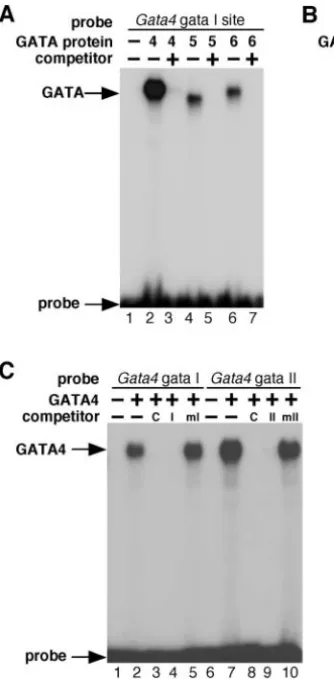
We also tested the three conserved, putative GATA sites contained within the CR2 region of the enhancer to determine if they were bound in EMSA by GATA4, GATA5 or GATA6. The gata I and gata II sites were each efficiently bound by GATA factors (Fig. 7), while the gata III site exhibited only very weak binding by GATA factors in vitro (data not shown). GATA4 bound very robustly to the gata I site in the Gata4
[image:7.612.44.338.68.380.2]lateral mesoderm enhancer (Fig. 7A, lane 2), whereas GATA5 and GATA6 bound more weakly to that site (Fig. 7A, lanes 4 and 6, respectively). Binding to the gata I site by each of the three GATA factors was specific because the binding of each Fig. 6.The Gata4lateral mesoderm enhancer contains a high-affinity Forkhead-binding site. (A) Recombinant FOXF1 protein was transcribed and translated in vitro and used in EMSA with a radiolabeled double-stranded oligonucleotide encompassing the Gata4Fox I site (lanes 2-5). Lane 1 contains reticulocyte lysate without recombinant FOXF1 protein (represented by a minus sign). FOXF1 efficiently bound to the Gata4Fox I site (lane 2) and this binding was efficiently competed by a range of excess unlabeled Gata4Fox I
oligonucleotides (lane 3-5). A nonspecific lysate-derived band is denoted. (B) Recombinant FOXF1 protein was transcribed and translated in vitro, and used in EMSA with a radiolabeled double-stranded
oligonucleotide representing a canonical FOXF1 site (Control FoxFI site). Lane 1 contains reticulocyte lysate without recombinant FOXF1 protein (represented by a minus sign). FOXF1 efficiently bound to the control Fox I site (lane 2). This binding was efficiently competed by an excess of unlabeled control probe (lane 3) and by an excess of Gata4Fox I probe (lanes 4-6), even when only a 10-fold excess of the competitor was present (lane 6). A mutant version of the Gata4Fox I site failed to compete for FOXF1 binding even at a 100-fold excess (lane 7). (C) Recombinant FOXA2 protein was transcribed and translated in vitro, and used in EMSA with a radiolabeled double-stranded
oligonucleotide representing a canonical control FOXA2 site (lanes 2- 5) or the Gata4Fox I site (lanes 7-10). Lanes 1 and 6 contain reticulocyte lysate without recombinant FOXA2 protein (represented by a minus sign). FOXA2 efficiently bound to the control FOXA2 site (lane 2) and to the Gata4 Fox I site (lane 7), and this binding was efficiently competed by an excess of unlabeled control FOXA2 probe (lanes 3 and 8) and by an excess of Gata4Fox I probe (lanes 4 and 9), but not by an excess of unlabeled mutant Gata4Fox I probe (lanes 5 and 10).
De
was efficiently competed by the addition of excess unlabeled control gata I site (Fig. 7A, lanes 3,5,7). Similarly, GATA4 also bound very robustly to the Gata4G2 enhancer gata II site (Fig. 7B, lane 2), whereas the binding of GATA5 and GATA6 to that site was considerably weaker (Fig. 7B, lanes 4 and 6, respectively). As with the gata I site, binding of all three factors to the gata II site was specifically inhibited by the addition of excess unlabeled gata II probe (Fig. 7B, lanes 3, 5 and 7). Because the binding of GATA4 to both the gata I and gata II sites appeared to be stronger than GATA5 or GATA6, we examined the binding of GATA4 in additional detail by competing with a bona fide control GATA-binding site and with mutant versions of the Gata4gata I and gata II sites (Fig. 7C). Binding of GATA4 to the gata I and II sites in the Gata4
enhancer was efficiently competed in both cases by a 100-fold excess of the Nkx2.5gs1 control site (Fig. 7C, lanes 3,8), which is a bona fide GATA4-binding site and has been described previously (Lien et al., 1999). The gata I site was also efficiently competed by excess self probe (Fig. 7C, lane 4) but not by mutant version of itself (Fig. 7C, lane 5). Similarly, the gata II site was competed by excess unlabeled self probe (Fig. 7C, lane 9) but not by a mutant form of itself (Fig. 7C, lane 10). These results demonstrate that the gata I and gata II sites in the Gata4 lateral mesoderm enhancer both represent extremely robust and specific GATA-binding sites.
In addition to the FOX and gata sites in the enhancer, we also tested two candidate SMAD sites in the enhancer (Fig. 5) to determine if these putative sites might be targets for SMAD
protein binding and direct regulation by BMP signaling. Because SMAD proteins bind to DNA as oligomers with SMAD4 (Derynck and Zhang, 2003; von Bubnoff and Cho, 2001), we tested the ability of SMAD1/SMAD4, SMAD4/SMAD5, SMAD4/SMAD8, and SMAD4 alone to bind to either of the two candidate SMAD-binding sites in the enhancer. In no case were we able to detect any binding by SMAD factors to either of the sites in the enhancer under conditions in which a control SMAD site was bound by SMAD4 oligomers in EMSA (data not shown). Furthermore, we introduced mutations predicted to disrupt SMAD protein binding into the two candidate SMAD elements in the G2 lateral mesoderm enhancer and tested the effect of those mutations on enhancer function in transgenic embryos. Simultaneous mutation of both putative SMAD sites in the enhancer had no effect on transgene expression at any time point examined, including early time points at 7.75 dpc and 8.5 dpc (data not shown). Taken together, these data suggest that the two putative elements in the G2 enhancer are not bona fide SMAD-binding sites and that the Gata4G2 lateral mesoderm enhancer is not a direct target of BMP signaling.
[image:8.612.46.213.73.412.2]Gata4lateral mesoderm enhancer activity is dependent on Forkhead- and GATA-binding sites To test the function of the Gata4FOX and GATA sites in vivo, we introduced mutations into the Fox I site and both of the functional GATA sites (gata I and gata II), in the context of the G2∆2-lacZ transgene (Fig. 4), and determined the effect of
Fig. 7.The Gata4lateral mesoderm enhancer contains two high-affinity GATA-binding sites.
(A,B) Recombinant GATA4, GATA5 and GATA6 proteins were transcribed and translated in vitro, and used in EMSA with radiolabeled double-stranded
oligonucleotides encompassing the Gata4gata I site (A, lanes 2-7) or the gata II site (B, lanes 2-7). In A and B, lane 1 contains reticulocyte lysate without recombinant protein (represented by a minus sign). GATA4 efficiently bound to both the gata I and gata II sites (A and B, lane 2) and this binding was specifically competed by excess unlabeled gata I (A, lane 3) and gata II probes (B, lane 3). GATA5 and GATA6 proteins also bound, although more weakly than GATA4 protein, to the gata I (A, lanes 4,6) and gata II sites (B, lanes 4,6), and binding by GATA5 and GATA6 was specifically competed by excess unlabeled gata I (A, lanes 5,7) and gata II probes (B, lanes 5,7). Approximately equivalent amounts of GATA4, GATA5 and GATA6 proteins were used in each sample. (C) Recombinant GATA4 protein was transcribed and translated in vitro and used in EMSA with a radiolabeled double-stranded oligonucleotide encompassing the Gata4gata I site (lanes 1-5) or the gata II site (lanes 6-10). Lanes 1 and 6 contain reticulocyte lysate without recombinant GATA4 protein (represented by a minus sign). GATA4 efficiently bound to both the gata I and gata II sites (lanes 2,7). Binding of GATA4 to the Gata4gata I site was competed by an excess of unlabeled gata I site (I, lane 4) and by an excess of an unlabeled control GATA site from the Nkx2.5gene (C, lane 3), but not by an excess of a mutant version of the Gata4gata I site (mI, lane 5). Likewise, the binding of GATA4 to the Gata4gata II site was specifically competed by excess unlabeled gata II probe (II, lane 9) and by excess unlabeled control probe (C, lane 8), but not by an excess of a mutant version of the gata II probe (mII, lane 10).
De
those mutations on enhancer function in transgenic embryos collected at 7.75 dpc, 9.5 dpc and 11.5 dpc (Fig. 8). The introduced mutations were identical to those used in the EMSA analyses, which were shown in Figs 6 and 7 to completely ablate FOXF1 and GATA4 binding, respectively. The wild-type G2∆2-lacZ transgene directed robust expression throughout the lateral mesoderm and allantois at 7.75 dpc (Fig. 8A). The wild-type construct continued to direct expression broadly in the visceral mesoderm and septum transversum mesoderm at 9.5 dpc (Fig. 8D) and directed expression restricted to mesenchyme surrounding the liver at 11.5 dpc (Fig. 8G). Mutation of the Fox I site in the enhancer resulted in a complete disruption of enhancer activity in 15 out of 15 F0 transgenic embryos at all time points (Fig. 8B,E,H), indicating a key role for Forkhead factors in the activation of the enhancer. Likewise, double mutation of the two bona fide GATA sites in the enhancer completely ablated enhancer activity in 14 out of 14 independent F0 transgenic embryos at all time points (Fig. 8C,F,I). These results demonstrate that Gata4expression in the lateral mesoderm is subject to auto- and/or cross-regulation by GATA4 and other GATA factors.
Activation of the Gata4 lateral mesoderm enhancer is dependent on BMP4
[image:9.612.347.560.251.510.2]Although we did not observe any bona fide SMAD-binding sites in the enhancer, several lines of evidence suggested the possibility that the Gata4G2 lateral mesoderm enhancer might be an indirect downstream target of BMP4. Previous studies indicated an interrelationship among Forkhead and GATA factors and BMP4 during the development of the endoderm (Rossi et al., 2001). Furthermore, the broad expression of the G2-lacZ transgene in the mesoderm and in the allantois, combined with the largely overlapping expression of Bmp4in those lineages or in nearby lineages where signaling could occur from one region to another, prompted us to test whether BMP4 might be an upstream regulator of Gata4 enhancer activity (Fig. 9). To test this hypothesis, we crossed mice
[image:9.612.59.275.319.552.2]Fig. 8.The Gata4lateral mesoderm enhancer is dependent on conserved Forkhead and GATA sites for its function in vivo. The wild-type Gata4enhancer transgene construct G2∆2 (wt; A,D,G) and transgenes containing mutations either in the Fox I site (mFox; B,E,H), or in both the gata I and gata II sites (mGATA; C,F,I), in the context of G2∆2, were used to generate transgenic embryos. Representative transgenic embryos are shown at 7.75 dpc (A-C), 9.5 dpc (D-F) and 11.5 dpc (G-I). The wild-type construct directed strong expression in the lateral mesoderm (arrowhead) and allantois (al) at 7.75 dpc (A), in the septum transversum (ST) and visceral mesoderm (arrowhead) at 9.5 dpc (D), and in the mesenchyme surrounding the liver (L) at 11.5 dpc (G). Mutations in the Fox I site completely eliminated transgene expression in all of the fifteen independent transgenic embryos analyzed (B,E,H). Similarly, mutation of both GATA sites completely eliminated lacZexpression in the fourteen independent transgenic embryos analyzed (C,F,I). Arrowheads in A-C indicate lateral mesoderm; arrowheads in D-F indicate visceral mesoderm.
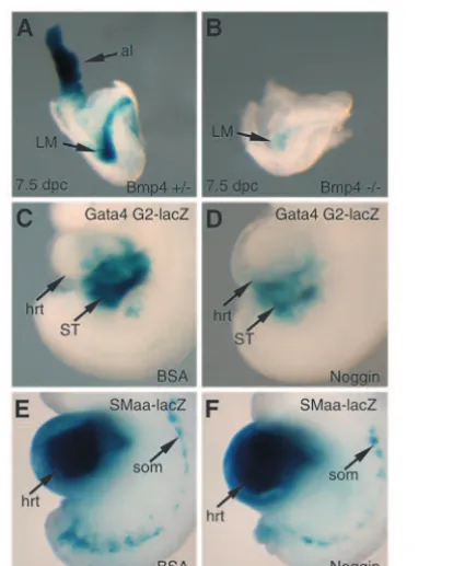
Fig. 9.Activity of the Gata4lateral mesoderm enhancer requires BMP4. (A,B) The G2-lacZtransgene was crossed into either a Bmp4+/–or a Bmp4–/–background, and embryos were collected at 7.5
dpc and stained with X-gal. Heterozygous Bmp4+/–; G2-lacZTg/0
embryos displayed strong expression of lacZin the lateral mesoderm (LM) and allantois (al; A). By contrast, expression directed by the Gata4lateral mesoderm enhancer in Bmp4–/–; G2-lacZTg/0 embryos
was dramatically attenuated in all embryos examined (B), indicating that transgene activity is dependent on BMP4. (C-F) Explanted tissue containing the heart (hrt) and septum transversum (ST) of embryos from a single G2-lacZstable transgenic line (C,D) or a single SMaa-lacZstable transgenic line (E,F) was collected at 9.5 dpc and cultured for 48 hours in the presence of BSA (C,E) or recombinant Noggin (D,F). Following incubation, explants were X-gal stained. Gata4lateral mesoderm enhancer activity was significantly reduced in all 15 viable embryo explants treated with Noggin when compared with the 15 viable embryo explants treated with BSA. By contrast, no differences were observed in X-gal staining in the heart or somites in SMaa-lacZexplants treated with BSA or Noggin (compare E and F), indicating that embryo explants were not in general crisis in the presence of Noggin. Viability of embryo explants was assessed by the observation of a continually beating heart.
De
harboring a stably integrated G2-lacZtransgene with Bmp4+/–
mice to generate mice that were G2-lacZTg/0; Bmp4+/–and crossed these mice to Bmp4+/–mice to generate embryos that were positive for the Gata4 enhancer transgene and null for
Bmp4. Approximately half of the Bmp4-null embryos died prior to gastrulation because of the role of BMP4 in gastrulation (Winnier et al., 1995). However, about half of the
Bmp4 null embryos survived gastrulation and were clearly alive at 7.5 dpc, as has been previously reported (Winnier et al., 1995). At this stage, the activity of the Gata4enhancer was easily detectable in heterozygous Bmp4embryos, with strong expression of lacZ evident in the lateral mesoderm and allantois (Fig. 9A). By contrast, the activity of the Gata4
enhancer was barely detectable in the lateral mesoderm of
Bmp4null embryos (Fig. 9B), indicating that the Gata4lateral mesoderm enhancer requires BMP4 for activity in vivo. It was difficult to determine the requirement of BMP4 for activation of Gata4G2-lacZin the allantois, as that structure was missing or severely reduced in Bmp4null embryos, as has been reported previously (Winnier et al., 1995). However, in Bmp4–/– embryos where some allantoic tissue was present, we never observed transgene expression (data not shown), suggesting that BMP4 is required for Gata4activation in the allantois as well.
The BMP antagonist Noggin inhibits Gata4 lateral mesoderm enhancer activity
Mice lacking Bmp4 often die prior to gastrulation and those that do survive past gastrulation have defects in mesodermal development, although mesoderm is present in those embryos (Winnier et al., 1995). Therefore, we considered the possibility that the severe reduction in Gata4 transgene expression in
Bmp4null embryos (shown in Fig. 9B) might be secondary to an overall mesodermal defect. Although we considered this possibility unlikely because Bmp4 mutants that survive gastrulation do have embryonic mesoderm, we wanted to define further the relationship between BMP signaling and the
Gata4mesodermal enhancer. As an independent approach to examine the role of BMP signaling on Gata4 enhancer activation, we tested whether the activity of the enhancer was sensitive to the BMP inhibitor Noggin.
Transgenic embryos from a single stable G2-lacZtransgenic line were explanted at 9.5 dpc and the region of the embryos containing the heart, septum transversum and portions of the visceral mesoderm were maintained in culture in the presence of BSA (Fig. 9C) or recombinant Noggin (Fig. 9D). As a control, transgenic embryos from a single stable transgenic smooth muscle α-actin line, SMaa-lacZ, which expresses β -galactosidase in the heart and somites at 9.5 dpc, were also explanted in the presence of BSA (Fig. 9E) or Noggin (Fig. 9F). Embryo explants were maintained in culture for 48 hours and the hearts continued to beat throughout the time course of the experiment, indicating the explants were viable for the duration of the experiment. A total of fifteen G2-lacZ
transgenic embryo explants from six different litters were treated with BSA and fifteen were treated with Noggin, and we compared X-gal staining in the septum transversum of embryo explants from the two groups. In each case, Noggin-treated
Gata4 G2-lacZ transgenic embryos exhibited a clear attenuation of β-galactosidase activity (Fig. 9D) when compared with control-treated embryos (Fig. 9C). By contrast,
no change in β-galactosidase activity in SMaa-lacZ explants was observed in the presence of Noggin (Fig. 9F) when compared with BSA-treated control explants (Fig. 9E), indicating that embryo explants were not in general crisis as a result of Noggin treatment. Thus, these results show that the activity of the Gata4 enhancer is inhibited by Noggin and further demonstrate that Gata4is a downstream target of BMP in the septum transversum and lateral mesoderm. Taken together, the results presented in Fig. 9 indicate that the Gata4
mesodermal enhancer requires BMP signaling for activation, and suggest that GATA4 may serve as a downstream effector of BMP4 in the lateral mesoderm and septum transversum mesenchyme.
Discussion
Gata4is among the earliest markers of the endoderm and the lateral mesoderm following gastrulation in the mouse embryo (Arceci et al., 1993; Heikinheimo et al., 1994; Nemer and Nemer, 2003), and targeted inactivation of the Gata4 gene in mice results in delayed ventral morphogenesis and subsequent cardia bifida as a result of severe endodermal defects (Kuo et al., 1997; Molkentin et al., 1997; Narita et al., 1997). However, despite the importance of Gata4 during development, it has been unclear how Gata4 expression is activated. In this manuscript, we identified the first transcriptional enhancer from the mouse Gata4 gene, and we show that this novel enhancer is active throughout the lateral mesoderm beginning at 7.5 dpc. As development proceeds, expression directed by the Gata4 lateral mesoderm enhancer becomes successively restricted to the visceral mesoderm and to the mesodermal component of the septum transversum (Fig. 2), and finally becomes restricted to the mesenchyme surrounding the liver at 11.5 dpc (Fig. 3).
The Gata4 lateral mesoderm enhancer requires an evolutionarily conserved Forkhead-binding site for activity in vivo (Fig. 8), and FOXF1 binds to the Forkhead-binding site in the enhancer with high affinity (Fig. 6). Thus, these results demonstrate that Gata4 is a direct transcriptional target of Forkhead factors, and we believe that FOXF1 is a likely candidate based on the overlapping expression pattern of Foxf1
and the G2-lacZ transgene (Fig. 2). The Gata4 lateral mesoderm enhancer also requires two high-affinity GATA-binding sites for function in vivo (Fig. 8), suggesting an essential role for auto- and cross-regulation of Gata4
expression by itself and other GATA factors.
GATA4 as a downstream effector of BMP signaling
BMP proteins are key mediators of tissue specification and of patterning in multiple developmental lineages. BMPs are known to play essential roles in bone and cartilage development, neural patterning, heart development, development of the extraembryonic mesoderm and in signaling from the mesoderm to the endoderm during endodermal specification (Hogan, 1996; Shivdasani, 2002; Tam et al., 2003; Zaret, 2001; Zhao, 2003). As members of the TGFβ superfamily of signaling molecules, BMP signaling modulates gene expression, at least in part, through the action of SMAD transcription factors (Derynck and Zhang, 2003; von Bubnoff and Cho, 2001). It has been proposed that BMP factors are able to exert such a diverse array of downstream effects through the
De
interactions of SMAD proteins with other transcription factors and through the resulting downstream activation of numerous transcription factor networks (Derynck and Zhang, 2003). In this regard, several studies have suggested that GATA transcription factors are downstream targets of BMP signaling. In response to BMP signaling, Gata4expression is upregulated in the precardiac mesoderm in the chick (Schultheiss et al., 1997). Similarly, BMP4 signaling from the mesoderm positively induces the expression of Gata4 in prehepatic endoderm during liver development, demonstrating activation of Gata4by BMP4 over a range from mesoderm to endoderm (Rossi et al., 2001). However, the pathways through which
Gata4is activated in response to BMP4 have not been defined in the endoderm or the mesoderm. Here, we show that the activity of the Gata4lateral mesoderm enhancer is attenuated by the BMP antagonist Noggin and is significantly inhibited in the absence of BMP4 (Fig. 9). Taken together with our analyses of the SMAD-binding sites in the enhancer, these observations demonstrate that Gata4 is probably an indirect downstream target of BMP4 in the lateral mesoderm via the enhancer described here, and suggest that GATA4 functions as a downstream effector of BMP signaling in the mesoderm.
A reinforcing transcriptional pathway dependent on GATA4
In this study, we demonstrate that the Gata4lateral mesoderm enhancer requires an essential Forkhead-binding site that is efficiently bound by FOXF1 (Figs 6, 8), supporting the possibility that BMP activation of Gata4could be mediated by FOXF1 or other Forkhead proteins. Indeed, a recent study performed in Xenopus embryos has shown that Foxf1
expression is reduced when BMP4 signaling is reduced, indicating that FOXF1 is also a downstream target of BMP4 (Tseng et al., 2004). Interestingly, however, Bmp4 has been shown to be a potential target of FOXF1 during mouse development, as the level of Bmp4 mRNA was significantly reduced in the posterior primitive streak, the lateral plate and the allantois in FOXF1 null embryos (Mahlapuu et al., 2001b). Taken together, these results suggest that FOXF1 and BMP4 may function in a reinforcing regulatory circuit designed to amplify a transcriptional response downstream of BMP4 signaling, possibly via GATA and other transcription factor families. GATA transcription factors also appear to activate
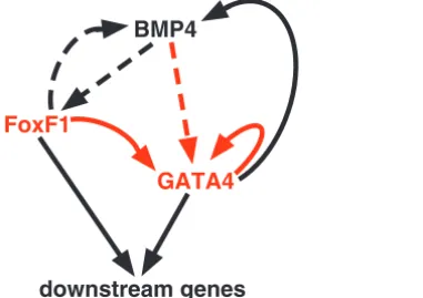
Bmp4expression, further supporting the notion of a reinforcing transcriptional network (Nemer and Nemer, 2003; Peterkin et al., 2003). The mouse and Xenopus Bmp4regulatory sequences contain GATA-binding sites that are functional in studies performed in vitro, suggesting that Bmp4expression may be activated or maintained by GATA factors (Nemer and Nemer, 2003; Peterkin et al., 2003). A reinforcing transcriptional model is consistent with the work presented here, which suggests a model forGata4activation in the lateral mesoderm in which BMP4 activates the expression of Gata4 indirectly via FOXF1, and GATA4 functions in a positive-feedback loop to reinforce its own expression through essential GATA sites in its enhancer (Fig. 10).
Modular regulation of Gata4 transcription
During embryonic development, Gata4 is expressed in multiple tissues and exhibits a dynamic and distinct pattern of expression in each different lineage where it is expressed
(Arceci et al., 1993; Heikinheimo et al., 1994; Parmacek and Leiden, 1999). Notably, the Gata4 enhancer described here directs expression only to a subset of the endogenous pattern of Gata4 expression, suggesting that other distinct modular enhancers sufficient to direct expression to other lineages are also likely to be present within the Gata4 locus. This type of modular regulation has been observed for other key transcriptional regulators of tissue specification and differentiation, including Nkx2.5(Schwartz and Olson, 1999) and mef2c(De Val et al., 2004; Dodou et al., 2004; Dodou et al., 2003), and modular regulation has been suggested as a mechanism for regulatory diversity (Firulli and Olson, 1997). Given the role of GATA4 as an early regulator of cardiac and endodermal development, it will be important to identify enhancers sufficient to direct expression to those lineages also. It will be particularly interesting to determine whether Gata4
is a target of Forkhead proteins and is subject to auto-regulation in other lineages, as it is in the lateral mesoderm, or whether regulation in other lineages is via distinct transcriptional hierarchies.
The expression directed by the Gata4 enhancer described in these studies shows a progressive restriction from an initially broad pattern within the mesoderm to a narrow pattern restricted only to the mesenchymal cells surrounding the liver. The presence of a distinct enhancer for this expression domain suggests a role for GATA4 in the specification of mesodermal cells and their derivatives early in mesoderm development, and it also suggests a potential role for GATA4 in regulating gene expression in the cells of the liver mesenchyme. Thus, it will be important to define the function of GATA4 in the early lateral mesoderm and in the development of the mesenchyme surrounding the liver. The rapid and progressive restriction of enhancer activity from 7.5 dpc to 11.5 dpc suggests that the activators of the Gata4lateral mesoderm enhancer also become restricted during development. Alternatively, the Gata4
BMP4
FoxF1
GATA4
[image:11.612.369.560.77.211.2]downstream genes
Fig. 10.A reinforcing transcriptional network for gene activation in the lateral mesoderm dependent on GATA4. In this model, GATA4 expression is activated indirectly by BMP4 (indicated by a dashed arrow) and directly by Forkhead factors, such as FOXF1. BMP4 and FOXF1 reciprocally activate the expression of one another, which further reinforces GATA4 expression. GATA4 then further reinforces the program by activating its own transcription and the transcription of downstream mesodermal genes. Red arrows represent evidence provided in the current study. Black arrows represent evidence provided by previously published studies. Dashed arrows indicate either a direct or indirect activation; solid arrows represent direct activation through direct enhancer binding.
De
enhancer may contain cis-acting elements that are responsive to active repression and that ultimately restrict the activation of the enhancer to the mesoderm of the septum transversum and, finally, to the liver mesenchyme.
While the importance of GATA4 during embryogenesis is clearly established, an understanding of its transcriptional regulation has remained elusive. Here, we show that the expression of Gata4 in the lateral mesoderm and septum transversum is mediated by an enhancer located ~40 kb upstream of the start of transcription. Detailed analyses of this enhancer show functional FOX and GATA sites that are essential for transcriptional activation in vivo. These data suggest that transcriptional regulation of Gata4is modular, and that enhancer elements may be remote from the coding sequences of the gene. The use of deep evolutionary sequence conservation allowed the identification of its early mesodermal enhancer and suggests that enhancers driving cardiac and endodermal expression might be identified through a similar process. Indeed, we have recently identified a separate enhancer from the Gata4 gene that is sufficient to direct expression to the endoderm (A.R. and B.L.B., unpublished). It will be interesting to determine whether pathways similar to those described here also regulate Gata4 expression in the endoderm and other lineages where it is expressed.
We thank Robert Costa, Jeff Molkentin, Rik Derynck and Pao-Tien Chuang for providing plasmids, and Vina Lu, Stephanie Greene and Eric Ho for assistance with these studies. We are grateful to Ken Zaret and Robert Costa for helpful discussions, and to Gail Martin and Jim Martin for providing mice. We also appreciate the critical comments on the manuscript provided by Jim Martin. We thank Eddy Rubin and Jan-Fang Cheng for BAC sequence and VISTA analyses, which were supported by the Berkeley PGA from the National Heart, Lung, and Blood Institute. A.B.H. was supported by a predoctoral fellowship from the Howard Hughes Medical Institute and A.R. was supported in part by a postdoctoral fellowship from the Spanish Ministerio de Educación, Cultura y Deportes. This work was supported by grant HL64658 from the NHLBI and by a grant from the Sandler Family Supporting Foundation to B.L.B.
References
Altschul, S. F., Gish, W., Miller, W., Myers, E. W. and Lipman, D. J.(1990). Basic local alignment search tool. J. Mol. Biol.215, 403-410.
Anderson, J. P., Dodou, E., Heidt, A. B., De Val, S. J., Jaehnig, E. J., Greene, S. B., Olson, E. N. and Black, B. L.(2004). HRCis a direct transcriptional target of MEF2 during cardiac, skeletal, and arterial smooth muscle development in vivo. Mol. Cell. Biol. 24, 3757-3768.
Arceci, R. J., King, A. A., Simon, M. C., Orkin, S. H. and Wilson, D. B. (1993). Mouse GATA-4: a retinoic acid-inducible GATA-binding transcription factor expressed in endodermally derived tissues and heart. Mol. Cell Biol.13, 2235-2246.
Bossard, P. and Zaret, K. S. (1998). GATA transcription factors as potentiators of gut endoderm differentiation. Development125, 4909-4917. Burch, J. B.(2005). Regulation of GATA gene expression during vertebrate
development. Semin. Cell Dev. Biol.16, 71-81.
Carlsson, P. and Mahlapuu, M.(2002). Forkhead transcription factors: key players in development and metabolism. Dev. Biol.250, 1-23.
De Val, S., Anderson, J. P., Heidt, A. B., Khiem, D., Xu, S. M. and Black, B. L.(2004). Mef2cis activated directly by Ets transcription factors through an evolutionarily conserved endothelial cell-specific enhancer. Dev. Biol. 275, 424-434.
Denson, L. A., McClure, M. H., Bogue, C. W., Karpen, S. J. and Jacobs, H. C. (2000). HNF3beta and GATA-4 transactivate the liver-enriched homeobox gene, Hex. Gene246, 311-320.
Derynck, R. and Zhang, Y. E. (2003). dependent and Smad-independent pathways in TGF-beta family signalling. Nature425, 577-584.
Divine, J. K., Staloch, L. J., Haveri, H., Jacobsen, C. M., Wilson, D. B., Heikinheimo, M. and Simon, T. C.(2004). 4, 5, and GATA-6 activate the rat liver fatty acid binding protein gene in concert with HNF-1alpha. Am. J. Physiol. Gastrointest. Liver Physiol.287, G1086-G1099. Dodou, E., Xu, S. M. and Black, B. L.(2003). mef2cis activated directly by
myogenic basic helix-loop-helix proteins during skeletal muscle development in vivo. Mech. Dev.120, 1021-1032.
Dodou, E., Verzi, M. P., Anderson, J. P., Xu, S. M. and Black, B. L.(2004). Mef2cis a direct transcriptional target of Isl-1 and GATA factors in the anterior heart field during mouse embryonic development. Development 131, 3931-3942.
Durocher, D., Charron, F., Warren, R., Schwartz, R. J. and Nemer, M. (1997). The cardiac transcription factors Nkx2-5 and GATA-4 are mutual cofactors. EMBO J.16, 5687-5696.
Firulli, A. B. and Olson, E. N.(1997). Modular regulation of muscle gene transcription: a mechanism for muscle cell diversity. Trends Genet.13, 364-369.
Graves, J. A.(1996). Mammals that break the rules: genetics of marsupials and monotremes. Annu. Rev. Genet.30, 233-260.
Grepin, C., Nemer, G. and Nemer, M.(1997). Enhanced cardiogenesis in embryonic stem cells overexpressing the GATA-4 transcription factor. Development124, 2387-2395.
Heikinheimo, M., Scandrett, J. M. and Wilson, D. B.(1994). Localization of transcription factor GATA-4 to regions of the mouse embryo involved in cardiac development. Dev. Biol.164, 361-373.
Hogan, B. L. (1996). Bone morphogenetic proteins in development. Curr. Opin. Genet. Dev.6, 432-438.
Hogan, B., Beddington, R., Costantini, F. and Lacy, E. (1994). Manipulating The Mouse Embryo. Plainview, NY: Cold Spring Harbor Laboratory Press.
Hu, T., Yamagishi, H., Maeda, J., McAnally, J., Yamagishi, C. and Srivastava, D. (2004). Tbx1 regulates fibroblast growth factors in the anterior heart field through a reinforcing autoregulatory loop involving forkhead transcription factors. Development131, 5491-5502.
Jones, C. M., Lyons, K. M. and Hogan, B. L.(1991). Involvement of Bone morphogenetic protein-4 (BMP-4) and Vgr-1 in morphogenesis and neurogenesis in the mouse. Development111, 531-542.
Kalinichenko, V. V., Zhou, Y., Bhattacharyya, D., Kim, W., Shin, B., Bambal, K. and Costa, R. H.(2002). Haploinsufficiency of the mouse Forkhead Box f1gene causes defects in gall bladder development. J. Biol. Chem.277, 12369-12374.
Kalinichenko, V. V., Gusarova, G. A., Kim, I. M., Shin, B., Yoder, H. M., Clark, J., Sapozhnikov, A. M., Whitsett, J. A. and Costa, R. H.(2004). Foxf1 haploinsufficiency reduces Notch-2 signaling during mouse lung development. Am. J. Physiol. Lung Cell Mol. Physiol.286, L521-L530. Klinedinst, S. L. and Bodmer, R.(2003). Gata factor Pannier is required to
establish competence for heart progenitor formation. Development 130, 3027-3038.
Kothary, R., Clapoff, S., Darling, S., Perry, M. D., Moran, L. A. and Rossant, J.(1989). Inducible expression of an hsp68-lacZhybrid gene in transgenic mice. Development105, 707-714.
Kuo, C. T., Morrisey, E. E., Anandappa, R., Sigrist, K., Lu, M. M., Parmacek, M. S., Soudais, C. and Leiden, J. M. (1997). GATA4 transcription factor is required for ventral morphogenesis and heart tube formation. Genes Dev.11, 1048-1060.
Lee, Y., Shioi, T., Kasahara, H., Jobe, S. M., Wiese, R. J., Markham, B. E. and Izumo, S.(1998). The cardiac tissue-restricted homeobox protein Csx/Nkx2.5 physically associates with the zinc finger protein GATA4 and cooperatively activates atrial natriuretic factorgene expression. Mol. Cell Biol.18, 3120-3129.
Liang, Q., De Windt, L. J., Witt, S. A., Kimball, T. R., Markham, B. E. and Molkentin, J. D.(2001). The transcription factors GATA4 and GATA6 regulate cardiomyocyte hypertrophy in vitro and in vivo. J. Biol. Chem.276, 30245-30253.
Lien, C. L., Wu, C., Mercer, B., Webb, R., Richardson, J. A. and Olson, E. N.(1999). Control of early cardiac-specific transcription of Nkx2-5by a GATA-dependent enhancer. Development126, 75-84.
Liu, W., Selever, J., Wang, D., Lu, M. F., Moses, K. A., Schwartz, R. J. and Martin, J. F.(2004). Bmp4signaling is required for outflow-tract septation and branchial-arch artery remodeling. Proc. Natl. Acad. Sci. USA101, 4489-4494.
Mahlapuu, M., Enerback, S. and Carlsson, P.(2001a). Haploinsufficiency of the forkhead gene Foxf1, a target for sonic hedgehog signaling, causes lung and foregut malformations. Development128, 2397-2406.