INTRODUCTION
Segmental organization of the anterior-posterior (AP) body axis is observed in diverse animal phyla, including arthropods, annelids and chordates. However, there has been debate regarding whether segmentation among phyla has a common origin and shares common genetic programs (Damen, 2007; Peel et al., 2005; Tautz, 2004). Segmentation mechanisms in Drosophila occur via hierarchical interactions between transcription factor gradients that subdivide embryos into progressively smaller units, eventually resulting in a repetitive pattern of segments along the entire AP axis of the embryo (Ingham, 1988; Nüsslein-Volhard and Wieschaus, 1980; Pankratz and Jackle, 1993; St. Johnston and Nüsslein-Volhard, 1992). An important feature of this segmentation mode, called long-germ segmentation, is that the syncytial state of the early Drosophilaembryo allows diffusion of transcription factors to form gradients throughout the egg, enabling simultaneous specification of all segments.
Although the explication of Drosophila segmentation has provided a paradigm for investigating the evolution of the segmentation process, recent studies in other arthropods have demonstrated that long-germ segmentation is evolutionarily derived and not representative of arthropods. Indeed, most arthropods
(chelicerates, myriapods, crustaceans and insects) show short- or intermediate-germ segmentation (Davis and Patel, 2002; Liu and Kaufman, 2005; Tautz et al., 1994). Because posterior segments are formed sequentially in an anterior-to-posterior progression from a posterior cellularized region, referred to as a ‘growth zone’ in these arthropods, the free diffusion of transcriptional factors that occurs in Drosophila is not possible in the posterior growth zone. Therefore, it has been speculated that there might have been a drastic change in the molecular mechanisms underlying posterior patterning and segmentation during arthropod evolution (Sander, 1976; Patel, 1994; Davis and Patel, 1999; Peel and Akam, 2003; Tautz, 2004; Peel et al., 2005).
The segmentation of short- and intermediate-germ arthropods is reminiscent of somitogenesis in vertebrates, suggesting involvement of a cell-cell signaling system. In vertebrates, somites are formed sequentially in an anterior-to-posterior progression from the presomitic mesoderm (PSM) (Pourquie, 2001). This process involves a molecular oscillator, called the somitogenesis clock, which drives the periodic expression of genes in the PSM. The formation of each somite is accomplished by a wave of oscillatory gene expression. Several genes of the Notch pathway, including Delta, lunatic fringe, and hairy, exhibit oscillatory expression in the PSM. Therefore, Notch signaling is a crucial component of the somitogenesis clock.
Delta/Notch signaling has been shown to be involved in the segmentation of one short-germ arthropod, the wandering spider Cupiennius salei.In C. salei, Delta is expressed in the posterior growth zone as well as in segmental stripes anterior to the growth zone (Stollewerk et al., 2003). In addition, RNAi silencing of components of the Delta/Notch signaling pathway (i.e. Notch, Delta, Presenilinand Suppressor of Hairless) produces severe segmentation defects (Schoppmeier and Damen, 2005; Stollewerk et al., 2003). Furthermore, in a short-germ insect, the cockroachPeriplaneta
Development 138, 3823-3833 (2011) doi:10.1242/dev.060681 © 2011. Published by The Company of Biologists Ltd
Department of Life Systems, Institute of Technology and Science, The University of Tokushima, 2-1 Minami-Jyosanjima-cho, Tokushima City, 770-8506 Japan.
*These authors contributed equally to this work
†Present address: Division of Developmental Neurobiology, Graduate School of Medical Sciences, Kumamoto University, Kumamoto 860-8556, Japan ‡Author for correspondence (mito@bio.tokushima-u.ac.jp)
Accepted 28 June 2011 SUMMARY
Delta/Notch signaling controls a wide spectrum of developmental processes, including body and leg segmentation in arthropods. The various functions of Delta/Notch signaling vary among species. For instance, in Cupiennius spiders, Delta/Notch signaling is essential for body and leg segmentation, whereas in Drosophilafruit flies it is involved in leg segmentation but not body
segmentation. Therefore, to gain further insight into the functional evolution of Delta/Notch signaling in arthropod body and leg segmentation, we analyzed the function of the Delta (Gb⬘Delta) and Notch (Gb⬘Notch) genes in the hemimetabolous,
intermediate-germ cricket Gryllus bimaculatus. We found that Gb⬘Deltaand Gb⬘Notch were expressed in developing legs, and that RNAi silencing of Gb⬘Notch resulted in a marked reduction in leg length with a loss of joints. Our results suggest that the role of Notch signaling in leg segmentation is conserved in hemimetabolous insects. Furthermore, we found that Gb⬘Deltawas expressed transiently in the posterior growth zone of the germband and in segmental stripes earlier than the appearance of winglesssegmental stripes, whereas Gb⬘Notchwas uniformly expressed in early germbands. RNAi knockdown of Gb⬘Deltaor Gb⬘Notchexpression resulted in malformation in body segments and a loss of posterior segments, the latter probably due to a defect in posterior growth. Therefore, in the cricket, Delta/Notch signaling might be required for proper morphogenesis of body segments and posterior elongation, but not for specification of segment boundaries.
KEY WORDS: Cricket, Intermediate-germ insect, Delta, Notch, Segmentation, Growth zone, RNA interference
Ancestral functions of Delta/Notch signaling in the
formation of body and leg segments in the cricket
Gryllus
bimaculatus
Taro Mito*,‡, Yohei Shinmyo*,†, Kazuki Kurita, Taro Nakamura, Hideyo Ohuchi and Sumihare Noji
D
E
V
E
LO
P
M
E
N
americana,Delta is expressed in segmental stripes anterior to the growth zone and knockdown of the Notchgene causes posterior segmentation defects (Pueyo et al., 2008). These observations suggest that the involvement of Delta/Notch signaling in body segmentation might be an ancestral feature of arthropods and that Notch-mediated segmentation might be inherited from a common ancestor of arthropods and vertebrates (Couso, 2009). The apparent lack of involvement of Delta/Notch signaling in body segmentation in Drosophilaand Tribolium(Richards et al., 2008; Campos-Ortega and Knust, 1990) suggests that this mechanism might have been lost in the common ancestor of beetles and flies, and possibly of all higher or holometabolous insects. However, such a scenario is controversial because data from paleontology and comparative morphology research seem to suggest that a segmented common ancestor for all bilaterians is unlikely (Chipman, 2010). In addition, the role of Notch signaling in arthropod segmentation process remains ambiguous. Therefore, further exploration of the role of Notch signaling in arthropod segmentation is needed.
In this study, we examined the expression patterns and functions of the Delta and Notch genes in the intermediate-germ cricket Gryllus bimaculatus, focusing on formation of body and leg segments. We found that Deltawas expressed transiently in the posterior growth zone and in segmental stripes before the emergence of wingless segmental stripes, whereas Notch was uniformly expressed in early G. bimaculatus embryos. Our functional analyses ofGryllus Delta andNotchgenes indicate that Notch signaling is required for proper morphogenesis of segments and formation of posterior segments through regulation of germband elongation, but is not required for the specification of segment boundaries. By contrast, our data demonstrate that Notch signaling is essential for the specification of segment boundaries and normal growth during leg formation.
MATERIALS AND METHODS Animals
G. bimaculatus(two-spotted cricket) nymphs and adults were reared at
28-30°C at 70% humidity under 10 hour/14 hour light/dark photoperiod as previously described (Niwa et al., 1997). Fertilized eggs were collected with wet paper towels and incubated at 28°C in a plastic dish. Embryos were staged according to the scheme of Zhang et al. (Zhang et al., 2005).
Cloning
Total RNA was extracted from cricket embryos at various stages using Isogen (Nippon-Gene). cDNA was synthesized using the Superscript First Strand Synthesis Kit (Invitrogen) with random hexamers. To clone
Gb⬘Delta, we first performed a degenerate polymerase chain reaction
(PCR) using primers targeting the VCLKHYQA (forward) and NQDLNYCT (reverse) motifs of a conserved domain including the Delta-Serrate-Lag2 (DSL) domain. 5⬘and 3⬘RACE PCRs were performed using gene-specific primers and anchor primers supplied in a SMART RACE cDNA Amplification Kit (Clontech). The Gb⬘DeltacDNA sequences were deposited at the DNA Data Bank of Japan (DDBJ) (accession number: AB635584). To isolate two Gb⬘Notch PCR fragments, we used two pairs of degenerate primers targeting the CEGDINE (forward)/CDYGCNN (reverse) and AAVAA (forward)/WAAAVNN (reverse) motifs. We then isolated a 2280-bp fragment using specific primers directed against these PCR fragments. The Gb⬘NotchcDNA sequences were deposited at the DDBJ (accession number: AB635585).
Embryo fixation and whole-mount in situ hybridization
Embryo fixation and whole-mount in situ hybridization with digoxigenin (DIG)-labeled antisense RNA probes were performed as previously described (Niwa et al., 2000; Zhang et al., 2005). Double labeled in situ hybridization was performed as previously described (Mito et al., 2007).
Parental RNAi experiments
Double-stranded RNA (dsRNA) directed against Gb⬘Delta, Gb⬘Notch and
DsRed2was prepared as described previously (Miyawaki et al., 2004). In
all RNAi experiments, DsRed2dsRNA [660 bp, derived from pDsRed2-N1 (Clontech)] was used as a negative control. To exclude potential off-target effects in RNAi experiments, we used two different dsRNAs corresponding to two non-overlapping regions in each of the Gb⬘Deltaand
Gb⬘Notchtranscripts and confirmed that both of the dsRNAs derived from
the two different regions resulted in the same phenotype. The dsRNAs were designated Gb⬘Delta1(508 bp; nucleotide positions 1225-1732),
Gb⬘Delta2(321 bp; nucleotide positions 901-1221), Gb⬘Notch1(677 bp;
nucleotide positions 61-737) and Gb⬘Notch2(461 bp; nucleotide positions 1678-2138) (Fig. 1A,B). The final concentration of dsRNA was adjusted to 20 M and dsRNAs were injected as described previously (Mito et al., 2005). Eggs were collected 5 to 10 days after injection.
Immunocytochemistry
The ventral nerve cord was visualized by incubating embryos with a goat anti-HRP antibody (Jackson ImmunoResearch) that reacts specifically with the N-linked carbohydrate epitope found on insect neurons (Seppo et al., 2003). After fixation, cricket embryos were washed in KTBT [50 mM Tris-HCl (pH 7.5), 150 mM NaCl, 10 mM KCl, 0.1% Triton X-100]. The embryos were then blocked with 1.5% blocking reagent (Roche) in KTBT and incubated with FITC-conjugated anti-HRP antibody diluted 1:5 in 1.5% blocking reagent in KTBT for 3 hours at 4°C. Blocking was followed by three washes with KTBT.
Cell proliferation assay
The cell proliferation assay was carried out using the Click-iT EdU Alexa Fluor 488 Imaging Kit (Invitrogen) (Salic and Mitchison, 2008). 5-Ethynyl-2⬘-deoxyuridine (EdU) was injected into the posterior of the cricket egg at the appropriate stages. Embryos were incubated for 4 hours at 28°C after EdU injection and then fixed for 15 minutes. EdU-incorporating cells were detected according the manufacturer’s instructions. To counterstain nuclei, embryos were held in a 20 ng/ml solution of Hoechst 33342 in PBT (PBS with 0.1% Tween-20) for 15 minutes at room temperature after EdU incorporation. Embryos were then washed in PBT and mounted on slides for visualization.
RESULTS
Isolation of Gryllus Deltaand Notchorthologs
Using degenerate primers directed against the conserved regions of Drosophila Delta– a conserved DSL domain and nine epidermal growth factor (EGF) repeats (Fig. 1A) – a short Gb⬘Deltaclone was isolated and used to design specific primers for 5⬘ and 3⬘
RACE and isolate fragments of the gene. TheGb⬘Deltagene is predicted to encode 455 amino acids with a conserved DSL domain and five EGF repeats (Fig. 1A).
To clone Gb⬘Notch, two pairs of degenerate primers were designed against the extracellular and intracellular domains, and two PCR fragments from both domains were obtained. Using specific primers directed against these PCR fragments, we isolated a 2280-bp fragment encoding 760 amino acids and containing several conserved domains, EGF repeats, Notch/Lin-12 repeats, a transmembrane domain, a RAM 23 domain and ankyrin repeats (Fig. 1B).
Gb⬘Delta and Gb⬘Notchexpression in early stage
Gryllus embryos
In Gryllus, the anterior segments, the gnathal to mesothoracic (T2) segments, are specified almost simultaneously in the early embryo, whereas the remaining posterior segments, the metathoracic (T3) and abdominal segments, are formed sequentially through elongation of the posterior growth zone of the germband
D
E
V
E
LO
P
M
E
N
(Miyawaki et al., 2004). We examined whether Gb⬘Delta and Gb⬘Notch are expressed during this segmentation process by whole-mount in situ hybridization (WISH) (Fig. 2).
Intense expression of Gb⬘Deltawas observed in the prospective mandible segment and the posterior region of the embryo 30-34 hours after egg-laying (hAEL) (Fig. 2A,B). As can be observed in sagittal sections (Fig. 2C,C⬘), the posterior domain was localized to the ectoderm cells in the most posterior region of the embryo, overlapping with the posterior growth zone. Interestingly, Gb⬘Delta expression receded from the growth zone and remained in the terminal region of the embryo at 36 hAEL (Fig. 2D, arrowhead). Segmental expression of Gb⬘Deltawas observed in the prospective maxilla, labium and prothoracic (T1) segments at 34 hAEL (Fig. 2B), and in the prospective T2 and T3 segments at 36 hAEL (Fig. 2D).
To investigate the location and timing of the Gb⬘Delta stripes, we used expression of a segment polarity gene Gb⬘wg as a segmental marker. Gb⬘wgis expressed in stripes in an anterior-to-posterior sequence during segmentation and seems to be involved in the specification of segment boundaries in Gryllus embryos (Miyawaki et al., 2004). Double staining for Gb⬘Deltaand Gb⬘wg revealed that the Gb⬘Deltastripes overlap with Gb⬘wgstripes (Fig. 2E,F) and precede the corresponding Gb⬘wgstripe (arrow in Fig. 2G). During subsequent posterior elongation, Gb⬘Deltastripes in the gnathal and thoracic regions faded out in an AP progression, whereas Gb⬘Deltastripes appeared sequentially in the prospective abdominal segments (arrows in Fig. 2H,I). Additionally, spots of Gb⬘Delta expression were observed in the head region (Fig. 2B,D,E,G-I) and trunk (Fig. 2G-I). These spots probably correspond to proneuronal cells, as observed in proneural regions during neural cell fate specification in Drosophila(Baker, 2000)
and other arthropods (Dove and Stollewerk, 2003; Kadner and Stollewerk, 2004; Stollewerk, 2002). In contrast to the specific expression pattern of Gb⬘Delta, Gb⬘Notchwas uniformly expressed during early embryogenesis (compare Fig. 2J,K with 2L,M).
Gb⬘Delta and Gb⬘Notchexpression in late stage
Gryllusembryos
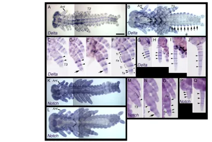
[image:3.612.54.294.57.287.2]Next, we observed the expression patterns of Gb⬘Delta and Gb⬘Notch during late embryogenesis by WISH (Fig. 3). In Drosophila, Delta and Notch are expressed during leg development, where they are required for correct segmentation (Parody and Muskavitch, 1993; Shellenbarger and Mohler, 1975). In stage-8 Gryllusembryo legs, Gb⬘Delta was expressed in two rings (arrowheads in Fig. 3C), corresponding to the proximal sides Fig. 1. Delta and Notch orthologs in Gryllus bimaculatus.
(A,B)Comparison of the structures of the Gryllus Delta and Notch proteins with those of Drosophila. (A)GryllusDelta contains the conserved DSL domain and five EGF repeats (EGF-rep). (B)The partial
[image:3.612.313.561.58.354.2]GryllusNotch protein contains several conserved domains, EGF repeats, Notch lin 12 repeats (N/lin 12), transmembrane domain (trans), a RAM 23 domain (RAM) and ankyrin repeats (Ank). Regions used to synthesize Gb⬘Delta(A) and Gb⬘Notch(B) dsRNAs are shown. Pest, a region rich in proline, glutamine, serine and threonine residues.
Fig. 2. Expression patterns of Gb⬘Delta and Gb⬘Notchduring early embryogenesis of Gryllus bimaculatus.(A-I)Expression patterns of Gb⬘Deltaat 30 (A), 34 (B,C), 36 (D), 40 (E), 44 (F) and 52 (G) hours after egg laying (hAEL). (C,C⬘) Sagittal sections of an embryo at 34 hAEL stained for Gb⬘Delta(C) and stained with DAPI to visualize nuclei (C⬘). The posterior region of the embryo (e) is shown. Amnion (a) is growing with forming the amniotic cavity (ac). Arrowheads in B and C indicate the posterior end of the embryo. (D)The posterior Gb⬘Delta
domain is retained only in the terminal region of the embryo (arrowhead). (E-G)Embryos double stained for Gb⬘Delta (blue) and
Gb⬘wg(brown). (F)High magnification view of the boxed area in E.
Gb⬘Deltawas expressed in segmental stripes during anterior (B,D,E) and posterior (G-I, arrows) segmentation. (G)The Gb⬘Deltastripe, but not the Gb⬘wgstripe, was observed in the first abdominal segment (A1; arrow). (J,K)Expression patterns of Gb⬘Notchat 36 (J) and 50 (K) hAEL. (L,M)Embryos at 36 (L) and 50 (M) hAEL hybridized with a sense control. Gb⬘NotchRNA was ubiquitously distributed during anterior and posterior segmentation. Mn, mandible; Mx, maxilla; Lb, labium; T1-3, thoracic segments 1 to 3; A1, abdominal segment 1. Scale bar: in A, 250m for A,B,D,E,G-M.
D
E
V
E
LO
P
M
E
N
of the femur/tibia boundary and the tibia/tarsus boundary (see Fig. 3F), and small spots (Fig. 3C). During stage 9, an additional broad ring appeared in the prospective tarsal region (arrows in Fig. 3D,E). By stage 10, the broad tarsal ring divided into two narrow rings (arrows in Fig. 3F) and additional rings appeared in the prospective trochanter and tarsus (asterisks in Fig. 3F). In addition, faint and broad expression was observed in the prospective femur and tibia (brackets in Fig. 3F).
A number of Gb⬘Deltarings were also observed in the developing antenna of the late stage embryos (Fig. 3A,B,G-J). In stage-8 antennae, three intense rings of Gb⬘Delta expression (arrowheads in Fig. 3G) were observed in the middle region and a few weak rings were observed in the proximal and distal regions (Fig. 3G). By stage 10, additional rings were intercalated between the intense rings and also appeared in the proximal and distal regions (Fig. 3H-J).
Gb⬘Notchwas expressed throughout the whole embryo, with upgraded expression in rings in the legs and antennae during late embryogenesis (Fig. 3K-Q). In stage-8 legs, intense expression of Gb⬘Notchwas observed as a broad ring in the prospective femur (bracket in Fig. 3M) and as a smaller domain in a more distal region (arrowhead in Fig. 3M). By stage 10, these intense domains became restricted to the femur/tibia boundary and the tibia/tarsus
boundary, consequently forming intense ring-like domains in these segment boundaries (Fig. 3N,O, arrowheads). In addition, a new intense ring appeared in the prospective tarsus (arrow in Fig. 3N) and divided into two narrow rings (Fig. 3O, arrows), similar to the Gb⬘Deltatarsal ring formation. In antennae, narrow rings were intercalated between three intense rings (arrowheads in Fig. 3P,Q) and were also added in the distal region by stage 10 (Fig. 3P,Q), similar to the rings of Gb⬘Deltaexpression observed in the antenna. It is known that fringe, a negative regulator of Notch signaling, is expressed in the epidermis of the trunk segments after segmentation in the short-germ insect Schistocerca gregaria, suggesting the involvement of Notch signaling in segment morphogenesis, but not in the establishment of segmental boundaries (Dearden and Akam, 2000). Similarly, Gb⬘Delta was expressed in the trunk epidermis of embryos during late embryogenesis (Fig. 3B, arrows).
Morphological analyses of Gb⬘Delta and Gb⬘Notch RNAi-treated embryos
[image:4.612.53.477.56.340.2]To examine the functions of Gb⬘Delta and Gb⬘Notch in the formation of body and leg segments, we injected dsRNA directed against these genes into adult females to determine knockdown Fig. 3. Expression patterns of Gb⬘Delta and Gb⬘Notchduring late embryogenesis of Gryllus bimaculatus.(A,B)Expression patterns of
Gb⬘Deltain whole embryos at stage 9 (A) and 10 (B). (C-F)Expression patterns of Gb⬘Deltain developing leg buds at late stage 8 (C), mid stage 9 (D), late stage 9 (E) and stage 10 (F). Arrowheads indicate Gb⬘Delta rings on the proximal sides of the femur/tibia boundary and the tibia/tarsus boundary. Arrows in D and E indicate an emerging broad Gb⬘Deltaring, which is divided into two narrow rings indicated by arrows in F. Asterisks indicate emerging new Gb⬘Deltarings in the trochanteric and tarsal regions. Bracket indicates faint and broad expression in the femoral and tibial regions. (G-J)Expression patterns of Gb⬘Deltain developing antennae at late stage 8 (G), mid stage 9 (H), late stage 9 (I) and stage 10 (J). Arrowheads indicate intense rings of Gb⬘Deltaexpression. (K,L)Expression patterns of Gb⬘Notchin whole embryos at stage 9 (K) and 10 (L). (M-O)Expression patterns of Gb⬘Notchin developing leg buds at late stage 8 (M), late stage 9 (N) and stage 10 (O). Bracket and arrowhead in M indicate intense expression of Gb⬘Notchin a broad ring and in a smaller domain, respectively. Arrowheads in N and O indicate intense domains at the femur/tibia boundary and the tibia/tarsus boundary. Arrow in N indicates an emerging intense ring, which is divided into two narrow rings indicated by arrows in O. (P,Q)Expression patterns of Gb⬘Notchin developing antennae at late stage 8 (P) and stage 10 (Q). Arrowheads indicate intense Gb⬘Notchrings. See text for details. Ant, antenna; T3, metathoracic leg; Tr, trochanter; Fe, femur; Ti, tibia; Ta, tarsus. Scale bar: in A, 250m for A,B,K,L.
D
E
V
E
LO
P
M
E
N
phenotypes (parental RNAi) (Bucher et al., 2002; Mito et al., 2005). We categorized Gb⬘Deltaand Gb⬘NotchdsRNA-injected embryos into three classes based on the phenotype severity of embryos after segmentation (Table 1), and observed a higher degree of ‘undeveloped embryos’ in Gb⬘Delta and Gb⬘Notch RNAi-treated embryos. The impeded development of RNAi-treated embryos could be due to an effect on oogenesis as these embryos were more numerous and the number of eggs laid decreased by the day in RNAi-treated embryos. In the present study, we focused on effects on embryonic morphology. Compared with late-stage control embryos (Fig. 4A,A⬘), most Gb⬘Delta RNAi-treated embryos were classified as class III, showing various morphological abnormalities in the formation or size and/or shape of their segments (52.6%, n154/293, Fig. 4B,B⬘). In most cases, the size of segments was enlarged in the thoracic region and reduced in the abdominal region (Fig. 4B,B⬘). Segmental boundaries were irregularly arranged and not well defined in some class III embryos (Fig. 4D,D⬘). We also observed a shortening of the distance between the right and left appendages or proximal fusion of the right and left appendages, suggesting that the development of midline structures was defective in Gb⬘Delta RNAi-treated embryos (Fig. 4B⬙, arrow).
Phenotype class II embryos, which had more severe morphological defects than those observed in class III embryos, showed a loss of the posterior terminal structures and some abdominal segments (8.5%, n25/293, Fig. 4C,C⬘). In class I, the most severe phenotypic category, embryos were missing some thoracic segments and all abdominal segments (2.7%, n8/293, Fig. 4E). The appendages in Gb⬘Delta RNAi-treated embryos were frequently curved and somewhat thin, but maintained correctly formed segment junctions, unlike those in the Notch RNAi experiments (see below).
Gb⬘NotchRNAi-treated embryos showed similar body segment abnormalities (53.8%, n119/221, Fig. 4F,F⬘). In these embryos, the segments were irregularly shaped with an abnormal (reduced or enlarged) size. In addition, segmental boundaries were not properly arranged and were less well defined (Fig. 4F,F⬘) than in control embryos (Fig. 4A,A⬘), and the cuticle frequently failed to form (Fig. 4F-H,F⬘-H⬘). We also observed fusion of the left and right appendages, as observed in Gb⬘Delta RNAi-treated embryos (Fig. 4F⬙, arrow). Moreover, more severely affected embryos, class II embryos (10.0%, n22/221, Fig. 4G,G⬘) and class I embryos (2.3%, n5/221, Fig. 4H,H⬘), were present. In addition to body segment malformations, Gb⬘NotchRNAi-treated embryos exhibited shortening of all appendages (compare Fig. 4A⬙
with 4F⬙) with fusion of segments (see below).
RNAi knockdown ofGb⬘Deltaor Gb⬘Notchresults
in defects in formation of the central nervous system
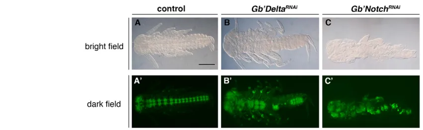
Because Delta/Notch signaling is known to be involved in neurogenesis in other species, such as Drosophila andCupiennius (Baker, 2000; Stollewerk, 2002), and both Gb⬘Delta and Gb⬘Notch
RNAi-treated embryos showed defects in the midline region (Fig. 4B,F), we examined the role of Delta/Notch signaling in the formation of the central nervous system (CNS). Anti-HRP staining (Fig. 5) revealed disorganization of the CNS with partial loss or hyperplasia of neurons in RNAi-treated embryos (Fig. 5B⬘,C⬘), suggesting that the neurogenic role for Delta/Notch signaling is conserved in Gryllus.
Gb⬘Notch RNAi treatment causes fusion and shortening of leg segments
Compared with control legs with distinguishable segments, including the coxa, trochanter, femur, tibia and tarsus, with the tibial spurs in the distal end of the tibia (arrow in Fig. 6A) and the tarsal spur in the distal end of tarsal segment 1 (arrowhead in Fig. 6A), Gb⬘NotchRNAi-treated embryos displayed shortened legs and a loss of joints, resulting in fusion of the femoral and tibial segments (Fig. 6B,C). In the RNAi-affected legs, tibial spur- and tarsal spur-like structures were observed (arrow and arrowhead, respectively, in Fig. 6C).
InDrosophila legs, activated epidermal growth factor receptor (EGFR) signaling is present in all segments and is necessary for joint formation (Galindo et al., 2005). A similar periodic pattern is observed for Gryllus Egfr (Gb⬘Egfr) expression during leg development (Nakamura et al., 2008). To evaluate the effects of Gb⬘Notch depletion on leg formation, we investigated Gb⬘Egfr expression in Gb⬘Notch RNAi-treated legs at stage 11. In control T3 legs, Gb⬘Egfrwas expressed in the femur/tibia and tibia/tarsus joints (Fig. 6D, arrows) and as three tarsal rings (Fig. 6D, arrowheads). Expression of Gb⬘Egfr was severely reduced in Gb⬘NotchRNAi-treated T3 legs (Fig. 6E). In the RNAi legs, the most distal ring was retained (Fig. 6E, red arrowhead), and another tarsal ring (Fig. 6E, black arrowhead) and a more proximal domain (Fig. 6E, white arrowhead) were observed, probably corresponding to a region of spur-like structures in later embryos. Hence, Gb⬘Notch depletion appeared to result in loss of leg joints, accompanied by a reduction of EGFR signaling activity. By contrast, the Gb⬘Egfrrings were not affected in Gb⬘Delta RNAi-treated embryos at this stage (n>20, data not shown).
[image:5.612.49.561.74.129.2]In addition, we also investigated the expression patterns of orthologs of the Drosophilaleg patterning genes, dachshundand Distal-less, in Gb⬘Notch RNAi-treated legs at stage 11. In T3 legs of Gryllus embryos, two broad rings of the Gb⬘dachshund (Gb⬘dac) expression were apparent in the distal femur and distal tibia/proximal tarsus regions at this stage (Inoue et al., 2002) (Fig. 6F, arrowheads). In Gb⬘Notch RNAi-treated legs, only a single broad expression domain was observed (Fig. 6G, bracket), consistent with the observed morphological defects of reduced segment size and fusion of femoral to tarsal segments. In wild-type embryo legs, Gb⬘Distal-less(Gb⬘Dll) was strongly expressed in the distal tip and more proximal tarsal domains (Fig. 6H, arrowheads), as well as in the region from the proximal tarsus to the distal tibia, but weakly expressed in the femur to the distal trochanter (Inoue et al., 2002) (Fig. 6H). Gb⬘Dllwas expressed strongly in the distal Table 1. Effect of parental RNAi for Gb⬘Deltaand Gb⬘Notch
Number of embryos of given phenotypic class (percentage)
Total number pRNAi Class III Class II Class I Wild type Undeveloped of embryos
Gb⬘Delta 154 (52.6) 25 (8.5) 8 (2.7) 43 (14.7) 63 (21.5) 293
Gb⬘Notch 119 (53.8) 22 (10.0) 5 (2.3) 4 (1.8) 71 (32.1) 221
DsRed2 0 (0) 0 (0) 0 (0) 149 (84.2) 28 (15.8) 177
D
E
V
E
LO
P
M
E
N
region of the legs of Gb⬘Notch RNAi-treated embryos (Fig. 6I, bracket) and weakly in a more proximal region adjacent to the strong expression domain (Fig. 6I, arrowhead). Such an alteration of the Gb⬘Dll expression pattern is consistent with the morphological fusion and shortening of the leg segments in RNAi-treated embryo legs. These results indicate that, in Gryllus leg development, Notch signaling acts downstream of the proximodistal axis patterning genes to form the leg joints and regulate leg growth.
Delta-Notch signaling is required for AP axis elongation
As Deltaand NotchRNAi phenotypes exhibited segmentation defects, we examined further the effects of RNAi treatment during the segment patterning process. We observed that
Gb⬘Deltawas expressed in segmental stripes earlier than Gb⬘wg stripes (Fig. 2F), suggesting that Gb⬘Delta/Notch signaling could function upstream of segment polarity genes, such as Gb⬘wg, in segmentation.
[image:6.612.59.502.55.426.2]If the body segment malformations observed in Gb⬘Delta or Gb⬘NotchRNAi-treated embryos were caused by defects in the segmentation process, silencing of Gb⬘Delta or Gb⬘Notchshould affect the Gb⬘wgstriped pattern. To examine this possibility, we analyzed expression of Gb⬘wgin Gb⬘Delta and Gb⬘Notch RNAi-treated embryos during early germband stages (Fig. 7). In control embryos at 36 hAEL, Gb⬘wgwas expressed as six anterior (gnathal and thoracic) segmental stripes with specific domains in the anterior head and posterior terminal region (Miyawaki et al., 2004) (Fig. 7A). We detected a reduction of the number of stripes in Gb⬘Delta RNAi-treated embryos (61.9%, n13/21) andGb⬘Notch
Fig. 4. Gb⬘Deltaand Gb⬘NotchRNAi phenotypes.(A,A⬘) Lateral (A) and dorsal (A⬘) views of a control embryo (DsRed RNAi) at 14 days after egg laying (AEL), immediately before hatching. (A⬙)Wild-type embryo (stage 8) stained for Gb⬘wg. (B,B⬘) Lateral (B) and dorsal (B⬘) views of a Gb⬘Delta
RNAi-treated class III embryo. The size and shape of the segments were affected in this embryo. Note the enlarged thoracic segments and reduced abdominal segments. (B⬙)Class III embryo stained for Gb⬘wg.Appendages were proximally fused (arrow). (C,C⬘) Lateral (C) and dorsal (C⬘) views of a Gb’DeltaRNAi-treated class II embryo. (C⬙)Class II embryo stained for Gb⬘wg. Note a severe defect of posterior segments. (D,D⬘) Lateral (D) and dorsal (D⬘) views of other body segment abnormalities in a Class III embryo. The segmental boundaries were not well defined in this embryo. (E)Gb⬘DeltaRNAi-treated class I embryo. Thoracic and abdominal segments were missing. (F,F⬘) Lateral (F) and dorsal (F⬘) views of a Gb⬘Notch
RNAi-treated class III embryo. A malformation of segments was observed in this embryo, as seen in Gb⬘DeltaRNAi-treated embryos. Note the failure of cuticle formation in each segment. (F⬙)Class III embryo stained for Gb⬘wg. Appendages were proximally fused (arrow). (G,G⬘) Lateral (G) and dorsal (G⬘) views of a Gb’NotchRNAi-treated class II embryo. (G⬙)Class II embryo stained for Gb⬘wg. (H,H⬘) Lateral (H) and dorsal (H⬘) views of a Gb⬘NotchRNAi-treated class I embryo. (H⬙)Class I embryo stained for Gb⬘wg.He, head; Tho, thorax; Abd, abdomen. Scale bar: in A, 400m for A-H,A⬘-H⬘; in A⬙, 250m for A⬙-C⬙,F⬙-H⬙.
D
E
V
E
LO
P
M
E
N
RNAi-treated embryos (63.6%, n7/11) (Fig. 7B-E). In the most severely affected Gb⬘Delta RNAi (28.6%, n6/21) andGb⬘Notch RNAi-treated embryos (27.3%, n3/11), only two segmental stripes were observed at this stage and the posterior terminal domain was retained (Fig. 7C,E). Given that at least four segmental stripes were observed in subsequent stages (Fig. 7G-J), these reductions in stripe number might be due, at least in part, to a segment patterning delay. In more mature embryos, at 45 hAEL, which normally have eight segmental stripes of Gb⬘wgin the anterior head and posterior terminal domains (Fig. 7F), Gb⬘Delta and Gb⬘Notch RNAi treatment resulted in a reduction of stripe number, disorganization of the stripe pattern, loss or severe reduction of the posterior terminal domain and loss of the midline separation between the left and right stripes (Fig. 7G-J). In addition, severely affected embryos had reduced posterior growth zone and arrested AP axis elongation at this stage (Fig. 7H,J, arrows). These effects appear to be related to defects observed in later stage Gb⬘Delta andGb⬘Notch RNAi-treated embryos (Fig. 4E,H), namely the loss of thoracic and abdominal segments, fusion of left and right legs, and segment malformation. Thus, inhibition of Notch signaling affects segmentation gene expression in early germbands producing a defective segment morphology phenotype.
To explore further the germband elongation defect, we analyzed expression of Gb⬘caudal (Gb⬘cad) in Gb⬘Delta and Gb⬘Notch RNAi-treated embryos during posterior elongation. Given that Gb⬘cadis required for formation of the posterior region and Gb⬘cad RNAi-treated embryos show a severe defect of posterior segments including all thoracic segments (Shinmyo et al., 2005), we predicted that Gb⬘cadexpression would be affected in Gb⬘Delta and Gb⬘Notch RNAi-treated embryos during posterior elongation. Contrary to our expectations, the posterior domain of Gb⬘cad was retained in RNAi-treated embryos (100%, n22/22, Gb⬘DeltaRNAi; 100%, n18/18, Gb⬘Notch RNAi) (Fig. 8A-F). Conversely, we also examined Gb⬘Deltaexpression in Gb⬘cadRNAi-treated embryos. In Gb⬘cad RNAi-treated embryos, Gb⬘Deltastripes were reduced whereas the posterior expression of Gb⬘Deltawas retained in a reduced domain compared with that in control embryos (100%, n6/6) (Fig. 8G,H), suggesting that Gb⬘cadis necessary for expression of Gb⬘Deltain anterior segments, but not in the posterior region.
To investigate the cause of the posterior elongation defect, we examined proliferation and cell death in Gb⬘Notch RNAi-treated embryos. An EdU incorporation assay for cell proliferation
assessment (Salic and Mitchison, 2008) showed a pronounced reduction of mitotic cells in the posterior region of Gb⬘Notch RNAi-treated embryos (33%; Fig. 8J,K) relative to control embryos (70%; Fig. 8I,K). There was no significant difference in the rate of cell death between control and Gb⬘NotchRNAi-treated embryos (data not shown). Therefore, the defect in posterior elongation inGb⬘Notch RNAi-treated embryos might be caused by inhibition of posterior cell proliferation. We were unable to assess the RNAi effects on cell proliferation in anterior regions owing to the presence of significant changes in anterior morphology in RNAi-affected embryos that prevented a precise comparison of cell proliferation rates in specific regions relative to wild-type embryos. Thus, it remains unclear whether cell proliferation is inhibited specifically in the posterior region. Taken together, these results suggest that Delta/Notch signaling might be involved in AP axis elongation during early germband development via control of cell proliferation, independently of the Wg/Cad pathway (Shinmyo et al., 2005).
DISCUSSION
We isolated Gb⬘Deltaand Gb⬘Notchgenes from the intermediate-germ cricket Gryllus bimaculatusand investigated their expression and function during embryogenesis. Our data suggest that Notch signaling is involved in segment morphogenesis, posterior elongation, neural differentiation and leg segmentation.
Role of Gb⬘Delta/Notch signaling in the leg formation
Gb⬘Deltaand Gb⬘Notch were expressed in developing legs in a periodic pattern of concentric rings. RNAi knockdown of Gb⬘Notch expression markedly reduced leg length with a loss of joints and disrupted leg patterning gene expression. These results suggest that Notch signaling is essential for the correct specification of segment boundaries and normal leg growth in Gryllus.
[image:7.612.52.478.58.189.2]In flies and spiders, Notch and its ligands are expressed in developing legs, as observed in Gryllus, and depletion of Notch causes segment fusion and leg shortening (de Celis et al., 1998; Bishop et al., 1999; Rauskolb and Irvine, 1999; Prpic and Damen, 2009). de Celis et al. (de Celis et al., 1998) have proposed that Notch activity is required for the establishment of boundaries that act as organizing centers for subsequent growth within leg segments, based on their mosaic analyses of developing legs lacking Notch, as well as data from experiments involving several Fig. 5. Effect on the formation of the CNS in Gb⬘Deltaand Gb⬘NotchRNAi-treated embryos. (A,A⬘) Bright field (A) and dark field (A⬘) images of a control embryo stained with anti-HRP antibody. (B,B⬘) Bright field (B) and dark field (B⬘) images of a Gb⬘DeltaRNAi-treated embryo stained with anti-HRP antibody. (C,C⬘) Bright field (C) and dark field (C) images of a Gb⬘NotchRNAi-treated embryo stained with anti-HRP antibody. Disorganized CNS with partial loss or hyperplasia of neurons was observed in Gb⬘Deltaand Gb⬘NotchRNAi-treated embryos. Scale bar: in A, 400m for all panels.
D
E
V
E
LO
P
M
E
N
mutants that affect leg segment lengths and/or the formation of intervening joints. Notch activity might also play such a role in Gryllusleg development.
Compared with Drosophila Notch mutants, Drosophilamutants of the Notch ligands Deltaand Serrateshow mild phenotypes in leg formation (Bishop et al., 1999). Drosophilawild-type legs contain five tarsal segments in a distal portion, whereas Delta mutant legs are shortened with fused tarsal segments 2-4. Ultimately, the Notch mutant phenotype looks like a composition
[image:8.612.56.488.57.313.2]of the Drosophila Deltaand Serratemutant phenotypes (Bishop et al., 1999). We could not detect obvious abnormalities in leg formation in Gb⬘Delta RNAi-treated embryos by our morphological analysis or WISH analysis with a Gb⬘Egfr probe (data not shown), implying that clearly discernible trochanter/femur, femur/tibia, and tibia/tarsus joints were formed normally in these embryos. Thus, it is possible that there are other Notch ligand(s) that function redundantly with Gb⬘Delta in developing legs. However, it is impossible to evaluate correctly
Fig. 7. Effect of Gb⬘Deltaand
Gb⬘NotchRNAi in segment
patterning.(A-E)Control (A),
Gb⬘DeltaRNAi-treated (B,C) and
[image:8.612.49.420.597.741.2]Gb⬘NotchRNAi-treated (D,E) embryos at 36 hours after egg laying (hAEL) stained for Gb⬘wg. (F-J)Control (F), Gb⬘Delta RNAi-treated (G,H) andGb⬘Notch RNAi-treated (I,J) embryos at 45 hAEL stained for Gb⬘wg. Arrows in H and J indicate the defect in the posterior growth zone. Scale bar: in A, 250m for all panels.
Fig. 6. Expressions of leg patterning genes in Gb⬘Notch RNAi-treated legs.(A-C)Mesothoracic (T3) legs of control (A) and Gb⬘Notch RNAi-treated embryos (B,C) at 14 days after egg laying (AEL), immediately before hatching. Control leg shows distinguishable segments including the coxa, trochanter, femur, tibia, tarsus and claw. Arrow and arrowhead indicate the spurs on the tibia/tarsus and tarsus1/tarsus2, respectively. Compared with control, in Gb⬘Notch RNAi-treated embryos the size of leg was decreased and joint structure was invisible, and putative femoral and tibial segments were fused. The coxa and trochanter were also indistinguishable. C is a high magnification view of the boxed area in B. Putative coxa, trochanter-tibia, tarsus and claw exist. Arrow and arrowhead indicate the tibial spur- and tarsal spur-like structures, respectively. (D,E)Gb⬘Egfr expression in control (D) and Gb⬘NotchRNAi-treated (E) legs at stage 11. (D)Five concentric rings of Gb⬘Egfrexpression in the femur/tibia and tibia/tarsus joints (arrows) and three tarsal rings (arrowheads) are observed. (E)In the RNAi legs, Gb⬘Egfr expression levels were reduced, whereas the most distal ring (red arrowhead), another tarsal ring (black arrowhead) and a more proximal domain (white arrowhead) were observed. (F,G)Gb⬘dac expression in control (F) and
Gb⬘NotchRNAi-treated (G) legs at stage 11. (F)Two broad rings of Gb⬘dacexpression (arrowheads) are observed in the distal femur and tibia/tarsus region. (G)Only one broad ring of expression was observed (bracket) in the RNAi legs. (H,I)Gb⬘Dllexpression in control (H) and Gb⬘NotchRNAi-treated (G) embryos at stage 11. (H)The distal tip and more proximal tarsal domains of Gb⬘Dllexpression are indicated by arrowheads. (I)Expression of Gb⬘Dll
was retained in the Gb⬘NotchRNAi-treated legs. Arrowhead indicates the distal intense expression and the bracket the proximal expression. See text for details. co, coxa; tr, trochanter; fe, femur; ti, tibia; ta, tarsi; cl, claw. Scale bar: in A, 300m for A,B; in D, 200m for D-I.
D
E
V
E
LO
P
M
E
N
whether more distal regions (i.e. the tarsus and pre-tarsus) of the legs of Gb⬘Delta RNAi-treated animals are normally segmented in these analyses because most Gb⬘Delta RNAi-treated embryos ceased to develop prior to showing distinguishable morphologies or Gb⬘Egfrexpression in these structures. Nevertheless, though it is unclear whether Gb⬘Deltais necessary for tarsal segmentation, the role of Delta/Notch signaling in leg development seems to be highly conserved among Drosophila, Cupiennius and Gryllus species.
Role of Gb⬘Delta/Notch signaling in the formation of body segments
In the present experiments, RNAi knockdown of either Gb⬘Deltaor Gb⬘NotchRNAi disrupted trunk segment formation. The RNAi effects on segmentation are a composite of segmentation disturbance/malformation and posterior segment loss. Because Gb⬘wgstripes were less disorganized in early RNAi-treated embryos than at later stages (Figs 4, 7), Gb⬘Delta/Notch signaling might not be necessary for specification of segment boundaries but rather might be critical for segment morphogenesis. Administration of RNAi directed against components of the Delta/Notch signaling pathway, including Notch, Delta, Presenilinand Suppressor of Hairless, in Cupienniusspiders also resulted in severe segment malformation accompanied by disorganization of segmentation gene expression (Schoppmeier and Damen, 2005; Stollewerk et al., 2003). Hence, the function of Notch signaling in segment morphogenesis might be conserved between Gryllusand Cupiennius, though the causes of segment malformation in these species remain to be explored further for more detailed comparisons.
The segmentation disturbance observed in the RNAi-treated Gryllus embryos is also reminiscent of a defect seen in Drosophila Notch and Delta mutant embryos, in which a segmentation disturbance was attributed to abnormal determination of the mesectoderm (ventral midline cells) (Menne and Klambt, 1994; Martin-Bermudo et al., 1995). Whether Notch signaling plays similar roles in mesoderm patterning and ventral midline specification in Gryllusas in Drosophilashould be the focus of future studies.
Involvement of Gb⬘Delta/Notch signaling in
posterior elongation
The loss of sequentially formed segments in Gb⬘Deltaand Gb⬘Notch RNAi-treated Gryllusembryos is likely to be at least partially due to a reduction in cell proliferation in the growth zone. Expression of Gb⬘Delta in the posterior growth zone in a specific domain at early stages might be essential for control of posterior elongation and regulation of the region of Notch signaling activation. As the early posterior domains of Gb⬘cad and Gb⬘wg are retained in Gb⬘Delta and Gb⬘NotchRNAi-treated embryos, Notch signaling might not be important for establishing the posterior growth zone in early germbands, but rather might be involved in maintaining a functional growth zone during subsequent posterior elongation processes. Furthermore, it should be noted that neither expression of Gb⬘cadin the terminal region of Gb⬘Delta and Gb⬘Notch RNAi-treated embryos nor expression of Gb⬘Delta in the terminal region of Gb⬘cad RNAi-treated embryos was eliminated. We showed previously that Gb⬘Wg/Arm signaling and Gb⬘cadare essential for posterior elongation (Miyawaki et al., 2004; Shinmyo et al., 2005). Therefore, the function of Gb⬘Delta/Notch signaling in posterior elongation might be parallel to the cascade for posterior elongation mediating Gb⬘wgand Gb⬘cad.
[image:9.612.52.291.57.417.2]In another basal, short-germ insect, the cockroachPeriplaneta americana,Notch RNAi treatment has been shown to result in a loss of abdominal segments and reduced cell proliferation in the terminal region (Pueyo et al., 2008), though RNAi effects on segment formation appeared to be weaker than those observed for Gryllus. Thus, the function of Notch signaling in posterior elongation might be conserved among short/intermediate-germ insects. In the cockroach, RNAi directed againstNotchaffects hairysegmental expression, which was thought to be a consequence of disruption of a Notch-mediated segmentation mechanism. However, RNAi effects on posterior segmentation could include secondary defects as a result Fig. 8. Analysis of the germband elongation defect caused by
Gb⬘Deltaand Gb⬘NotchRNAi. (A-F)Effect ofGb⬘Delta and Gb⬘Notch
RNAi on expression of Gb⬘cadin embryos during posterior elongation. The posterior domain ofGb⬘cadseen in control embryos (A,B) was retained in Gb⬘Delta (C,D) and Gb⬘Notch (E,F) RNAi embryos. (G,H)Effect of Gb⬘cadRNAi on Gb⬘Deltaexpression in early germbands. Control (G) and Gb⬘cadRNAi-treated (H) embryos at 36 hours after egg laying (hAEL) are shown. In the RNAi embryos,
Gb⬘Deltasegmental stripes were reduced but the posterior expression (arrow in G) was retained in a reduced domain (arrow in H). (I,J)Effect of Gb⬘NotchRNAi treatment on the proliferation of growth zone cells. EdU was injected into 48 hAEL eggs and then the embryos were incubated for 4 hours before fixation. Posterior region of control (I) and
Gb⬘NotchRNAi-treated (J) embryos are shown. Nuclei were stained with Hoechst (blue), and proliferating cells were detected by EdU incorporation (green). (K)Quantification of cell proliferation in the posterior region. The longitudinal axis represents the ratio of
proliferating cells at 0-20% region of the embryo length (where 0% is the posterior pole). Error bars represent s.d. Scale bars: in A, 250m for A-H; in I, 100m for I,J.
D
E
V
E
LO
P
M
E
N
of a failure to maintain a functional growth zone. Thus, the existence of a Notch-mediated segmentation mechanism remains ambiguous in insects. However, the situation for the spider seems to be different from that of the cockroach. That is, RNAi directed against Deltaand Notch was shown to affect hairy segmental expression in Cupiennius, without any observable reduction in cell proliferation in the growth zone (Stollewerk et al., 2003). Nevertheless, malformation of the posterior growth zone was observed. Thus, the cause of the segment defect remains unclear.
Previous work in the spider Achaearanea tepidariorumrevealed that Delta/Notch signaling is essential for caudal lobe formation preceding initiation of segmentation (Oda et al., 2007). In Achaearaneaembryos,Deltaand NotchRNAi treatment results in a severe reduction of the posterior region (opisthosoma) due to a failure to generate cad-expressing caudal ectoderm cells, suggesting that Delta/Notch signaling promotes caudalexpression and caudal ectoderm fate specification (Oda et al., 2007). Considering the findings of Oda et al. in Achaearanea, our findings in Gryllus suggest that Notch signaling involvement in the posterior elongation process might be conserved between Gryllus and Achaearanea, though the genetic interactions between Notch signaling and cad might differ between them. In Cupiennius, however, the posterior region was not reduced in Deltaand NotchRNAi-treated embryos, though it was malformed without a reduction in cell proliferation in the growth zone (Stollewerk et al., 2003). Hence, the function of Notch signaling in posterior patterning seems to be divergent between these arthropod species. However, it should be noted that in Cupiennius, dsRNA was administered to embryos at a relatively later stage than in Gryllusand Achaearanea, possibly after segmentation had already been initiated (Schoppmeier and Damen, 2001). Thus, the phenotypic discrepancy between these species might arise from differences in RNAi methods and/or RNAi efficiency. Intriguingly, RNAi silencing of Presenilinand Suppressor of Hairless causes a defect in posterior growth and a breakdown of posterior segmentation in Cupiennius (Schoppmeier and Damen, 2005), implying that Notch signaling in Cupienniusis also involved in the posterior elongation process.
In light of analogous findings in other arthropod species, the present findings suggest that involvement of Delta/Notch signaling in posterior elongation might be an ancestral mode among arthropods. Further analyses of the posterior growth zone are needed to fully understand Delta/Notch involvement in arthropod embryogenesis.
Acknowledgements
We thank Takashi Matsushita, Tomohiro Uda and Chieko Ishifune for their technical assistance. This work was supported by a grant from the Japanese Ministry of Education, Culture and Sports, Science and Technology to T.M., H.O. and S.N.
Competing interests statement
The authors declare no competing financial interests.
References
Baker, N. E.(2000). Notch signaling in the nervous system. Pieces still missing from the puzzle. BioEssays 22, 264-273.
Bishop, S. A., Klein, T., Martinez Arias, A. and Couso, J. P.(1999). Composite signaling from Serrateand Deltaestablishes leg segments in Drosophila through
Notch. Development126, 2993-3003.
Bucher, G., Scholten, J. and Klingler, M.(2002). Parental RNAi in Tribolium
(Coleoptera). Curr. Biol.12, R85-R86.
Campos-Ortega, J. A. and Knust, E.(1990). Molecular analysis of a cellular decision during embryonic development of Drosophila melanogaster: epidermogenesis or neurogenesis. Eur. J. Biochem.190, 1-10. Chipman, A. D.(2010). Parallel evolution of segmentation by co-option of
ancestral gene regulatory networks. BioEssays 32, 60-70.
Couso, J. P.(2009). Segmentation, metamerism and the Cambrian explosion.Int. J. Dev. Biol.53, 1305-1316.
Damen, W. G. M.(2007). Evolutionary conservation and divergence of the segmentation process in arthropods. Dev. Dyn. 236, 1379-1391.
Davis, G. K. and Patel, N. H.(1999). The origin and evolution of segmentation.
Trends Cell Biol.9, M68-M72.
Davis, G. K. and Patel, N. H.(2002). Short, long, and beyond: molecular and embryological approaches to insect segmentation. Annu. Rev. Entomol. 47, 669-699.
Dearden, P. and Akam, M.(2000). A role for Fringe in segment morphogenesis but not segment formation in the grasshopper, Schistocerca gregaria. Dev. Genes Evol. 210, 329-336.
de Celis, J. F., Tyler, D. M., de Celis, J. and Bray, S.(1998). Notch mediates segmentation of the Drosophila leg. Development125, 4617-4626.
Dove, H. and Stollewerk, A.(2003). Comparative analysis of neurogenesis in the myriapod Glomeris marginata(Diplopoda) suggests more similarities to chelicerates than to insects. Development130, 2161-2171.
Galindo, M. I., Bishop, S. A. and Couso, J. P.(2005). Dynamic EGFR-Ras signaling in Drosophilaleg development. Dev. Dyn. 233, 1496-1508. Ingham, P. W.(1988). The molecular genetics of embryonic pattern formation in
Drosophila. Nature335, 25-34.
Inoue, Y., Mito, T., Miyawaki, K., Matsushima, K., Shinmyo, Y., Heanue, T. A., Mardon, G., Ohuchi, H. and Noji, S.(2002). Correlation of expression patterns of homothorax, dachshund, and Distal-lesswith the proximodistal segmentation of the cricket leg bud. Mech. Dev. 113, 141-148.
Kadner, D. and Stollewerk, A.(2004). Neurogenesis in the chilopod Lithobius forficatussuggests more similarities to chelicerates than to insects. Dev. Genes Evol. 214, 367-379.
Liu, P. Z. and Kaufman, T. C.(2005). Short and long germ segmentation: unanswered questions in the evolution of developmental mode. Evol. Dev. 7, 629-646.
Martín-Bermudo, M. D., Carmena, A. and Jiménez, F.(1995). Neurogenic genes control gene expression at the transcriptional level in early neurogenesis and in mesectoderm specification. Development121, 219-224.
Menne, T. V. and Klämbt, C.(1994). The formation of commissures in the
DrosophilaCNS depends on the midline cells and on the Notchgene.
Development120, 123-133.
Mito, T., Sarashina, I., Zhang, H., Iwahashi, A., Okamoto, H., Miyawaki, K., Shinmyo, Y., Ohuchi, H. and Noji, S.(2005). Non-canonical functions of
hunchbackin segment patterning of the intermediate germ cricket Gryllus bimaculatus. Development132, 2069-2079.
Mito, T., Kobayashi, C., Sarashina, I., Zhang, H., Shinahara, W., Miyawaki, K., Shinmyo, Y., Ohuchi, H. and Noji, S.(2007). even-skippedhas gap-like, pair-rule-like, and segmental functions in the cricket Gryllus bimaculatus, a basal, intermediate germ insect (Orthoptera). Dev. Biol. 303, 202-213. Miyawaki, K., Mito, T., Sarashina, I., Zhang, H., Shinmyo, Y., Ohuchi, H. and
Noji, S.(2004). Involvement of Wingless/Armadillo signaling in the posterior sequential segmentation in the cricket, Gryllus bimaculatus(Orthoptera), as revealed by RNAi analysis. Mech. Dev. 121, 119-130.
Nakamura, T., Mito, T., Miyawaki, K., Ohuchi, H. and Noji, S.(2008). EGFR signaling is required for re-establishing the proximodistal axis during distal leg regeneration in the cricket Gryllus bimaculatusnymph. Dev. Biol. 319, 46-55. Niwa, N., Saitoh, M., Ohuchi, H., Yoshioka, H. and Noji, S.(1997). Correlation
between Distal-less expression patterns and structures of appendages in development of the two-spotted cricket, Gryllus bimaculatus. Zool. Sci. 14, 115-125.
Niwa, N., Inoue, Y., Nozawa, A., Saito, M., Misumi, Y., Ohuchi, H., Yoshioka, H. and Noji, S.(2000). Correlation of diversity of leg morphology in Gryllus bimaculatus(cricket) with divergence in dpp expression pattern during leg development. Development127, 4373-4381.
Nüsslein-Volhard, C. and Wieschaus, E.(1980). Mutations affecting segment number and polarity in Drosophila. Nature287, 795-801.
Oda, H., Nishimura, O., Hirao, Y., Tarui, H., Agata, K. and Akiyama-Oda, Y. (2007). Progressive activation of Delta-Notch signaling from around the blastopore is required to set up a functional cad lobe in the spider Achaearanea tepidariorum. Development134, 2195-2205.
Pankratz, M. J. and Jackle, H.(1993). Blastoderm segmentation. InThe Development of Drosophila melanogaster (ed. M. Bate and A. Martinez Arias), pp. 467-516. Cold Spring Harbor, NY: Cold Spring Harbor Laboratory Press. Parody, T. R. and Muskavitch, M. A.(1993). The pleiotropic function of Delta
during postembryonic development of Drosophila melanogaster.Genetics 135, 527-539.
Patel, N. H.(1994). Evolution of arthropod segmentation: insights from comparisons of gene expression patterns. Development120(Suppl.), 201-207. Peel, A. D. and Akam, M.(2003). Evolution of Segmentation: Rolling Back the
Clock. Curr. Biol. 13, 708-710.
Peel, A. D., Chipman, A. D. and Akam, M.(2005). Arthropod segmentation: beyond the Drosophilaparadigm. Nat. Rev. Genet. 6, 905-916.
Pourquie, O.(2001). Vertebrate somitogenesis. Annu. Rev. Cell Dev. Biol. 17,
Prpic, N. M. and Damen, W. G. M.(2009). Notch-mediated segmentation of the appendages is a molecular phylotypic trait of the arthropods. Dev. Biol. 326, 262-271.
Pueyo, J. I., Lanfear, R. and Couso, J. P.(2008). Ancestral Notch-mediated segmentation revealed in the cockroach Periplaneta Americana. Proc. Natl. Acad. Sci. USA105, 16614-16619.
Rauskolb, C. and Irvine, K. D.(1999). Notch-mediated segmentation and growth control of the Drosophila leg. Dev. Biol. 210, 339-350.
Richards, S., Gibbs, R. A., Weinstock, G. M., Brown, S. J., Denell, R., Beeman, R. W., Gibbs, R., Bucher, G., Friedrich, M., Grimmelikhuijzen, C. J. et al. (2008). The genome of the model beetle and pest Tribolium castaneum. Nature
452, 949-955.
Salic, A. and Mitchison, T. J.(2008). A chemical method for fast and sensitive detection of DNA synthesis in vivo. Proc. Natl. Acad. Sci. USA 105, 2415-2420. Sander, K.(1976). Specification of the basic body pattern in insect embryogenesis.
Adv. Insect Physiol. 12, 125-238.
Schoppmeier, M. and Damen, W. G.(2001). Double-stranded RNA interference in the spider Cupiennius salei: the role of Distal-less is evolutionarily conserved in arthropod appendage formation. Dev. Genes Evol. 211, 76-82.
Schoppmeier, M. and Damen, W. G. M.(2005). Suppressor of Hairlessand
Presenilin phenotypes imply involvement of canonical Notch-signaling in segmentation of the spiderCupiennius salei. Dev. Biol. 280, 211-224.
Seppo, A., Matani, P., Sharrow, M. and Tiemeyer, M.(2003). Induction of neuron-specific glycosylation by Tollo/Toll-8, a DrosophilaToll-like receptor expressed in non-neural cells. Development130, 1439-1448.
Shellenbarger, D. L. and Mohler, J. D.(1975). Temperature-sensitivemutations of the Notchlocus in Drosophila melanogaster. Genetics 81, 143-162. Shinmyo, Y., Mito, T., Matsushita, T., Sarashina, I., Miyawaki, K., Ohuchi, H.
and Noji, S.(2005). cad is required for gnathal and thoracic patterning and for posterior elongation in the intermediate-germband cricket Gryllus bimaculatus. Mech. Dev. 122, 231-239.
St. Johnston, D. and Nüsslein-Volhard, C.(1992). The origin of pattern and polarity in the Drosophilaembryo. Cell68, 201-219.
Stollewerk, A.(2002). Recruitment of cell groups through Delta/Notch signaling during spider neurogenesis. Development129, 5339-5348.
Stollewerk, A., Schoppmeier, M. and Damen, W. G. M.(2003). Involvement of
Notch and Delta genes in spider segmentation. Nature423, 863-865. Tautz, D.(2004). Segmentation. Dev. Cell7, 301-312.
Tautz, D., Friedrich, M. and Schroder, R.(1994). Insect embryogenesis: what is ancestral and what is derived? Development120 (Suppl.), 193-199.
Zhang, H., Shinmyo, Y., Mito, T., Miyawaki, K., Sarashina, I., Ohuchi, H. and Noji, S.(2005). Expression patterns of the homeotic genes Scr, Antp, Ubx, and
abd-A during embryogenesis of the cricket Gryllus bimaculatus. Gene Expr. Patterns 5, 491-502.