INTRODUCTION
Stem cells with the capacity to generate all epidermal lineages have been identified in hair follicles, sebaceous glands and the interfollicular epidermis (IFE) (Fuchs, 2007; Watt et al., 2006). These cells have unique adhesive characteristics that distinguish them from their more differentiated progeny, including increased extracellular matrix (ECM) adhesion and cell-cell cohesion (Estrach et al., 2007; Fuchs et al., 1997; Halbleib and Nelson, 2006; Molès and Watt, 1997; Watt, 2002). As in other tissues, epidermal cell adhesion molecules, including integrins and cadherins, influence both intrinsic cellular signalling and the interaction of stem cells with their surrounding environment (Watt, 2002). Perturbation of integrin-mediated ECM adhesion or cadherin-mediated intercellular adhesion results in a range of epidermal abnormalities, including transient skin hyperplasia, abnormal wound healing, tissue thinning and loss of hair follicles (Brakebusch et al., 2000; Grose et al., 2002; Jamora et al., 2003; Lopez-Róvira et al., 2005; Perez-Moreno et al., 2003; Tinkle et al., 2004; Young et al., 2003). In some cases these changes are associated with depletion of the epidermal stem cell compartment (e.g. Benitah et al., 2005).
In recent years, several microarray studies have been performed on the hair follicle stem cell compartment known as the bulge. These have identified a range of bulge markers that are involved in cell adhesion, including nectin-like molecule 2 (Necl2; also known as Igsf4, RA175, SgIGSF, TSLC1, SynCAM1 and CADM1) (Morris et al., 2004; Ohyama et al., 2006; Tumbar et al., 2004). Necl2 belongs to the family of nectins and nectin-like (Necl) molecules, which are intercellular adhesion molecules characterised by their
calcium insensitivity and immunoglobulin (Ig)-like extracellular binding domains (Takai et al., 2008). Mutations in the gene encoding nectin-1 (PVRL1 for poliovirus receptor-like 1) cause the autosomal recessive form of cleft lip/palate ectodermal dysplasia [CLPED1 (Suzuki et al., 2000)], and Pvrl1-knockout mice exhibit abnormal epidermal differentiation (Wakamatsu et al., 2007).
Studies in various tissues and cell types indicate that Necl2 expression influences epithelial cell scattering, proliferation and differentiation (Ito et al., 2007; Ito et al., 2003; Mao et al., 2003; Masuda et al., 2005; Shingai et al., 2003). Necl2 mediates homotypic intercellular adhesion and heterotypic adhesion via nectin-3, Necl1 and class I-restricted T-cell-associated molecule (CRTAM) (Galibert et al., 2005; Takai et al., 2008). Necl2 has been shown to enhance E-cadherin-dependent adherens junction stability and directly interact with actin-binding proteins to regulate cytoskeletal organisation and Rho GTPase activity (Masuda et al., 2005). The cytoplasmic PDZ domain of Necl2 binds membrane-associated guanylate kinase (MAGuK) proteins including CASK, MPP3, Pals2 and syntenin (Takai et al., 2008). These interactions form a molecular scaffold that affects actin cytoskeletal organisation and cell polarity.
In this report, we have examined the expression and function of Necl2 in mammalian epidermis. We show that Necl2 is highly expressed in bulge stem cells and influences epidermal adhesion, proliferation and wound healing. Our results provide evidence for a novel link between cell adhesion, stem cell quiescence and tissue repair.
MATERIALS AND METHODS Sample preparation and immunostaining
Frozen tissue sections were prepared using OCT and fixed in 1% paraformaldehyde before staining. Sections of paraffin-embedded tissues
Necl2 regulates epidermal adhesion and wound repair
Adam Giangreco1,*, Kim B. Jensen2, Yoshimi Takai3, Jun Miyoshi4and Fiona M. Watt1,2,‡
Differential expression of cell adhesion molecules regulates stem cell location, self-renewal and lineage selection under steady state conditions and during tissue repair. We show that the intercellular adhesion protein nectin-like molecule 2 (Necl2) is highly
expressed in bulge stem cells of adult human and mouse hair follicles. Overexpression of Necl2 in cultured human keratinocytes led to upregulation of calcium/calmodulin-associated Ser/Thr kinase (CASK), increased calcium-independent intercellular adhesion, and inhibition of cell motility and in vitro wound healing. Although the rate of cell proliferation was reduced, terminal differentiation was unaffected. To assess the role of Necl2 in vivo, we examined the epidermis of Necl2-null mice and developed transgenic mice that expressed Necl2 in the basal layer of murine epidermis. Necl2 overexpression led to a reduction in S-phase cells and an increase in quiescent cells retaining DNA label in the bulge. Although epidermal homeostasis appeared normal in both transgenic and knockout mice, wound healing was markedly delayed. Necl2 overexpression resulted in reduced proliferation and increased levels of CASK and E-cadherin at the leading edge of healing wounds, consistent with its effects in culture. Our results demonstrate that Necl2 is involved in regulating epidermal stem cell quiescence and location.
KEY WORDS: Epidermis, Intercellular adhesion, Stem cell, Wound healing Development 136, 3505-3514 (2009) doi:10.1242/dev.038232
1Cancer Research UK Cambridge Research Institute, Li Ka Shing Centre, Robinson
Way, Cambridge CB2 0RE, UK. 2Wellcome Trust Centre for Stem Cell Research,
[LL002 (Jensen and Watt, 2006)], rat anti-CD34 (BD Biosciences), rabbit anti-Ki67 (Novocastra), rat anti-FLAG (Sigma), mouse anti-involucrin [SY5 (Sevilla et al., 2008)], rat anti-α6 integrin (BD Pharmingen), mouse anti-E-cadherin (Invitrogen), mouse anti-keratin-15 [LHK15 (Braun et al., 2003)], sheep anti-BrdU (Abcam), rabbit anti-keratin-6 (Covance). Species-specific secondary antibodies conjugated to Alexa Fluor 488, 555 or 633 were purchased from Molecular Probes. Images were acquired using a Leica TCS Tandem confocal with ⫻10 or ⫻20 objectives. E-cadherin levels at cell-cell borders were quantified as described previously (Sevilla et al., 2008).
Keratinocyte culture and retroviral transduction
Primary human neonatal foreskin epidermal keratinocytes were isolated and cultured on a J2 3T3 feeder layer in FAD medium supplemented with 10% FCS and HICE cocktail, as described previously (Jensen and Watt, 2006). To determine colony-forming efficiency, cells were seeded at 100-500 cells per 60 mm dish and cultured for 14 days before fixation and staining (Jensen and Watt, 2006). Colony-forming efficiency was determined as described previously (Jensen and Watt, 2006). The area of individual colonies was measured using Volocity software.
Keratinocyte growth rate assays were performed using a CellTiter 96 AQ kit (Promega) according to the manufacturer’s instructions. When cells were grown under low-calcium conditions, they were seeded without feeders in
complete KSFM (Invitrogen); to raise the level of Ca2+, the medium was changed to complete FAD 2 hours before fixation. Keratinocytes were cultured on de-epidermised dermis (DED) for 14 days at the air-medium interface, as described previously (Bagutti et al., 2001).
Full-length Necl-2-FLAG (Shingai et al., 2003) was cloned into pBabe-puromycin (pBp) and sequence verified. pBp-EV and pBp-GFP retroviral vectors were used as previously described (Jensen and Watt, 2006). Keratinocyte infection was performed via Phoenix cell transfection, AM12 transduction and supernatant transfer, as described previously (Lowell et al., 2000).
Flow and imaging cytometry
Cells were isolated and stained for Necl2, α6 integrin and CD34 using previously reported methods (Morris et al., 2004; Silva-Vargas et al., 2005). Dead cells were excluded and unstained controls used to set negative gates. Keratinocyte DNA content and involucrin expression were determined as previously described (Gandarillas and Watt, 1997; Morris et al., 2004). Cells retaining DNA label were quantified using a commercially available BrdU detection system (BD Biosciences). Data were collected using an LSR II flow cytometer (BD Biosciences).
Imaging cytometry analysis was performed using an Amnis ImageStream. Briefly, transduced human keratinocytes were isolated and stained with
[image:2.612.48.350.306.737.2]anti-α6-integrin-FITC or anti-α6-integrin-RPE (BD Biosciences). Cells were
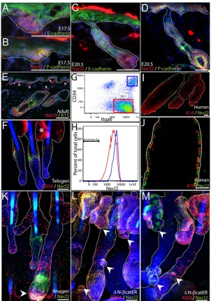
Fig. 1. Necl2 expression in developing and adult epidermis.(A-D) E17.5 (A,B) or E20.5 mouse skin (C,D) was stained for Necl2 (red) and E-cadherin (green, A,C) or P-cadherin (green, B,D) with DAPI nuclear counterstain (blue). (E,F) Adult mouse back skin section (E) and tail epidermal whole-mount (F) stained with antibodies against Necl2 (red, E; green, F) and keratin-15 (green, E) or CD34 (red, F) with DAPI nuclear counterstain (blue). Note staining of sebaceous glands (asterisk, F) is nonspecific. (G,H) Flow cytometric analysis of Necl2 expression (H) in adult integrin-α6 (Itga6)-positive basal cells (red box, G) and CD34- and Itga6-positive bulge cells (blue box, G). (I,J) Adult human hair follicles stained for Necl2 (green) and keratin-14 (red, I) or keratin-15 (red, J). (K) Necl2 (green) and Ki67 (red) immunostaining of adult mouse anagen follicles (tail epidermal whole mount) with DAPI nuclear counterstain (blue). Arrowhead indicates Necl2 expression in hair follicle bulb. (L,M) Adult tail whole mounts of K14ΔNβ-cateninER epidermis treated with 4-hydroxy-tamoxifen for 2 weeks and stained for Necl2 (green) and CD34 (red, L) or Ki67 (red, M) with DAPI nuclear counterstain (blue). Arrowheads indicate ectopic hair follicles arising from sebaceous glands and existing follicles. White lines (A-F, I-M) indicate position of basement membrane or delineate hair follicles and sebaceous glands. Scale bars: 100μm in A-F,I-M.
V
E
LO
P
M
E
N
allowed to adhere in suspension for 3 hours at 37°C, and 12,000 images were collected per sample. Two independent transduction experiments were performed for quantification of keratinocyte doublets.
Cell motility and in vitro wound-healing assays
To measure cell motility, primary human keratinocytes were cultured in complete KSFM buffered with 100 mM HEPES pH 7.5 for 18 hours. Six image fields per culture condition per cell type (containing 10-20 cells each) were collected every 6 minutes, and individual cell tracks were plotted using Metamorph software. For each cell, the position at each time point and maximum migration distance were plotted relative to the starting position (time=0 minutes). The experiments were repeated four times.
For in vitro wound-healing assays, keratinocytes were grown to confluence in KSFM. A scratch was made across the cultures using a Gilson pipette tip and the rate of wound closure was recorded using an IncuCyte automated imaging system.
Western blotting
Cells were incubated with hepatocyte growth factor (HGF; 10 U/ml) before washing in PBS and lysing in RIPA buffer supplemented with complete protease inhibitors cocktail (Roche) and phosSTOP phosphatase inhibitor (Roche). Equal amounts of protein were analysed by western blotting using antibodies against CASK (Ojeh et al., 2008), FLAG tag (M2, Sigma), phosphorylated ERK (rabbit polyclonal, Cell Signaling) and β-tubulin (Sigma), as previously described.
Mouse generation and husbandry
Transgenic mice expressing Necl2 under the control of the keratin-14 gene (Krt14) promoter (K14Necl2 transgenics) were prepared by cloning full-length murine Necl2-FLAG into the BamHI site of a keratin-14 expression cassette (Lo Celso et al., 2004). The construct was sequence verified and used to produce transgenic mice as previously described (Silva-Vargas et al., 2005). Three germline-transmitting, FVB/n strain founders were generated (7526, 7248A.4, 7248A.5). Of these, founder 7248A.5 exhibited highest transgene expression and transmitted the transgene at the expected
mendelian ratio. Heterozygous offspring from this line were used in all experiments. Necl2-knockout mice were generated on the C57Bl/6 background by homologous recombination as previously reported (Fujita et al., 2006).
Mice were housed in individually ventilated cages on a 12 hour light-dark cycle and allowed access to food and water ad libitum. Mice were examined between 2 and 4 months of age. Cells retaining BrdU label were generated by repeated BrdU injections into 10-day-old mice, as previously described (Braun et al., 2003). All in vivo experiments were performed under the terms of a UK Home Office license.
Skin wound healing
A single sterile 5 mm punch biopsy wound was administered along the central dorsal midline of 8-week-old female mice under general anaesthesia. Wound area was determined by measuring the saggital (x) and transverse (y) plane wound diameter and applying these to the ellipse area formula [area=π(radius x)(radiusy)]. Mice were allowed to recover for 7 or 14 days and wound area measured each day. Two hours before sacrifice, all wounded mice received a single injection of BrdU (10 mg/kg body weight). Wound tissue was bisected along the saggital plane; one half was processed for paraffin sections and the other for frozen sections.
RESULTS
Necl2 is upregulated in bulge stem cells
[image:3.612.48.336.421.738.2]At mouse embryonic day (E)17.5, when hair follicle placodes are forming, Necl2 was detectable throughout the basal layer of the epidermis but was most highly expressed in the developing placodes, which are characterised by reduced E-cadherin and elevated P-cadherin immunoreactivity (Fig. 1A,B) (Jamora et al., 2003). At E20.5 Necl2 expression remained enriched within hair follicles, with highest expression in the follicle bulb, where P-cadherin was also upregulated (Fig. 1C,D). In adult mouse telogen epidermis, Necl2 was most highly expressed in the hair follicle bulge, where it was
Fig. 2. Effects of Necl2 overexpression on growth and differentiation of cultured human keratinocytes. (A) Flow cytometry of keratinocytes transduced within empty vector (EV, red) or Necl2(blue) and labelled with anti-Necl2. (B) Necl2 immunoblots of Necl2-transduced or control (EV) AM12 packaging cells (A) and keratinocytes (K). Molecular mass markers (kDa) are indicated. (C,D) Representative dishes of control(C) and Necl2- (D) transduced keratinocyte colonies 2 weeks after plating. (E) Quantification of relative size of individual control (black) and Necl2transduced (red) clones. All clones (n>300) from one experiment are shown; data are representative of three separate experiments. (F) Keratinocyte growth was assessed by measuring mitochondrial activity (OD490; *P<0.05). (G) Flow cytometric determination of
proportion of involucrin-positive Necl2–transduced and control keratinocytes. (H,I) Quantification of basal cell density (H) and epidermal thickness (I) in epidermis reconstituted by control (EV) and Necl2-transduced cells cultured on de-epidermised dermis. (J,K) H&E-stained sections of control (J) and Necl2-transduced (K) keratinocytes grown on DED substrates for 2 weeks. (L) Western blot of control and Necl2-transduced keratinocytes cultured in the absence of HGF (0) or stimulated with HGF for the times indicated. Blot was
coexpressed with the bulge markers keratin-15 and CD34 (Fig. 1E,F). As reported previously (Morii et al., 2004), we also observed Necl2 expression in mast cells within the dermis (Fig. 1E, asterisks). Necl2 expression in adult bulge stem cells was confirmed by flow cytometric analysis of disaggregated cells that had been labelled with antibodies against CD34 and the α6-integrin subunit (Fig. 1G,H). We observed two discrete CD34+populations that differed in α6-integrin levels (Fig. 1G), as shown earlier (Blanpain et al., 2004; Silva-Vargas et al., 2005). When CD34+, α6-integrin ‘high’ keratinocytes were compared with CD34–, α6-integrin ‘high’ cells,
the CD34+population had higher levels of cell surface Necl2 (Fig. 1H), which is consistent with the immunofluorescence staining data (Fig. 1E,F). In addition, Necl2 expression was upregulated in the keratin-15-positive bulge compartment of human hair follicles (Fig. 1I,J) consistent with the results of previous microarray studies (Ohyama et al., 2006).
In anagen mouse skin, Necl2 continued to be highly expressed in the bulge. However, it was also highly upregulated in the hair follicle bulb; dual staining for Necl2 and the proliferation marker Ki67 revealed that whereas Necl2-expressing cells in the bulge were quiescent, Necl2-expressing bulb cells were actively proliferating (Fig. 1K, arrowhead).
Ectopic hair follicles can be induced in adult mouse epidermis by 4-hydroxy-tamoxifen-induced activation of a stabilised transgene encodingβ-catenin (K14ΔNβ-cateninER) (Lo Celso et al., 2004; Silva-Vargas et al., 2005). We found that Necl2 was expressed in CD34+ectopic follicles, regardless of whether they arose in the outer root sheath of existing follicles, the sebaceous glands or the interfollicular epidermis (Fig. 1L,M, arrowheads, and data not shown). We conclude that Necl2 expression is elevated in the reservoir of stem cells in the bulge of both mouse and human hair follicles, confirming previous microarray data (Morris et al., 2004; Ohyama et al., 2006; Tumbar et al., 2004). Necl2 undergoes dynamic
regulation during the mouse hair follicle cycle and is expressed in ectopic follicles that are induced in adult epidermis upon activation of β-catenin.
Necl2 influences keratinocyte growth and proliferation
To determine the functions of Necl2, we transduced primary human epidermal cells with full-length, FLAG-tagged murine Necl2. Necl2 protein overexpression was confirmed by flow cytometry and western blotting of cells transfected with empty vector or Necl2, using anti-FLAG tag antibodies (Fig. 2A,B).
Overexpression of Necl2 resulted in an increase of 160±10% (mean ± s.e.m.) in cells that formed colonies when compared with control cells (three independent experiments; three or more replicates per sample). Although Necl2 overexpression increased colony-forming efficiency, individual colonies were smaller than control colonies, as determined by plotting the area of individual colonies versus percentile rank (Fig. 2E). The reduction in colony size reflected a reduction in the growth rate of Necl2-overexpressing cells (Fig. 2F). There was no significant difference in the proportion of cells that initiated terminal differentiation in culture, as evidenced by involucrin expression (Fig. 2G). When keratinocytes transduced with empty vector or Necl2were seeded onto de-epidermised human dermis and cultured at the air-medium interface for 14 days, there were no differences in the degree of stratification (as measured by epidermal thickness) or differentiation of the epidermis that they reconstituted (Fig. 2H-K). The density of cells in the basal layer of reconstituted epidermis was also unaffected by Necl2 overexpression (Fig. 2H).
[image:4.612.48.328.439.736.2]Downregulation of CASK is associated with increased keratinocyte proliferation and migration (Ojeh et al., 2008). Since CASK is one of the MAGUK proteins that binds to the Necl2 cytoplasmic domain, we investigated whether Necl2 overexpression
Fig. 3. Necl2 overexpression influences keratinocyte adhesion and motility.(A) Identification of cell doublets based on Draq5 nuclear area/aspect ratio (left panel; blue gate) and characterisation of doublets by labelling with FITC- and RPE-conjugated anti-α6-integrin antibodies (right). Insert shows representative RPE-RPE (top), RPE-FITC (middle) and FITC-FITC (bottom) stained cell doublets. (B) Quantification of relative RPE-FITC labelled EV-EV, Necl2-EV and Necl2-Necl2 doublets. (C-G) Representative images (C-F) and quantification (G) of Necl2 expression in control (C,E) and Necl2-transduced (D,F) cells cultured under standard (high; C,D) and low-calcium (E,F) conditions. (H-L) Representative images (H-K) and quantification (L) of E-cadherin expression in control (H,J) and
Necl2-transduced (I,K) cells cultured under standard (high; H,I) and low-calcium (J,K) conditions. (G,L) Quantification of pixel intensity at cell-cell borders. (M-O) Control (M) and Necl2-transduced (N) cell motility tracks and maximum migration distances (O) relative to a starting position (x,y=0,0) at time=0 minutes. Each line in M,N shows motility of an individual cell. (B,G,L) Error bars represent s.e.m.; *P<0.05. Scale bars: 10μm in C-F,H-K.
V
E
LO
P
M
E
N
affected CASK levels (Fig. 2L-N). The level of CASK was higher in cells overexpressing Necl2 than in controls, both when cells were unstimulated and when treated with HGF, which stimulates keratinocyte motility (Birchmeier et al., 2003). By contrast, overexpression of Necl2 had no effect on levels of Erk MAPK phosphorylation (data not shown). As reported previously (Ojeh et al., 2008), localisation of CASK was predominantly nuclear in undifferentiated keratinocytes (Fig. 2M,N).
Necl2 regulates intercellular adhesion and keratinocyte motility
Necl2, like other nectin-like proteins, is believed to promote calcium-independent intercellular adhesion and adherens junction stabilisation by enhancing recruitment of cadherins to cell-cell borders (Takai et al., 2003). Consistent with this, Necl2-transduced keratinocytes were resistant to disaggregation by trypsin and EDTA. Under conditions in which nearly all control keratinocytes formed a single cell suspension, flow cytometric analysis revealed that a significant proportion of Necl2-transduced cells remained as aggregates of two or more cells (data not shown).
To examine the effect of Necl2 on homotypic and heterotypic intercellular adhesion, cells transduced with Necl2or empty vector (EV) were disaggregated into single cell suspensions. Each population was divided into two and labelled with FITC- or RPE-conjugated antibodies to the α6-integrin subunit, a pan basal cell marker (Silva-Vargas et al., 2005). Equal numbers of FITC- and RPE-labelled cells were combined homotypically (EV+EV or
Necl2+Necl2) or heterotypically (EV+Necl2) and incubated in suspension at 37°C for 3 hours on an orbital shaker. At the end of the incubation period, cells were labelled with Draq5 and imaging cytometry was used to distinguish cell singlets and doublets based on nuclear labelling (Fig. 3A, left panel). Doublets were segregated according to whether they represented cells labelled with RPE+RPE, FITC+FITC or RPE+FITC (Fig. 3A, right panel). RPE+FITC-labelled doublets, corresponding to cells that must have adhered during incubation in suspension, were quantified (Fig. 3B). The combination of Necl2+Necl2 cells formed significantly more doublets than EV+EV cells or EV+Necl2 cells, demonstrating that Necl2 overexpression promoted homotypic intercellular adhesion.
[image:5.612.48.364.371.737.2]To determine whether Necl2 promoted intercellular junction assembly, we cultured keratinocytes transduced with empty vector or Necl2 in standard and low-calcium medium (Fig. 3C-L), and examined expression of E-cadherin, as a marker of adherens junctions, and desmoplakin, which is a desmosome marker. Flag-tagged Necl2 was readily detected at cell-cell borders when cells were cultured in both standard (‘high’) and low-calcium medium (Fig. 3C-G). In standard calcium conditions, there were no detectable differences in E-cadherin localisation, with intense staining at all cellular borders in both control and Necl2-transduced keratinocytes (Fig. 3H,I,L). In low-calcium medium, control cells exhibited reduced or undetectable E-cadherin staining at cell-cell borders (Fig. 3J,L). By contrast, E-cadherin staining in Necl2-transduced keratinocytes was readily detectable at cell-cell borders under low-calcium conditions (Fig. 3K,L). Although Necl2 expression stabilised E-cadherin
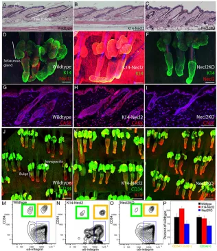
Fig. 4. Overexpression or deletion of Necl2 does not disturb epidermal homeostasis.(A-C) H&E-stained sections of dorsal skin of wild-type (A), K14Necl2 transgenic (B) and Necl2-knockout mice (C). (D-F) Tail epidermal whole mounts of wild-type (D), K14Necl2 transgenic (E) and Necl2-knockout (F) mice stained for keratin-14 (green) and Necl2 (red), with DAPI nuclear counterstain (blue). (G-I) Wild-type (G), K14Necl2 transgenic (H) and Necl2 null (I) dorsal skin sections stained with anti-CASK (red) and DAPI nuclear counterstain (blue). (J-L) Tail epidermal whole mounts of wild type (J), K14Necl2 transgenic (K) and Necl2-null (L) mice stained for keratin-14 (red) and CD34 (green). (M-P) Flow cytometric analysis of α 6-inegrin and CD34 double-positive (orange boxes) and
α6-integrin-positive, CD34-negative (green boxes) cells in wild-type (M), K14Necl2 transgenic (N) and Necl2-knockout (O) mouse epidermis. (P) Relative abundance of cells in M-O. IFE, interfollicular epidermis. Asterisk in J indicates nonspecific staining of sebaceous gland. Scale bars: 100μm in A-L.
N
membrane localisation, it did not affect the localisation of desmoplakin, which accumulated at cell-cell borders only under standard calcium concentrations (data not shown).
To ascertain whether increased Necl2-mediated adhesion affected keratinocyte motility, we cultured control and Necl2-transduced human keratinocytes in standard FAD medium (approximately 1.2 mM Ca2+) or low-calcium (KSFM, 0.09 mM Ca2+) medium and monitored their behaviour by time-lapse microscopy. Using standard culture conditions, there were no significant differences in keratinocyte motility (data not shown). By contrast, Necl2 -transduced keratinocytes cultured in KSFM displayed a marked reduction in cellular motility when compared with cells transduced with empty vector (Fig. 3M-O). This was quantified by measuring the overall maximum migration distance of Necl2-expressing keratinocytes and control cells (compare red and black, Fig. 3O).
We also examined whether Necl2 overexpression affected in vitro wound healing. Keratinocytes transduced with Necl2or empty vector were cultured to confluence in KSFM. The cultures were then wounded with a Gilson pipette tip and the time taken for wound closure was recorded by live cell imaging. Control cells closed the
wound in an average of 568±99 minutes (nine wounds). The average time to wound closure in four independent Necl2-transduced cultures was significantly slower at 1880±9 minutes (P<0.003). In a further four cases, Necl2-transduced cells failed to close the wound by the end of the recording period. From these experiments, we conclude that Necl2 overexpression promotes intercellular adhesion under low-calcium conditions, thereby reducing cell motility and in vitro wound healing.
Necl2 levels do not affect normal epidermal homeostasis
[image:6.612.50.335.294.739.2]To investigate the functions of Necl2 in vivo, we generated transgenic mice in which Necl2 was overexpressed via the keratin-14 gene (Krt14) promoter in the epidermal basal layer, undifferentiated cells of the sebaceous gland and hair follicle outer root sheath (K14Necl2 transgenics). Three independent transgenic lines were generated and the line with highest expression (7248A.5) was selected for detailed analysis. We also examined mice in which Necl2has been deleted by homologous recombination (Necl2 -knockout) (Fujita et al., 2006).
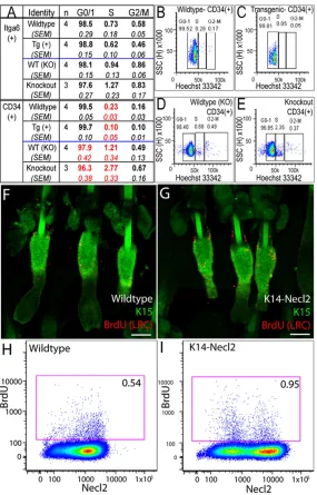
Fig. 5. Necl2 overexpression reduces epidermal proliferation. (A-E) Determination of proportion of cells in G0-G1, S, G2-M phases of the cell cycle by flow cytometry of cells labelled with Hoechst 33342. (A) Total undifferentiated cells (α6-integrin positive; Itga6+) or bulge cells (CD34 and α
6-integrin double positive; CD34+) are shown beneath the
appropriate wild-type controls. Data shown are means±s.e.m. from four transgenic and four matched wild-type controls and from three knockout and three wild-type control mice. Values in red are significantly different from controls (P≤0.05). (B-E) Representative flow cytometry profiles of CD34+bulge
populations, showing position of cell cycle gates in wild-type versus K14-Necl2 transgenic mice (B,C) and wild-type versus Necl2-knockout mice (D,E). (F,G) Tail epidermal whole mounts from wild-type (F) and transgenic (G) mice labelled with anti-BrdU (to detect LRC; red) and K15 (green). (H,I) Representative flow cytometry plots of BrdU-labelled LRC (red boxes) in wild-type (H) and K14Necl2 transgenic (I) epidermis. Numbers refer to percentage BrdU-positive cells. Cells were double labelled with anti-Necl2. Scale bars: 100μm in F,G.
V
E
LO
P
M
E
N
Analysis of haematoxylin and eosin (H&E)-stained sections of adult dorsal telogen and anagen skin and tail epidermal whole mounts did not reveal any abnormalities in K14Necl2 or Necl2-null mice (Fig. 4A-F). There were no effects on interfollicular epidermal thickness or differentiation and the sebaceous glands and hair growth cycle were grossly normal (Fig. 4A-F; and data not shown). We confirmed the presence or absence of Necl2 in wild-type, transgenic and knockout mice by antibody staining of tail epidermal whole mounts (Fig. 4D-F). CASK immunoreactivity was increased in K14Necl2 epidermis; however, Necl2 deletion did not significantly reduce CASK expression relative to wild-type epidermis (Fig. 4G-I). No differences in E-cadherin staining were observed (data not shown). CD34 expression in the hair follicle bulge was unaffected by Necl2 loss or overexpression, when evaluated both by whole-mount labelling (Fig. 4J-L) and by flow cytometry of disaggregated epidermal cells (Fig. 4M-P).
These results are consistent with the lack of an effect of Necl2 overexpression on terminal differentiation of primary human keratinocytes (Fig. 2G-K).
Overexpression of Necl2 results in decreased proliferation of bulge stem cells
Since overexpression of Necl2 reduced the growth rate of cultured keratinocytes (Fig. 2F), we examined the proliferative status of Necl2-knockout and K14Necl2 transgenic mouse epidermis. Data in Fig. 5A-E are representative of at least two experiments with independent cohorts of mice. The proportion of α6-integrin-positive epidermal cells (total basal cells) in each phase of the cell cycle was determined by DNA labelling with Hoechst 33342. There were no
statistically significant differences in the proportion of G0-G1, S or G2-M phase cells in K14Necl2 transgenic, wild-type and Necl2-null epidermis (Fig. 5A). However, when we analysed CD34 and α 6-integrin double-positive cells (bulge cells) we observed a statistically significant reduction in S-phase cells in K14Necl2 transgenics relative to strain-matched wild-type controls (Fig. 5A-C). Conversely, there was a significant increase in cells in S-phase and a reduction in G0-G1-phase cells in Necl2-null bulge cells relative to the appropriate wild-type controls (Fig. 5A,D,E).
To examine whether reduced proliferation in K14Necl2 transgenic bulge keratinocytes influenced BrdU label-retaining cell (LRC) abundance, neonatal mice were given repeated injections of BrdU and subsequently examined in adulthood (Braun et al., 2003). The number of LRCs was increased in Necl2-overexpressing mice, with labelled cells lying primarily in the bulge (Fig. 5F,G). These results were confirmed by flow cytometry (Fig. 5H,I).
We conclude that although Necl2 levels do not affect epidermal differentiation (Figs 2 and 4), Necl2 overexpression does result in reduced epidermal proliferation, both in culture and in vivo. Thus, although Necl2 expression is not confined to regions of low proliferation (Fig. 1), Necl2 might, in combination with other regulators (Horsley et al., 2008; Jensen and Watt, 2006; Jensen et al., 2009), promote quiescence in hair follicle keratinocytes.
Overexpression of Necl2 impairs epidermal wound healing
[image:7.612.50.362.397.737.2]Given the effects of Necl2 on keratinocyte motility and intercellular adhesion in culture (Fig. 3), we predicted that Necl2 would influence skin wound healing. We created 5-mm-diameter
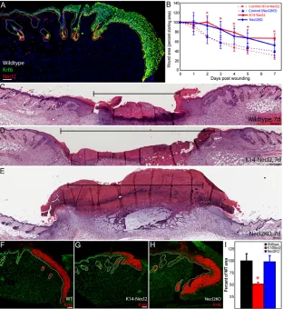
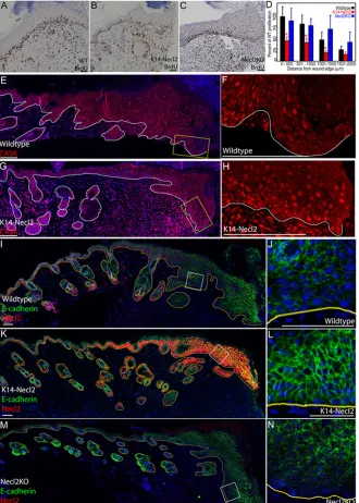
Fig. 6. Skin wound healing is modulated by Necl2.(A) Wild-type skin collected 7 days after wounding stained for Necl2 (red), keratin-6 (green) and DAPI (blue). (B) Wound size, expressed as a percentage of starting wound area, with time after wounding. Data are represented as mean values±s.e.m. (C-E) Representative H&E-stained sections of wild-type (C), K14Necl2 transgenic (D), and Necl2-knockout (E) mouse skin 7 days after wounding. Brackets show distance between wound margins. (F-H) Keratin-6 (red) and DAPI (green) labelling of the leading edge of re-epithelialisation 7 days after wounding wild-type (F), K14Necl2 transgenic (G) and Necl2-knockout (H) mice. (I) Quantification of average keratin-6-positive area per section prepared as in F-H, expressed as a percentage of wild-type controls. Data represent average of two sections per wound from a minimum of five animals. Error bars in B and I represent s.e.m.; *P<0.05. Scale bars: 100μm in A,C-E; 50μm in F-H.
N
full thickness wounds on the back skin of age- and sex-matched K14Necl2 and transgene-negative littermates as well as Necl2 -knockout and wild-type littermates and measured average wound size over time (Fig. 6). Necl2 was not observed in the hyperplastic epithelial front 7 days after wounding in wild-type mice (Fig. 6A). Relative to appropriate congenic wild-type controls (dashed blue and red lines, Fig. 6B), wound closure was significantly delayed both when Necl2 was overexpressed (red line) and when Necl2 was absent (blue line) (Fig. 6B). Both control mouse strains (FVB/n and C57/Bl6, red and blue dashed lines respectively) exhibited similar wound-healing kinetics.
The histological appearance of K14Necl2 and wild-type wounds was indistinguishable at all time points (Fig. 6C,D; and data not shown). However, the slow healing of Necl2-null wounds 7 days after wounding was associated with the formation of an abnormally large granulation tissue mass (Fig. 6E). Although no clear differences between wild-type and K14Necl2 wounds were
observed in H&E-stained sections, the upregulation of keratin-6 that is characteristic of hyperproliferative healing epidermis (Grose et al., 2002; Gurtner et al., 2008) was markedly reduced in K14Necl2 transgenic wounds (Fig. 6F-I).
To examine the mechanism by which Necl2 overexpression affected wound closure, S-phase cells were labelled by injecting mice with BrdU 7 days after wounding. There were significantly fewer labelled cells in the area immediately adjacent to the leading edge of wounds in transgenic compared with wild-type mice (Fig. 7A-D). By contrast, there was no significant difference between Necl2-null and wild-type epidermis (Fig. 7A-D).
[image:8.612.51.381.270.732.2]Although CASK expression was generally increased following wounding, expression was markedly reduced in the tongue of epithelial cells that formed the leading front of re-epithelialisation in wild-type mice (Fig. 7E,F), as has been previously reported (Ojeh et al., 2008). By contrast, CASK expression was typically maintained in the epithelial tongue in K14Necl2 transgenic mice
Fig. 7. Necl2 overexpression decreases proliferation and leads to upregulation of E-cadherin and CASK during wound healing. (A-C) BrdU-labelled S-phase cells (brown, A-C) at the leading edge of healing wild-type (A), transgenic (B) and Necl2-knockout (C) mouse epidermis. (D) Quantification of BrdU-positive cells as a function of distance from the wound edge in wild-type (black bars), transgenic (red bars), and knockout mice (blue bars). Data are presented relative to appropriate congenic wild-type controls (n=4 knockout mice/group; n=8 transgenic mice/group). Error bars show s.e.m.; *P<0.05. (E-H) Wounded wild-type (E,F) or K14Necl2 transgenic (G,H) epidermis stained with anti-CASK (red) and DAPI nuclear counterstain (blue; E,G). (I-N) E-cadherin (green, I-N) and Necl2 (red, I,K,M) expression at the leading edge of re-epithelialising wounds, 7 days after wounding. (E,G,I-N) DAPI nuclear counterstain (blue). Lines (A-C,E-N) denote dermal-epidermal boundary and boxes (E,G,I,K,M) denote regions shown at higher magnification in adjacent panels. Scale bars: 50μm in A-C; 100μm in E-N.
V
E
LO
P
M
E
N
(Fig. 7G,H). No differences in CASK expression between C57/Bl6 wild-type and Necl2-null mice following wounding were observed (data not shown).
To assess adherens junction stability after wounding, we stained epidermis of wild-type, transgenic and Necl2-null mice with anti-E-cadherin (Fig. 7I-N). E-anti-E-cadherin expression in the epidermal basal layer was reduced in conjunction with downregulation of Necl2 at the leading edge of healing wounds in wild-type mice (Fig. 7I,J). By contrast, Necl2 transgenic mice, but not knockout mice, maintained E-cadherin expression throughout the hyperplastic wounded area (Fig. 7K-N). These results suggest that Necl2 modulates epidermal E-cadherin and CASK stability during wound healing.
DISCUSSION
We show that the cell adhesion molecule Necl2 is expressed in embryonic hair follicles and adult bulge stem cells. We propose that within the hair follicle, Necl2 functions both as an inhibitor of stem cell proliferation and as an adhesion molecule that limits departure from the bulge by reducing motility and promoting intercellular adhesion. Necl2 expression does not autonomously influence differentiation or lineage selection, but does modulate wound healing.
From our experiments on cultured keratinocytes, we conclude that the major effect of overexpression of Necl2 is to promote intercellular adhesion, thereby reducing cell migration and overall motility. Necl2 overexpression also has modest effects on cell growth, but these do not affect the ability of cultured epidermis to stratify and undergo terminal differentiation.
Our observation that Necl2 is enriched within human and murine stem cells suggests that Necl2-dependent adhesion might provide keratinocytes with enhanced resistance to calcium fluctuations resulting from physiological or environmental stress. These findings might partially explain previous observations that bulge stem cells are more resistant to epidermal injury (Huelsken et al., 2001). The upregulation of Necl2 in the bulb of anagen follicles (see Fig. 1K) might also regulate the invasion of the dermis as the follicle elongates (Fuchs, 2009). Notwithstanding our observations, the lack of an overt skin phenotype in uninjured Necl2-knockout mice probably reflects partial or complete functional redundancy among nectin and nectin-like proteins (Fujita et al., 2006; van der Weyden et al., 2006; Yamada et al., 2006).
Although it is known that bulge stem cells contribute to epidermal wound healing, the mechanisms by which this occurs have largely remained elusive (Ito et al., 2005). We found that endogenous Necl2 expression was reduced in healing wounds and that loss or overexpression of Necl2 impaired wound healing. Our in vitro and in vivo results suggest that the effect of Necl2 overexpression was due to reduced cell proliferation and migration, correlating with enhanced adherens junction stability and maintenance of CASK expression (Ojeh et al., 2008). Our results are consistent with previous studies which demonstrate that Necl2 overexpression in kidney cells suppresses HGF-dependent cell scattering and motility because of retention of E-cadherin-based intercellular adhesion (Masuda et al., 2005).
The reduced skin wound healing of Necl2-null mice did not
et al., 2004; Weller et al., 2006). It is also possible that Necl2-CRTAM-dependent T cell interactions influence wound healing by modulating tissue inflammation, extracellular matrix deposition and macrophage or neutrophil-dependent granuloma clearance (Jameson and Havran, 2007).
Our data are consistent with previous observations regarding Necl2 function in other cell types and tissues (Ito et al., 2007; Masuda et al., 2005) and with the known functions of E-cadherin and CASK in epidermal development and homeostasis (Jamora et al., 2003; Jamora et al., 2005; Ojeh et al., 2008; Tinkle et al., 2004; Young et al., 2003). Our findings highlight the dual role of adhesion molecules as regulators of the location and proliferation of adult tissue stem cells.
Acknowledgements
We gratefully acknowledge Kristin Braun and Dawn Mazzatti for critical reading of the manuscript and the core facilities of the CRUK London and Cambridge Research Institutes for expert technical assistance. A.G. was the recipient of an NIH Postdoctoral Fellowship. These studies were funded by Cancer Research UK, the Wellcome Trust, the Medical Research Council and the European Union. We gratefully acknowledge the support of Hutchison-Whampoa and Cambridge University. Deposited in PMC for release after 6 months.
References
Bagutti, C., Hutter, C., Chiquet-Ehrismann, R., Fässler, R. and Watt, F. M.
(2001). Dermal fibroblast-derived growth factors restore the ability of β1 integrin-deficient embryonal stem cells to differentiate into keratinocytes. Dev. Biol. 231, 321-333.
Benitah, S. A., Frye, M., Glogauer, M. and Watt, F. M.(2005). Stem cell depletion through epidermal deletion of Rac1. Science309, 933-935.
Birchmeier, C., Birchmeier, W., Gherardi, E. and Vande Woude, G. F.(2003). Met, metastasis, motility and more. Nat. Rev. Mol. Cell Biol. 4, 915-925.
Blanpain, C., Lowry, W. E., Geoghegan, A., Polak, L. and Fuchs, E.(2004). Self-renewal, multipotency, and the existence of two cell populations within an epithelial stem cell niche. Cell118, 635-648.
Brakebusch, C., Grose, R., Quondamatteo, F., Ramirez, A., Jorcano, J. L., Pirro, A., Svensson, M., Herken, R., Sasaki, T., Timpl, R. et al.(2000). Skin and hair follicle integrity is crucially dependent on beta 1 integrin expression on keratinocytes. EMBO J. 19, 3990-4003.
Braun, K. M., Niemann, C., Jensen, U. B., Sundberg, J. P., Silva-Vargas, V. and Watt, F. M.(2003). Manipulation of stem cell proliferation and lineage commitment: visualisation of label-retaining cells in wholemounts of mouse epidermis. Development130, 5241-5255.
Estrach, S., Legg, J. and Watt, F. M.(2007). Syntenin mediates Delta1-induced cohesiveness of epidermal stem cells in culture. J. Cell Sci. 120, 2944-2952.
Fuchs, E.(2007). Scratching the surface of skin development. Nature445, 834-842.
Fuchs, E.(2009). The tortoise and the hair: slow-cycling cells in the stem cell race.
Cell137, 811-819.
Fuchs, E., Dowling, J., Segre, J., Lo, S. H. and Yu, Q. C.(1997). Integrators of epidermal growth and differentiation: distinct functions for β1 and β4 integrins.
Curr. Opin. Genet. Dev. 7, 672-682.
Fujita, E., Kouroku, Y., Ozeki, S., Tanabe, Y., Toyama, Y., Maekawa, M., Kojima, N., Senoo, H., Toshimori, K. and Momoi, T.(2006). Oligo-astheno-teratozoospermia in mice lacking RA175/TSLC1/SynCAM/IGSF4A, a cell adhesion molecule in the immunoglobulin superfamily. Mol. Cell. Biol. 26, 718-726.
Galibert, L., Diemer, G. S., Liu, Z., Johnson, R. S., Smith, J. L., Walzer, T., Comeau, M. R., Rauch, C. T., Wolfson, M. F., Sorensen, R. A. et al.(2005). Nectin-like protein 2 defines a subset of T-cell zone dendritic cells and is a ligand for class-I-restricted T-cell-associated molecule. J. Biol. Chem. 280, 21955-21964.
Gandarillas, A. and Watt, F. M.(1997). c-Myc promotes differentiation of human epidermal stem cells. Genes Dev. 11, 2869-2882.
Grose, R., Hutter, C., Bloch, W., Thorey, I., Watt, F. M., Fassler, R.,
Huelsken, J., Vogel, R., Erdmann, B., Cotsarelis, G. and Birchmeier, W.(2001). β-Catenin controls hair follicle morphogenesis and stem cell differentiation in the skin. Cell105, 533-545.
Ito, A., Nishikawa, Y., Ohnuma, K., Ohnuma, I., Koma, Y., Sato, A., Enomoto, K., Tsujimura, T. and Yokozaki, H.(2007). SgIGSF is a novel biliary-epithelial cell adhesion molecule mediating duct/ductule development.
Hepatology45, 684-694.
Ito, M., Liu, Y., Yang, Z., Nguyen, J., Liang, F., Morris, R. J. and Cotsarelis, G.
(2005). Stem cells in the hair follicle bulge contribute to wound repair but not to homeostasis of the epidermis. Nat. Med. 11, 1351-1354.
Ito, T., Shimada, Y., Hashimoto, Y., Kaganoi, J., Kan, T., Watanabe, G., Murakami, Y. and Imamura, M.(2003). Involvement of TSLC1 in progression of esophageal squamous cell carcinoma. Cancer Res. 63, 6320-6326.
Jameson, J. and Havran, W. L. (2007). Skin γδT-cell functions in homeostasis and wound healing. Immunol. Rev. 215, 114-122.
Jamora, C., DasGupta, R., Kocieniewski, P. and Fuchs, E.(2003). Links between signal transduction, transcription and adhesion in epithelial bud development. Nature422, 317-322.
Jamora, C., Lee, P., Kocieniewski, P., Azhar, M., Hosokawa, R., Chai, Y. and Fuchs, E.(2005). A signaling pathway involving TGF-b2 and snail in hair follicle morphogenesis. PLoS Biol. 3, e11.
Jensen, K. B. and Watt, F. M.(2006). Single-cell expression profiling of human epidermal stem and transit-amplifying cells: Lrig1 is a regulator of stem cell quiescence. Proc. Natl. Acad. Sci. USA 103, 11958-11963.
Jensen, K. B., Collins, C. A., Nascimento, E., Tan, D. W., Frye, M., Itami, S. and Watt, F. M.(2009). Lrig1 expression defines a distinct multipotent stem cell population in mammalian epidermis. Cell Stem Cell4, 427-439.
Lo Celso, C., Prowse, D. M. and Watt, F. M.(2004). Transient activation of b-catenin signalling in adult mouse epidermis is sufficient to induce new hair follicles but continuous activation is required to maintain hair follicle tumours.
Development131, 1787-1799.
Lopez-Róvira, T., Silva-Vargas, V. and Watt, F. M.(2005). Different consequences of β1 integrin deletion in neonatal and adult mouse epidermis reveal a context-dependent role of integrins in regulating proliferation, differentiation, and intercellular communication. J. Invest. Dermatol. 125, 1215-1227.
Lowell, S., Jones, P., Le Roux, I., Dunne, J. and Watt, F. M.(2000). Stimulation of human epidermal differentiation by Delta-Notch signalling at the boundaries of stem-cell clusters. Curr. Biol. 10, 491-500.
Mao, X., Seidlitz, E., Ghosh, K., Murakami, Y. and Ghosh, H. P.(2003). The cytoplasmic domain is critical to the tumor suppressor activity of TSLC1 in non-small cell lung cancer. Cancer Res. 63, 7979-7985.
Masuda, M., Kikuchi, S., Maruyama, T., Sakurai-Yageta, M., Williams, Y. N., Ghosh, H. P. and Murakami, Y.(2005). Tumor suppressor in lung cancer (TSLC)1 suppresses epithelial cell scattering and tubulogenesis. J. Biol. Chem. 280, 42164-42171.
Molès, J. P. and Watt, F. M.(1997). The epidermal stem cell compartment: variation in expression levels of E-cadherin and catenins within the basal layer of human epidermis. J. Histochem. Cytochem. 45, 867-874.
Morii, E., Ito, A., Jippo, T., Koma, Y., Oboki, K., Wakayama, T., Iseki, S., Lamoreux, M. L. and Kitamura, Y.(2004). Number of mast cells in the peritoneal cavity of mice: influence of microphthalmia transcription factor through transcription of newly found mast cell adhesion molecule, spermatogenic immunoglobulin superfamily. Am. J. Pathol. 165, 491-499.
Morris, R. J., Liu, Y., Marles, L., Yang, Z., Trempus, C., Li, S., Lin, J. S., Sawicki, J. A. and Cotsarelis, G.(2004). Capturing and profiling adult hair follicle stem cells. Nat. Biotechnol. 22, 411-417.
Ohyama, M., Terunuma, A., Tock, C. L., Radonovich, M. F., Pise-Masison, C. A., Hopping, S. B., Brady, J. N., Udey, M. C. and Vogel, J. C.(2006). Characterization and isolation of stem cell-enriched human hair follicle bulge cells. J. Clin. Invest. 116, 249-260.
Ojeh, N., Pekovic, V., Jahoda, C. and Määttä, A.(2008). The MAGUK-family protein CASK is targeted to nuclei of the basal epidermis and controls keratinocyte proliferation. J. Cell Sci. 121, 2705-2717.
Perez-Moreno, M., Jamora, C. and Fuchs, E.(2003). Sticky business: orchestrating cellular signals at adherens junctions. Cell112, 535-548.
Sevilla, L. M., Nachat, R., Groot, K. R. and Watt, F. M.(2008). Kazrin regulates keratinocyte cytoskeletal networks, intercellular junctions and differentiation. J. Cell Sci. 121, 3561-3569.
Shingai, T., Ikeda, W., Kakunaga, S., Morimoto, K., Takekuni, K., Itoh, S., Satoh, K., Takeuchi, M., Imai, T., Monden, M. et al.(2003). Implications of nectin-like molecule-2/IGSF4/RA175/SgIGSF/TSLC1/SynCAM1 in cell-cell adhesion and transmembrane protein localization in epithelial cells. J. Biol. Chem. 278, 35421-35427.
Silva-Vargas, V., Lo Celso, C., Giangreco, A., Ofstad, T., Prowse, D. M., Braun, K. M. and Watt, F. M.(2005). β-Catenin and Hedgehog signal strength can specify number and location of hair follicles in adult epidermis without recruitment of bulge stem cells. Dev. Cell9, 121-131.
Suzuki, K., Hu, D., Bustos, T., Zlotogora, J., Richieri-Costa, A., Helms, J. A. and Spritz, R. A.(2000). Mutations of PVRL1, encoding a cell-cell adhesion molecule/herpesvirus receptor, in cleft lip/palate-ectodermal dysplasia. Nat. Genet. 25, 427-430.
Takai, Y., Irie, K., Shimizu, K., Sakisaka, T. and Ikeda, W.(2003). Nectins and nectin-like molecules: roles in cell adhesion, migration, and polarization. Cancer Sci. 94, 655-667.
Takai, Y., Miyoshi, J., Ikeda, W. and Ogita, H.(2008). Nectins and nectin-like molecules: roles in contact inhibition of cell movement and proliferation. Nat. Rev. Mol. Cell. Biol. 9, 603-615.
Tinkle, C. L., Lechler, T., Pasolli, H. A. and Fuchs, E.(2004). Conditional targeting of E-cadherin in skin: insights into hyperproliferative and degenerative responses. Proc. Natl. Acad. Sci. USA 101, 552-557.
Tumbar, T., Guasch, G., Greco, V., Blanpain, C., Lowry, W. E., Rendl, M. and Fuchs, E.(2004). Defining the epithelial stem cell niche in skin. Science303, 359-363.
van der Weyden, L., Arends, M. J., Chausiaux, O. E., Ellis, P. J., Lange, U. C., Surani, M. A., Affara, N., Murakami, Y., Adams, D. J. and Bradley, A.
(2006). Loss of TSLC1 causes male infertility due to a defect at the spermatid stage of spermatogenesis. Mol. Cell. Biol. 26, 3595-3609.
Wakamatsu, K., Ogita, H., Okabe, N., Irie, K., Tanaka-Okamoto, M., Ishizaki, H., Ishida-Yamamoto, A., Iizuka, H., Miyoshi, J. and Takai, Y.(2007). Up-regulation of loricrin expression by cell adhesion molecule nectin-1 through Rap1-ERK signaling in keratinocytes. J. Biol. Chem. 282, 18173-18181.
Watabe, K., Ito, A., Koma, Y., Wakayama, T., Iseki, S., Shinomura, Y. and Kitamura, Y.(2004). Distinct roles for the SgIGSF adhesion molecule and c-kit receptor tyrosine kinase in the interaction between mast cells and the mesentery. Biochem. Biophys. Res. Commun. 324, 782-788.
Watt, F. M.(2002). Role of integrins in regulating epidermal adhesion, growth and differentiation. EMBO J. 21, 3919-3926.
Watt, F. M., Lo Celso, C. and Silva-Vargas, V.(2006). Epidermal stem cells: an update. Curr. Opin. Genet. Dev. 16, 518-524.
Weller, K., Foitzik, K., Paus, R., Syska, W. and Maurer, M.(2006). Mast cells are required for normal healing of skin wounds in mice. FASEB J. 20, 2366-2368.
Yamada, D., Yoshida, M., Williams, Y. N., Fukami, T., Kikuchi, S., Masuda, M., Maruyama, T., Ohta, T., Nakae, D., Maekawa, A. et al.(2006). Disruption of spermatogenic cell adhesion and male infertility in mice lacking TSLC1/IGSF4, an immunoglobulin superfamily cell adhesion molecule. Mol. Cell. Biol. 26, 3610-3624.
Young, P., Boussadia, O., Halfter, H., Grose, R., Berger, P., Leone, D. P., Robenek, H., Charnay, P., Kemler, R. and Suter, U.(2003). E-cadherin controls adherens junctions in the epidermis and the renewal of hair follicles.
EMBO J. 22, 5723-5733.