Toxicity effects of monosodium glutamate (MSG) on
embryonic development of zebrafish (
Danio rerio
); a
promising model to study excitotoxins
A.S. Mahaliyana, M.F.A. Fasmina, A.M.T.B. Alahakoon and G.M.G.M.M. Wickrama
Department of Animal Science, Faculty of Animal Science and Export Agriculture, Uva Wellassa University of Sri Lanka
Abstract- Zebrafish (Danio rerio) is a widely used biological model to investigate different chemicals since it has certain similarities with human biology. Present study investigated the impact of monosodium glutamate (MSG); an excessively used food additive all over the world, on the embryonic development of zebrafish. Acute toxicity experiments were performed for a 4-day period using zebrafish eggs. Ten different test concentrations of monosodium glutamate (MSG) of 10, 30, 50, 100, 150, 200, 250, 300, 400, 500 mgL⁻1 were used as treatment concentrations. The results clearly indicated that with the increase of the MSG concentrations, different observable deformities are formed in zebrafish embryo. At the low concentrations of MSG such as 10, 30, 50 mgL⁻1 there were no observable malformations in zebrafish embryonic development. However, at high concentrations such as 100, 150, 200, 250, 300, 400, 500 mgL⁻1 there were distinguishable negative alterations such as growth retardation, shrinkage of chorion, yolk sac edema, lack of pigmentation, tail deformities and scoliosis in developing embryos. Zebrafish embryo can be successfully used to investigate excitotoxins such as MSG. However, the impacts of these concentrations on internal anatomical and physiological changes in zebrafish embryo should be comprehensively investigated.
Index Terms- Zebrafish, Danio rerio, Monosodium glutamate, MSG, Embryonic development
I. INTRODUCTION
he zebrafish (Danio rerio); a teleost belonging to the family Cyprinidae, has used extensively as a renowned vertebrate model (Jusuf and Harris, 2009) over the past twenty years. This has been investigated for studying genetics and developmental physiology, more recently for human disease and the screening of therapeutic drugs & chemical studies (Fishman, 2001). Zebrafish have been considered as favorable research models in biomedical and health research and well suited as a model system to perform chemical biology experiments efficiently in educational settings due to their several specialties (Grunwald and Eisen, 2002). They are easy to maintain in large numbers, readily reproducing under laboratory conditions, smaller in size, high in fecundity, contain transparent embryos, have rapid development and easy to culture (Völker, 2007; Lammer, 2009; Oliveira et al., 2009). The mechanisms that involved in differentiation of the tissues in this model are optically more visible. Availability of the whole genome sequence and
numerous mutant lines facilitate studies of development and physiology. Similarities between zebrafish and mammalian genetics suggest that the zebrafish is an excellent vertebrate model of human developmental and disease processes.
More recently, use of renowned or novel small molecules (i.e. chemicals) to study phenotypic changes and identify the corresponding cellular target(s) has gained popularity in the scientific community. Thus, zebrafish can be used as an effective tool for studying such type of chemical biology approaches. Zebrafish embryo is a model with high potential to investigate the effects of a wide range of chemical compounds including flavour enhancers, food additives and different controversial compounds (Abdelkader et al, 2012).
Monosodium glutamate (MSG) is a form of glutamate and is the sodium salt of glutamic acid; an amino acid, which exists as white crystals in physical nature. It does not give unique taste alone, however is able to provide distinct flavour to food when it is added as an additive, yet the mechanism has still not been properly clarified (Abdelkader et al., 2012). Hence, MSG is a widely used food additive with thousands of food that are prepared in both households and restaurants, and ingested to human as a part of their meals (Humphries et al., 2008). The Joint FAO/ WHO Expert Committee on Food Additives determined the Acceptable Daily Intake (ADI) limit for MSG as 120 mg/ kg body weight in 1970 (JECFA, 1971). This recommendation was later undergone to a revision (JECFA, 1971), and the Joint Expert Committee on Food Additives (JECFA) again claimed that it was not necessary to set a numerical ADI for MSG, which is also included in the FDA’s Generally Regarded As Safe (GRAS) list.
and hepatic toxicity (Olney, 1969; Simson et al., 1977; Farombi and Onyema, 2006; Diniz et al., 2004; Egbuonu et al., 2009). In addition, MSG has been classified as an exo-toxin, since it can lead to develop certain disorders including some endocrine disorders (Ikonomidou and Turski, 1995). The toxicity and teratogenicity of MSG have been investigated using different laboratory animals (Collison et al., 2012). Hence, this study investigated the effect of different concentrations of MSG on zebrafish embryonic development.
II. MATERIALS AND METHODS
Zebrafish maintenance and egg collection
Adult male and female zebrafish (domestic aquarium strain) were maintained in a separate glass tanks with aerators, filters in the Aquaculture Laboratory, Uva Wellassa University of Sri Lanka, Badulla. All the water quality parameters inside the tanks were maintained at optimum levels (Temperature at 26 ± 2 ͦ C and pH 5 to 6) and fish were fed with Artemia nauplii and commercial feed twice a day. Initially, suitable male and female brooders were selected for the breeding. Breeding tank was prepared with artificial plants and breeding traps because of their predatory activity on laid eggs. The bottom of the tank was covered with a stainless steel grid which helps to collect all the eggs on the bottom. In addition, artificial plants were used to stimuli spawning. Selected males and females were placed in the spawning tank day before the test in the ratio of 2:1, male to female. Mating and spawning were taken place 30 minutes after the onset of sunlight in the following morning. About 30- 60 minutes after spawning, spawn traps were removed and eggs were carefully collected using a pipette. Fertilized viable eggs were selected under the binocular microscope for further investigations.
Toxicity assessment
Acute toxicity experiment was performed for a 4-day period using zebrafish (Danio rerio) eggs. Ten different test concentrations of monosodium glutamate (MSG) of 10, 30, 50, 100, 150, 200, 250, 300, 400, 500 mgL⁻1 were prepared by diluting MSG (99% purity) in deionized water. Deionized water was used as the negative control and as the internal plate control. Fertilized zebrafish embryos were immersed in the test solutions before cleavage, by the 16 cell-stage. At least twice, the number of eggs needed per treatment group (40 eggs) were randomly selected and transferred into the respective concentrations and controls within the 90 minutes of post fertilization. Twenty four (24) well plates were filled with 2 mL per well with freshly prepared test solutions. In each plate, twenty wells were used for test concentrations and four were used as internal plate control. The eggs in standard 24- well plate were covered by shelf adhesive foil and maintain at a temperature of 26 ± 1 °C. Dead eggs were removed immediately since they can adversely affect on the other viable eggs. Three replicates were performed individually. In this study, the acute toxic effect of MSG on the zebrafish embryo was observed and identified using binocular stereomicroscope.
III. RESULT AND DISCUSSION
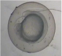
Embryonic development of the zebrafish (Danio rerio) has been described in several studies. Kimmel et al., 1995 have described seven broad periods of embryogenesis as the zygote, cleavage, blastula, gastrula, segmentation, pharyngula, and hatching periods. Figure 1 shows the normally developed embryo for four days period of time.
Figure 1: Normal development of zebra fish embryo for four days period of time
In zebrafish embryo toxicity test, several lethal, sub- lethal and teratogenic endpoints were observed. According to OECD guideline for testing chemicals -236, 2013, Fish Embryo Toxicity Assay, fish embryos are considered as dead when it is coagulated, lack of somite formation, non-detachment of tail and
lack of heart beat (Figure 2). In control, no dead embryos and deformities were observed. Growth retardation, shrinkage of chorion, yolk sac edema, lack of pigmentation, tail deformities and scoliosis are the major deformities observed during the study.
a
d
c
Figure 2: (a) coagulated embryo, (b) lack of somite formation, (c) non-detachment of tail
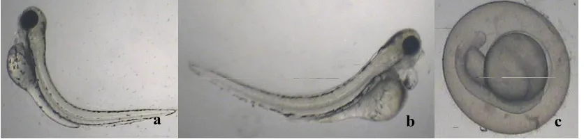
Fish embryo stage is highly sensitive to chemicals and pollutants. Since embryos are covered by semi permeable protective membrane chorion, it does not fully protect the embryo against all chemical penetration. In MSG low concentrations of 10, 30 and 50 mgL-1, there were no observable
[image:3.612.167.451.57.155.2]deformities and they showed normal embryonic development (Figure 3). In contrary with the finding of this study, Abdelkader
[image:3.612.113.526.248.386.2]et al., 2012 has reported that even 50 mgL-1 concentration level also caused to deviations of the zebrafish embryo from the normal growth.
Figure 3: Normal development of zebra fish embryo at MSG concentrations of 10, 30 and 50 mgL-1, respectively
Growth retardation is the only deformity observed in the concentration of 100 mgL-1 (Figure 4). Development of embryonic stages in embryos can be observed continuously with the days. But even after 4 days period of time there were no any development in some of embryos treated with 100 mgL-1 MSG. It may be due to the inhibition of growth hormone secretion by MSG and alterations of the metabolism as described by Hermanussen et al., 2006. Finding of the Abdelkader et al, 2012 on zebrafish embryo is in agreement with the finding of these observations. Furthermore, they have demonstrated the internal anatomical deformities such as elongated heart, cardiac sac edema, yolk-sac edema and spinal kyphosis.
Figure 4: Growth retardation in zebra fish embryo at MSG concentration of 100 mgL-1
In the concentration of 150 mgL-1 also growth retardation was observed, but the development was stopped at initial stage (Figure 5). In addition, lack of tail development also was recorded. In general, tail development can observed after 24 hours post fertilization. However, in this study certain tail deformities were observed (Figure 5).
a
b
c
b
[image:3.612.367.487.413.522.2]Figure 5: Growth retardation and lack of tail development in zebra fish embryo at MSG concentrations of 150 mgL-1
Edema and lack of pigmentations were recorded in the concentrations of 250 and 300 mgL-1 (Figure 6). Edema is caused by the accumulation of fluid inside pericardium and yolk sac. In this study, only yolk sac edema was observed which could be caused by accumulation of MSG inside the yolk sac. The onset of pigmentation in normal strains occurs shortly after 24-hours post-fertilization (Kimmel et al., 1995). Pigmentation changes in fish are often due to stress induced (Ozoh, 1980; Nguyen and
[image:4.612.162.505.316.449.2]Janssen, 2002). Pigmentation of the skin is controlled by Melanocyte Stimulating Hormone (αMSH) and Melanin-Concentrating Hormone (MCH), which are known to be up regulated during stress response. Reduction in pigmentation of zebrafish embryo treated with MSG may either result from stress at the cellular, organ or individual level or alter the function of αMSH and MCH directly.
Figure 6: Edema and lack of pigmentations in zebra fish embryo at MSG concentrations of 250 and 300 mgL-1
Edema, scoliosis and growth retardation were recorded in 400 and 500 mgL-1 MSG concentrations (Figure 7). Scoliosis might have resulted from impaired ionic regulation (Stominska and Jezierska, 2000; Khayatzaden and Abbasi, 2010) or damage of the vertebral column (Ozoh, 1979; Stouthart et al., 1994; Cheng et al., 2000; Jezierska et al., 2000; Nguyen and Janssen, 2002; Hallare et al., 2005) or fractured vertebrae through tetanic muscular contraction (Holcombe et al., 1976; Bengtsson, 1974).
In addition, MSG may lead to disrupt the endocrine function which is responsible for the proper maintenance of many systems in the body. This disruption may play a role in causing various malformations in the treated animals including stunted skeletal development and development of other malformations (Ele fteriou et al., 2003).
Figure 7: Edema, scoliosis and growth retardation in zebra fish embryo at MSG concentrations of 400 and 500 mgL-1
a
b
b
a
a
a
[image:4.612.130.548.590.690.2]As results indicate in this study, there is a negative impact on zebrafish embryonic development by MSG, especially when the concentrations are increased. Certain neurological disorders, abnormal neural development, certain endocrine disorders and neuropsychiatric disorders can be derived by the excessive intake of excitotoxin such as MSG (Ikonomidou and Turski, 1995). Different animal models have been used so far to demonstrate the toxicity of MSG and they have shown that these toxic concentrations can adversely alter the anatomical structures and the physiological functions, especially in early developmental stages of the life (Khadija et al., 2000; Bhattacharya et al., 2011; Al-Qudsi and Al-Jahdali, 2012). With the increased concentration of MSG caused by the excessive intake, can derive metabolic disruptions followed by biological malformations in embryonic development. Even, infant stages of human can also be at a risk of high concentrations of MSG into body through excessive consumption (Abdelkader et al., 2012).
There is an emerging trend in the world with the sophisticated lifestyles of human towards the fast food which contain lot of food additives such as MSG (Qudsi and Al-Jahdali). It has been estimated that in European population daily glutamate intake may be up to 10g/ day when considering consumption of glutamate from other food sources (Beyreuther et al., 2007). In Asian countries, the mean intake of added glutamate has been shown to be about 2.2g/ day (He et al., 2011). These high concentrations can be adversely affect on human health, especially the early developmental stages. Hence, this has to be comprehensively investigated using animal models. Because of exposure to nutritional and environmental challenges during critical stages of early development of life, the metabolism in later life can be remarkably affected (Jackson et al., 2010). Zebrafish embryonic development can provide an ideal model for these types of studies due to its easiness as well as more similarities with human genome.
IV. CONCLUSION
The results of this study clearly indicate that with the increase of the MSG concentrations, different observable deformities are formed in zebrafish embryo. Although at the low concentrations of MSG (i.e. 10, 30, 50 mgL⁻1) there were no observable malformations in zebrafish embryonic development, at high concentrations (i.e. 100, 150, 200, 250, 300, 400, 500 mgL⁻1) there were distinguishable negative alterations such as growth retardation, shrinkage of chorion, yolk sac edema, lack of pigmentation, tail deformities and scoliosis. Zebrafish embryo can be successfully used to investigate excititoxins such as MSG. However, further studies on internal anatomical and physiological changes can be recommended to carryout for more comprehensive understanding on this regards.
REFERENCES
[1] Abdelkader, T. S., Seo-Na, C., Tae-Hyun, K., Juha, S., Dongso, K. and Park, J. H., 2016. Teratogenicity brain aromatase-induction of monosodium glutamate in estrogen-responsive mosaic transgenic zebra fish Danio rerio. African Journal of Biotechnology, 11(48), 10816-10823.
[2] Al-Qudsi, F. and Al-Jahdali, A., 2012. Effect of monosodium glutamate on chick embryo development. Journal of American Science, 8(10).
[3] Bengtsson, B. E., 1974. Vertebral damage to minnows Phoxinus phoxinus exposed to zinc. Oikos, 134-139.
[4] Beyreuther, K., Biesalski, H. K., Fernstrom, J. D., Grimm, P., Hammes, W. P., Heinemann, U. and Walker, R., 2007. Consensus meeting: monosodium glutamate–an update. European journal of clinical nutrition, 61(3), 304-313. [5] Bhattacharya, T., Bhakta, A. and Ghosh, S. K., 2011. Long term effect of monosodium glutamate in liver of albino mice after neo-natal exposure. Nepal Med Coll J, 13(1), 11-16.
[6] Cheng, S. H., Wai, A. W. K., So, C. H. and Wu, R. S. S., 2000. Cellular molecular basis of cadmium‐induced deformities in zebrafish embryos. Environmental Toxicology Chemistry, 19(12), 3024-3031.
[7] Collison, K. S., Makhoul, N. J., Zaidi, M. Z., Al-Rabiah, R., Inglis, A., res, B. L. and Al-Mohanna, F. A., 2012. Interactive effects of neonatal exposure to monosodium glutamate aspartame on glucose homeostasis. Nutrition and metabolism, 9(1), 1.
[8] Diniz, Y. S., Fernes, A. A., Campos, K. E., Mani, F., Ribas, B. O. and Novelli, E. L., 2004. Toxicity of hypercaloric diet monosodium glutamate: oxidative stress metabolic shifting in hepatic tissue. Food Chemical Toxicology, 42(2), 313-319.
[9] Egbuonu, A. C. C., Obidoa, O., Ezeokonkwo, C. A., Ezeanyika, L. U., and Ejikeme, P. M., 2009. Low dose oral administration of monosodium glutamate in male albino rats may be nephroprotective. Bio-Research, 7(1). [10] Elefteriou, F., Takeda, S., Liu, X., Armstrong, D. and Karsenty, G., 2003.
Monosodium glutamate-sensitive hypothalamic neurons contribute to the control of bone mass. Endocrinology, 144(9), 3842-3847.
[11] Farombi, E. O. and Onyema, O. O., 2006. Monosodium glutamate-induced oxidative damage genotoxicity in the rat: modulatory role of vitamin C, vitamin E quercetin. Human and experimental toxicology, 25(5), 251-259. [12] Fishman, M. C., 2001. Zebrafish--the canonical vertebrate. Science,
294(5545), 1290-1291.
[13] Fourteenth Report of the Joint FAO/WHO Expert Committee on Food Additives (JECFA), 1971. FAO Nutrition Meetings Report Series No. 48, WHO Technical Report Series, No. 462.
[14] Grunwald, D. J. and Eisen, J. S., 2002. Headwaters of the zebrafish— emergence of a new model vertebrate. Nature reviews genetics, 3(9), 717-724.
[15] Hallare, A. V., Schirling, M., Luckenbach, T., Köhler, H. R. and Triebskorn, R., 2005. Combined effects of temperature cadmium on developmental parameters biomarker responses in zebrafish (Danio rerio) embryos.Journal of Thermal Biology, 30(1), 7-17.
[16] He, K., Du, S., Xun, P., Sharma, S., Wang, H., Zhai, F. and Popkin, B., 2011. Consumption of monosodium glutamate in relation to incidence of overweight in Chinese adults: China Health Nutrition Survey (CHNS). The American journal of clinical nutrition, 93(6), 1328-1336.
[17] Hermanussen, M., Garcia, A. P., Sunder, M., Voigt, M., Salazar, V. and Tresguerres, J. A. F., 2006. Obesity, voracity, short stature: the impact of glutamate on the regulation of appetite. European journal of clinical nutrition, 60(1), 25-31.
[18] Holcombe, G. W., Benoit, D. A., Leonard, E. N. and McKim, J. M., 1976. Long-term effects of lead exposure on three generations of brook trout (Salvelinus fontinalis). Journal of the Fisheries Board of Canada, 33(8), 1731-1741.
[19] Humphries, P., Pretorius, E. and Naude, H., 2008. Direct indirect cellular effects of aspartame on the brain. European journal of clinical nutrition, 62(4), 451-462.
[20] Ikonomidou, C. and Turski, L., 1995. Glutamate in neurodegenerative disorders. Neurotransmitters Neuromodulators: Glutamate. CRC Press, Boca Raton, 17, 253-272.
[21] Jackson, A. A., Burdge, G. C. and Lillycrop, K. A., 2010. Diet, nutrition modulation of genomic expression in fetal origins of adult disease. [22] Jezierska, B., 2000. The effect of heavy metals on postembryonic
development of common carp, Cyprinus carpio L. Archiwum Rybactwa Polskiego, 8(1), 119-128.
[23] Jezierska, B., Lugowska, K., Witeska, M. and Sarnowski, P., 2000. Malformations of newly hatched common carp larvae. Electronic Journal of Polish Agricultural Universities, 3(2).
[25] Khadija, A., Ati, A., Mohammed, S., Saad, A. M. and Mohamed, H. E., 2009. Response of broiler chicks to dietary monosodium glutamate. Pak. Vet. J, 29(4), 165-168.
[26] Khayatzaden, J. and Abbasi, E., 2010. The Effects of Heavy Metals on Aquatic Ecosystem. The 1st International Applied Geological Congress: 688-694.
[27] Kimmel, C. B., Ballard, W. W., Kimmel, S. R., Ullmann, B. and Schilling, T. F., 1995. Stages of embryonic development of the zebrafish. Developmental dynamics, 203(3), 253-310.
[28] Lammer, E., 2009. Refinement of the fish embryo toxicity test (FET) with zebrafish (Danio rerio): Is it a real replacement of the acute fish toxicity test?.
[29] Nguyen, L. T. H. and Janssen, C. R., 2002. Embryo-larval toxicity tests with the African catfish (Clarias gariepinus): comparative sensitivity of endpoints. Archives of environmental contamination toxicology, 42(2), 256-262.
[30] Oliveira, R., Domingues, I., Grisolia, C. K. and Soares, A. M., 2009. Effects of triclosan on zebrafish early-life stages adults. Environmental Science Pollution Research, 16(6), 679-688.
[31] Olney, J. W., Labruyere, J. and De Gubareff, T., 1979. Brain damage in mice from voluntary ingestion of glutamate aspartate. Neurobehavioral toxicology, 2(2), 125-129.
[32] Organization for Economic Cooperation Development- OECD., 2013. OECD Guidelines for testing of chemicals-Fish Embryo Acute Toxicity (FET) Test, OECD OCED. 236.
[33] Ozoh, P. T., 1979. Malformations inhibitory tendencies induced to Brachydanio rerio (Hamilton-Buchanan) eggs larvae due to exposures in low concentrations of lead copper ions. Bulletin of environmental contamination toxicology, 21(1), 668-675.
[34] Ozoh, P. T., 1980. Effect of lead on pigment pattern formation in zebrafish (Brachydanio rerio). Bulletin of environmental contamination toxicology, 24(1), 276-282.
[35] Simson, E. L., Gold, R. M., Stish, L. J. and Pellett, P. L. (1977). Axon-sparing brain lesioning technique: the use of monosodium-L-glutamate other amino acids. Science, 198(4316), 515-517.
[36] Storto, M., de Grazia, U., Battaglia, G., Felli, M. P., Maroder, M., Gulino, A. and Calogero, A., 2000. Expression of metabotropic glutamate receptors
in murine thymocytes thymic stromal cells. Journal of neuroimmunology, 109(2), 112-120.
[37] Stouthart, A. J. H. X., Spanings, F. A. T., Lock, R. A. C. and Bonga, S. W., 1994. Effects of low water pH on lead toxicity to early life stages of the common carp (Cyprinus carpio). Aquatic toxicology, 30 (2), 137-151. [38] Thirty-first Report of the Joint FAO/WHO Expert Committee on Food
Additives (JECFA), 1987. WHO Technical Report Series, No. 759. 29-31. [39] Völker, D., 2007. Chemical-sensitive genes in zebrafish (Danio rerio) early
development - identification characterisation of differential expression in embryos exposed to the model compound 3, 4-dichloroaniline. Ph.D. Dissertation, Department Zelltoxikologie. Helmholtz Centre for Environmental Research – UFZ, Germany 142p.
AUTHORS
First Author – A.S. Mahaliyana, BSc. Sp. Hons, Lecturer, Department of Animal Science, Faculty of Animal Science and Export Agriculture, Uva Wellassa University of Sri Lanka Second Author – M.F.A. Fasmina, BSc. Sp. Hons,
Demonstrator, Department of Animal Science, Faculty of Animal Science and Export Agriculture, Uva Wellassa University of Sri Lanka
Third Author – A.M.T.B. Alahakoon, BSc. Sp. Hons,
Demonstrator, Department of Animal Science, Faculty of Animal Science and Export Agriculture, Uva Wellassa University of Sri Lanka