Chemistry Theses Department of Chemistry
8-9-2016
Establishing the Relationship Between Function
and Dynamics Within the Gated Mechanism of
D-arginine Dehydrogenase
Michael Souffrant
Follow this and additional works at:https://scholarworks.gsu.edu/chemistry_theses
This Thesis is brought to you for free and open access by the Department of Chemistry at ScholarWorks @ Georgia State University. It has been accepted for inclusion in Chemistry Theses by an authorized administrator of ScholarWorks @ Georgia State University. For more information, please [email protected].
Recommended Citation
Souffrant, Michael, "Establishing the Relationship Between Function and Dynamics Within the Gated Mechanism of D-arginine Dehydrogenase." Thesis, Georgia State University, 2016.
ESTABLISHING THE RELATIONSHIP BETWEEN FUNCTION AND DYNAMICS WITHIN THE GATED MECHANISM OF D-ARGININE DEHYDROGENASE
by
MICHAEL SOUFFRANT
Under the Direction of Donald Hamelberg, PhD
ABSTRACT
Enzymes are ubiquitous in biological systems. They catalyze chemical reactions and are involved in many biochemical processes. The enzyme of interest is Pseudomonas aeruginosa D-arginine dehydrogenase (PaDADH). This flavin-dependent enzyme is composed of approximately 375 amino acid residues and has a broad substrate specificity with D-amino acids. A water recognition motif, observed in roughly 1200 non-redundant protein data bank (PDB) structures, was revealed to be embedded near the active site of PaDADH. This motif coincides with the conformational changes of the enzyme’s gated mechanism. Molecular dynamics simulations were carried out to study the gated properties and structural characteristics of PaDADH in solution. Single amino acid mutations were undertaken to further understand the dynamics of the gated mechanism of this enzyme. In addition, pKa,shift analyses were evaluated to probe for the basic catalytic amino acid residue that is suggested to trigger the catalytic mechanism of PaDADH.
ESTABLISHING THE RELATIONSHIP BETWEEN FUNCTION AND DYNAMICS WITHIN THE GATED MECHANISM OF D-ARGININE DEHYDROGENASE
by
MICHAEL SOUFFRANT
A Thesis Submitted in Partial Fulfillment of the Requirements for the Degree of Master of Science
in the College of Arts and Sciences Georgia State University
Copyright by
ESTABLISHING THE RELATIONSHIP BETWEEN FUNCTION AND DYNAMICS WITHIN THE GATED MECHANISM OF D-ARGININE DEHYDROGENASE
by
MICHAEL SOUFFRANT
Committee Chair: Donald Hamelberg
Committee: Stuart Allison Giovanni Gadda
Electronic Version Approved:
DEDICATION
To
Michelle & George Souffrant
ACKNOWLEDGEMENTS
Prior to joining Dr. Hamelberg’s lab, I was clueless in the research field of computational biophysical chemistry. My original intent was to attend medical school and start a career in health. During my time in his lab, not only I gained much further insight on some of the theoretical approaches of computational chemistry, but I also expanded my understanding in high computing applications. Dr. Hamelberg readily applies this concept of “self-motivation”,
which enabled me to discover more in regards to my personality and the type of scientist that I wanted to eventually become. He allowed me to freely indulge into my scientific ideas, and use his resources at moment’s notice. Thank you for your assistance, patience, and the daily morning lectures which consistently have a significant influence on my life’s outcome.
I also wanted to send many thanks to Arghya Barman, a post-doctoral fellow in our lab, for his support and aid on a day-to-day basis throughout my research assignments. Ever since his presence, my comprehension and abilities in the biophysical research field had evolved tenfold. Thank you for your encouragements and your willingness to help with no remorse.
In addition, thank you Dr. Gadda, and Dr. Allison for deciding to be part of my thesis committee on such short notice. Your critiques and philosophical approaches in regards to my work and my knowledge as a scientist is greatly appreciated.
Likewise, I also want to send many thanks to my lab mates for taking the time to criticize my work and becoming my support group in times of need. I truly believe that we are a team in which we compensate for each other’s strengths and weaknesses in several ways. Aside from
TABLE OF CONTENTS
ACKNOWLEDGEMENTS ... v
LIST OF TABLES ... ix
LIST OF FIGURES ... x
1 INTRODUCTION... 1
1.1 Enzymatic Properties of PaDADH ... 1
1.2 Molecular Dynamics ... 3
1.2.1 Molecular dynamics algorithms ... 4
1.2.2 Limitations of molecular dynamics ... 6
1.2.3 Thermodynamic cycle ... 7
1.2.4 Thermodynamic integration method ... 8
1.3 Motifs ... 11
1.3.1 Beta hairpin motif ... 11
1.3.2 Greek key motif ... 12
1.3.3 Zinc finger motif ... 13
1.3.4 EF-hand motif ... 14
1.3.5 Water recognition motif ... 15
2 WATER RECOGNITION MOTIF IN PaDADH ... 21
2.1 Introduction ... 21
2.3 Results & Discussions ... 31
2.3.1 PaDADH free in solution ... 31
2.3.2 PaDADH in complex with D-arginine ... 35
2.4 Conclusions ... 37
3 PaDADH Y53F, S45A, AND A46G MUTANT DYNAMICS ... 38
3.1 Introduction ... 38
3.2 Experimental Procedures ... 44
3.3 Results & Discussions ... 45
3.3.1 Y53F ... 45
3.3.2 S45A ... 47
3.3.3 A46G ... 49
3.4 Conclusions ... 51
4 PaDADH TYR53, AND GLU246 PKA SHIFT CALCULATIONS ... 52
4.1 Introduction ... 52
4.2 Experimental Procedures ... 55
4.3 Results & Discussions ... 57
4.3.1 Open conformation free in solution (A) ... 57
4.3.2 Close conformation in complex with D-arginine (B) ... 59
4.3.3 Individual trial thermodynamic integration values ... 61
5 CONCLUSIONS ... 63
5.1 Concluding Remarks ... 63
LIST OF TABLES
Table 2.1 Steady-state kinetics study of PaDADH with D-amino acids.1 ... 25
Table 3.1 Kinetic study of PaDADH Y53F mutant variant.53... 39
Table 3.2 Kinetic study of PaDADH S45A mutant variant.58 ... 40
Table 3.3 Kinetic study of PaDADH A46G mutant variant.58 ... 41
Table 4.1 Quantitative analysis of thermodynamic integration values. ... 61
LIST OF FIGURES
Figure 1.1 Thermodynamic cycle involving proton binding and release. ... 9
Figure 1.2 Beta hairpin motif in Pseudomonas aeruginosa D-arginine dehydrogenase.1 ... 11
Figure 1.3 Greek key motif in Pseudomonas aeruginosa D-arginine dehydrogenase.1 ... 12
Figure 1.4 Zinc finger motif in Ligand of Numb Protein-X2.21 ... 13
Figure 1.5 EF-hand motif in calmodulin.26 ... 14
Figure 1.6 Localized conserved water molecules found in Pin1.4 ... 16
Figure 1.7 Analysis of Conserved Wat1 and Wat2 in Pin1.4 ... 17
Figure 1.8 Unique properties of water recognition motif.4 ... 19
Figure 2.1 Distinct water recognition motif in PaDADH1. ... 21
Figure 2.2Schematic representation of the “active site lid” of PaDADH.1 ... 23
Figure 2.3 Distance betweenTyr53 (CZ) and FAD (N5) with water recognition motif.1... 27
Figure 2.4 Dynamics analyses of PaDADH free in solution. ... 31
Figure 2.5 Distribution of buried water molecules in the water recognition motif. ... 33
Figure 2.6 Schematic representation of the absence and presence of water. ... 34
Figure 2.7 Dynamics analyses of PaDADH in complex with D-arginine. ... 35
Figure 3.1 Distance betweenTyr53 (CZ) and FAD (N5) with site of mutations.1... 42
Figure 3.2 Dynamics analyses of PaDADH Y53F mutant variant. ... 45
Figure 3.3 Dynamics analyses of PaDADH S45A mutant variant... 47
Figure 3.4 Dynamics analyses of PaDADH A46G mutant variant. ... 49
Figure 4.1 Thermodynamic cycle involving PaDADH Tyr53 (A). ... 57
Figure 4.2 Thermodynamic cycle involving PaDADH Glu246 (A). ... 58
Figure 4.4 Thermodynamic cycle involving PaDADH Glu246 (B)... 60
1 INTRODUCTION
1.1 Enzymatic Properties of PaDADH
Pseudomonas aeruginosa D-arginine dehydrogenase (PaDADH) is a flavin-depdendent enzyme which converts D-amino acids into their corresponding imino acids.1-3 Typically, an enzyme has one defined substrate according to its biological function, however, PaDADH has the ability to catalyze every amino acid at different efficiencies with the exeption of D-aspartate, D-glutamate, and glycine. An X-ray crystal structure of this enzyme was reported at 1.06 Å resolution, in which case, two conformers of certain residues were established.1 The ligand-free configuration, with a 70% occupancy in electron density, was denoted to be the open conformation of the enzyme, whereas the product-bound conformer in complex with iminoarginine, with a 30% occupancy in electron density, was designated to be the close conformation based on a tyrosine over the active site. Consequently, the notion of a gate which enhanced substrate binding, enabled product release, and played a role in the broad substrate specificity of PaDADH became plausible. Using the molecular dynamics (MD) approach to study the two conformational states of the gated mechanism of this enzyme and reaching convergence with kinetic assessments became the objective of this investigation. Such study may help into gaining further insight on the catalytic properties of other biological protein structures.
generated an interest to further understand the water recognition motif structure, but also, the significant role of the gated mechanism in catalysis. MD methods were applied on the wild type enzyme structure to determine a correlation between the gated mechanism, and the water recognition motif. Single amino acid mutants of PaDADH were simulated to probe the different mechanical properties of the gated mechanism of the enzyme. Thus, motif properties, MD simulations, and kinetic measurements were explored to gain more insight on the strutural components of the two conformers of PaDADH in catalysis.
1.2 Molecular Dynamics
Molecular dynamics (MD) is a statistical mechanics method that takes into account the microscopic state ensemble to describe the physical quantities of a system at the macroscopic level. The concept of molecular dynamics evolved from the dynamics of liquids shown by Alder, Wainwright, and Rahman during the years of 1950s and 1960s.6 As the years passed, both the theoretical aspect of the method and computing technology have greatly improved rendering a viable tool for many areas in research. For example, MD simulations have been used to study different macromolecules such as enzymes, ligand binding receptor proteins, or nucleic acids.
Molecular dynamics methods can be separated into two main categories known as classical and quantum. The classical statistical mechanics approach to MD simulations treats the molecules as the ‘ball and stick’ model. The ball is a representation of a particular atom whereas
the stick is an analogy of a bond between two atoms.7 The quantum or first-principles statistical mechanics approach to MD simulations takes into account the presence of electrons, however the dynamics of a typical nucleus with its inner electrons is classically integrated. Quantum MD simulations show significant improvements in comparison to the classical approach, however, the method is computationally expensive.8 Thus, classical MD became the more practical approach for the simulation of large complex systems.
details as opposed to synthesizing the macromolecule and characterizing its properties, hence, the increase of high computing applications for drug design in pharmaceutical companies.
1.2.1 Molecular dynamics algorithms
The molecular dynamics approach follows the atomic force field model which defines a complex system as a set of atoms held together by interatomic forces.6 If one was to evaluate the potential energy of N interacting atoms as a function of their positions U(r1, … , rN), by taking the gradient (
∇)
of such function with respect to the displacements (xyz coordinates) of theatoms, the force acting upon each atom would be determined as shown in equation 1.10
𝐹
𝑖= −∇
𝑟𝑖𝑈(𝑟
1, ⋯ , 𝑟
𝑁) = − (
𝜕𝑈𝜕𝑥𝑖
,
𝜕𝑈 𝜕𝑦𝑖,
𝜕𝑈
𝜕𝑧𝑖
) (1)
Electrons delocalize while being shared by many different nuclei, which in turn displaces into different electric field environments. Thus, it is possible to determine the dynamics of the nuclei without explicitly considering the electrons, which is feasible by considering the potential energy surface. The physics and chemistry of MD simulations are based on the different potentials involving each atom of a particular system.11
𝑈(𝑟
1, ⋯ , 𝑟
𝑁) = ∑
𝑎𝑖2
(𝑙
𝑖− 𝑙
𝑖0)
2𝑏𝑜𝑛𝑑𝑠
+ ∑
𝑏𝑖
2
(𝜃
𝑖− 𝜃
𝑖0)
2𝑎𝑛𝑔𝑙𝑒𝑠
+
∑
𝐶𝑖2
𝑡𝑜𝑟𝑠𝑖𝑜𝑛𝑠
[1 + cos(𝑛𝑤
𝑖− 𝛾
𝑖] + ∑
4
𝜀𝑖𝑗[(
𝜎𝑖𝑗 𝑟𝑖𝑗
)
12
−
𝑎𝑡𝑜𝑚𝑠𝑝𝑎𝑖𝑟𝑠
(
𝜎𝑖𝑗𝑟𝑖𝑗
)
6] + ∑
𝑘
𝑞𝑖𝑞𝑗𝑟𝑖𝑗
𝑎𝑡𝑜𝑚𝑝𝑎𝑖𝑟𝑠
(2)
two terms establish the energies generated during the deformation of bond lengths (li) and bond
angles (𝜃𝑖) from their equilibrium values (li0) and (𝜃𝑖0). ai and bi represent the harmonic force
constants which prevent modelling chemical changes. Rotations around the chemical bond is define by the third term, in which case, n and ci represent the periodicity and heights of rotational
barriers respectively. The attractive and repulsive interatomic van der Waals forces in the form of the Lennard-Jones 12-6 potential is defined by the fourth term. Coulomb electrostatic potential is denoted in the fifth term where qi and qj represent the partial charges of atom i and j
respectively, and rij expresses the relative distance between both atoms.
𝐹
𝑖= 𝑚
𝑖𝑑2𝑟𝑖(𝑡)𝑑𝑡2
(3)
Upon solving for the force vector acting on each atom of a complex system, an algorithm is used in order to generate the MD trajectories. For instance, the Verlet algorithm is usually applied for computing simulations of macromolecules due to its simplicity and stability. Consequently, MD trajectories are described by position and velocity vectors, in which case, time is dependent on both the time step, and the total number of steps undertaken during simulation6.
𝑟
𝑖(𝑡 + Δ𝑡) ≅ 2𝑟
𝑖(𝑡) − 𝑟
𝑖(𝑡 − Δ𝑡) +
𝐹𝑖(𝑡)𝑚𝑖
Δ𝑡
2
(4)
sub-femtosecond time scale should be used for highest accuracy within each integrated step since the sum in potential is based on the localization of electrons surrounding the atoms. Though less accurate, a femtosecond time step is more efficient and detailed enough to thoroughly study a system of interest. However, by continuously increasing the magnitude of time at the femtosecond scale the margin of error increases, hence, a time step of more than 5 femtoseconds would not be a feasible approach for the application of an MD simulation.12
1.2.2 Limitations of molecular dynamics
In order to consistently update the positions and velocities of each atom in the system, the sum of the potentials, and the forces must be recomputed at each step. Such aspect of MD trajectories has created a need for better algorithms to compensate for the long-range forces, and new explicit models that would enhance trajectory assessments.6 For example, sampling methods of the TIP3P water model have decrease computational time of MD simulations more than three folds.13 High temperature simulations, accelerated molecular dynamics (aMD), and replica exchange molecular dynamics (REMD) were all methods developed to improve sampling.14 The particle meshed Ewald method enabled efficient computation of long-range interactions, in which case, atoms separated by a distance larger than a certain cut-off aren’t neglected.15
Classical atomic force field sampling cannot be applied to gain further insight on the acid-base chemistry of electrons, redox reactions, or even proton tunneling because the approach is based on treating the atoms, their valence electrons, and bonds as the “ball and stick”model.12 In theory, the atomic force field represents the forces that influence the atoms of a system under experimental conditions. Deriving empirical potential energies to be transferable and applicable to many systems of interests at various environmental parameters is challenging, and create a sense of abstraction in the field of computational chemistry. Even through the method is combining experimental data with theoretical calculations, there still exist a need for improvement of the atomic force fields, which would enhance higher timescale simulations, accuracy and stability.
Most biochemical processes occur at the microsecond or millisecond scale. In order to gain further understanding on the characteristics of a system, there has to be enough sampling at the femtosecond time scale to reach higher orders in time (microsecond, millisecond). Such approach has to take into account the atom count of a system since the integrated step is evaluated for each atom. Thus, processing simulations at the microsecond or millisecond time scale requires hundreds of millions or billions of steps, which creates limitations in both computation time and size of biochemical processes. MD methods are currently being explored to design better atomic force fields in order to generate simulations more efficiently.
1.2.3 Thermodynamic cycle
and key’ approach is more rigid, in which case, macromolecules are considered to be static
analogous to a key opening a door. The ‘induced fit’ model, however, considers the flexibility of the ligand(s) and its corresponding enzyme.16 During the formation of enzyme-substrate complex both structures changes in configurations for optimum free energy release. Due to the atomistic movements of the enzyme and substrate structures the ‘induce fit’ type models can now be
investigated using MD simulations. However, the change in Gibbs’s free energy related to an enzyme free in solution binding with different ligands is computationally expensive, because it requires the assessment of repeated trajectories, which show the stochastic interactions involved between the enzyme’s active site and the various ligands. Nevertheless, calculating the change in Gibb’s free energy of the enzyme-ligand complexes and the enzyme with different ligands free in
solution is significantly less expensive computationally. Since G is a state function, a thermodynamic cycle between these Gibb’s free energy properties can be established, in which case, the relative binding affinities of various ligands to the same protein is evaluated.
1.2.4 Thermodynamic integration method
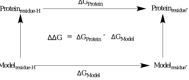
Figure 1.1 Thermodynamic cycle involving proton binding and release.
The change in Gibb’s free energy of the deprotonation process of a protein residue is compare to the deprotonated spontaneity of that same residue free in solution. The double free energy difference is denoted as∆∆G = ∆Gprotein - ∆Gmodel.
The molecular dynamics approach gives an ensemble of configurations, which would reflect both protein and solvent structures with their electrostatic and non-electrostatic effects. During a free energy simulation, the system of interest is slowly transformed from an initial state A to a final state B free in solution and within the microenvironment of the enzyme. The energy function U gradually changes with a ‘coupling parameter’ , where varies from 0 to 1 in
correspondence with UA to UB, respectively (equation 5). An intermediate value of would be in respect to an hybrid system, that is a mixture of A and B in such way that would be greater
than 0 but less than 1.17
𝑈(𝜆) = (1 − 𝜆)𝑈
𝐴+ 𝜆𝑈
𝐵(5)
Removing the proton partial charge by increments causes a change in its electrostatic potential. Subsequently, the average values of the electrostatic potentials are evaluated at their
respective values, which are then integrated to generate the total change in Gibbs free energy.
Protein
residue-HD
Protein
residue-G
ProteinD
G
ModelDD
G
=
D
G
Protein-
D
G
Model𝜕𝑈 𝜕𝜆
⁄
(𝜆) = ⟨
𝜕𝑈𝜕𝜆
⟩
𝜆= 〈𝑈
𝐵− 𝑈
𝐴〉
𝜆= ∑ 𝛿𝑞
𝑖 𝑖〈𝑉
𝑖〉𝜆
(6)
The large brackets in regards to equation 6 above defines an ensemble average related to
U(). The charge increments are written as δqi where i is the proton of interest going from state 0
to 1. The proton’s electrostatic potential is established as Vi and the sum operator sign is the
integrated value of δqi<Vi> after being calculated from each lambda. The average quantity of
UB -UA is defined as the energy required for a virtual ionization reaction while the system is fixed in its reference conformation, also known as the energy gap. By evaluating simulations for
a set of values between 0 and 1, the Gibbs free energy change of the process can be estimated, hence the name ‘thermodynamic integration’.
∆𝐺 = −𝑘𝑇𝑙𝑜𝑔𝐾
𝑎(7)
𝑝𝐾
𝑎= −𝑙𝑜𝑔
10𝐾
𝑎=
12.303𝑘𝑇
∆𝐺
(8)
𝑝𝐾
𝑎,𝑝𝑟𝑜𝑡𝑒𝑖𝑛= 𝑝𝐾
𝑎,𝑚𝑜𝑑𝑒𝑙+
12.303𝑘𝑇
∆∆𝐺
(9)
The standard Gibbs free energy is related to the natural log of the acid dissociation constant through Boltzmann’s constant ‘k’ and temperature ‘T’ (equation 7). By solving for the
1.3 Motifs
During several structural analyses of enzymes, scientists began to notice repeated patterns in terms of the secondary structures of proteins. These secondary properties were called structural motifs, also known as super-secondary structures. The different conformation of motifs is based on the connectivity between secondary structure elements. For instance, helices and strands have a multitude of ways to interact with one another. Based on such perspective, there is a limited number of ways secondary structure elements of proteins can combine to be biologically viable. In addition, two macromolecules may share the same super-secondary structure but have dissimilar primary sequences.
1.3.1 Beta hairpin motif
[image:24.612.271.371.486.687.2]One of the most common structural motifs revealed in proteins is known as the beta hairpin motif.18,19 Due to further understanding of its three-dimensional structure, this super-secondary structure is also referred to as beta-ribbon or beta-beta unit. In its basic form, a beta hairpin is a super-secondary structure with two beta strands similar to the conformation of a hairpin.
When present, the two strands are adjacent in primary sequence, however they are antiparallel in the secondary structure of the protein. Each antiparallel strand is then connected by a two to five amino acid loop (Figure 1.2). If a beta hairpin was removed from its protein, the structure will still form. When present, the beta hairpin motif is a necessity for the biological activity of the macromolecule.18,19
1.3.2 Greek key motif
[image:25.612.260.386.296.527.2]Another common super-secondary structure which may be considered an extrapolation of the beta hairpin motif is the Greek key motif as shown in Figure 1.3.20
Figure 1.3 Greek key motif in Pseudomonas aeruginosa D-arginine dehydrogenase.1
1.3.3 Zinc finger motif
[image:26.612.171.475.204.407.2]Aside from these super-secondary protein structures there exist other common motifs, which involve ligand binding. For instance, the zinc finger, shown in Figure 1.4, is a relatively small motif with one or more zinc ions in a coordinated covalent bond with surrounding histidine and cysteine residues to stabilize the fold.
Figure 1.4 Zinc finger motif in Ligand of Numb Protein-X2.21
1.3.4 EF-hand motif
Another super-secondary structure that is most commonly found in calcium binding proteins is the EF-hand motif.25 This particular motif is a helix-loop-helix structural domain that is sometimes present in the calcium binding protein family. Similar to the zinc finger, the calcium ion is coordinated to both helices included the core of the motif which involves a loop and a strand (Figure 1.5).
Figure 1.5 EF-hand motif in calmodulin.26
This specific motif is found within 66 subfamilies of calcium-binding proteins. The EF hand named was coined about 25 years ago by Kretsinger and Nockolds as a graphical representation of the calcium-binding motif in parvalbumin. In certain cases, the motif has been
Ca2+
E helix
shown to not only form coordination with calcium but also magnesium. This particular motif is very significant due to the usual calcium influx within the cytosol of a cell. Meaning, there is a diverse range of enzymes that contain this super-secondary structure which would enable them to bind to calcium in the cell cytosol for proper biological functions. For instance, signal transduction between cellular compartments and muscle contraction are mostly govern by the ability of protein structures to bind to calcium which in turn is related to their EF-hand motif.25, 27
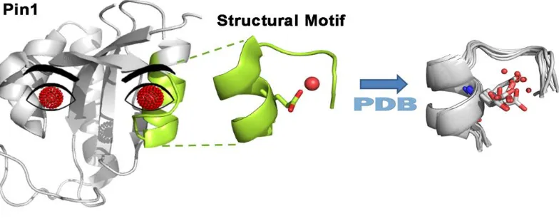
1.3.5 Water recognition motif
Peptidyl-propyl cis-trans isomerase NIMA-interacting 1 (Pin1) is an enzyme (PPIase) which catalyzes the cis/trans isomerization of peptide bonds preceding a proline residue.28,29 Pin1 is a Parvulin protein which has high affinity for the isomerization of the propyl ω-bond in the
presence of phosphorylated serine (pSer) or threonine (pThr) as precedents of the propyl
bond.29-32 The enzyme has WW domain and a catalytic domain connected by a flexible
loop.4 NMR experiments have shown the catalytic and WW domains to be allosterically
coupled.33 Due to its catalytic abilities, Pin1 is a subcellular signaling pathway regulator
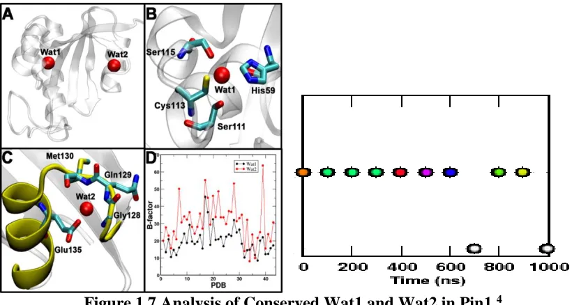
Figure 1.6 Localized conserved water molecules found in Pin1.4
Wat1 at site 1 was alluded to be highly localized in comparison to wat2 at site 2 which was suggested to exchange with the water molecules from the solvent. Wat2 at site 2 was found in a variety of non-redundant proteins and was characterized as a distinct water recognition motif.
Figure 1.7 Analysis of Conserved Wat1 and Wat2 in Pin1.4
Panel A displays the location of site 1 and 2 with their corresponding waters in Pin1. Panel B shows the significant hydrogen bonding involved at site 1 with wat1, which enabled such high localization. Panel C indicates the distinct water recognition motif including the significant interactions with wat2 at site 2. Panel D establishes a comparison of the recorded B factor values of the different PDB structures of Pin1 in regards to wat1 and wat2 at their corresponding sites. In addition, the exchangeable characteristics of wat2 at site2 are displayed over time.
Recently two conserved localized water molecules had been revealed in Pin1. Wat1 was revealed to be located at site 1 and Wat2 at site 2. Wat1 was suggested to be highly localized in
site 1 with a favorable binding free energy of -2.8 0.3 kcal/mol. Wat2 showed high
exchangeability with the bulk water solvent at site 2 with a lower favorable binding free energy
remaining 9 structures had water molecules surrounding the super-secondary structure or absent from the site. Wat1 formed hydrogen bonds with neighboring active site residues such as His59, Ser111, and Ser115. In addition, Wat1 is suggested to be involved in a critical interaction with Cys113 which is viable for protein function.32,36-38 Meaning, in the absence of Wat1 the hydrogen bond interactions would be disrupted and Cys113 would not be properly aligned for proper enzymatic function. Wat2 is involved in interactions with the surrounding residues such as Gln129, Met130, and Glu135.
All of the crystal structures of human Pin1 were downloaded from the PDB, in which case, files with multiple copies of Pin1 were separated into individual copies for a total of 47 crystal structures. The Debye-Waller factors (B factors) of Wat1 and Wat2 were then recorded to compare their localization in their corresponding protein structure. Based on the analysis of Panel 1D (Figure 1.7), Wat1 generally appeared to be less mobile than Wat2. Furthermore, as shown from Figure 1.7 (Panel 2), Wat2 exchanged with the water solvents multiple times during
Figure 1.8 Unique properties of water recognition motif.4
1A displays 16 water recognition motifs that were found in non-redundant proteins in the PDB database that included a glutamate residue at a similar location. 1B shows 43% of the 16 water recognition motifs, in which case, the water molecule was properly buried. 1C represent the only water recognition motif found where a glutamine residue was present as oppose to a glutamate. 2A represents the superposition of the water recognition motif found in Pin1 (green) and FKBP (cyan) structures. Trp from FKBP and Phe in Pin1, including their relative glutamate residues, occupied the same position on the water recognition motif. 2B shows the highly electropositive environment created by Lys132, and Arg127 residues from Pin1. A strong favorable electrostatic interaction with Glu135 led to a vacant pocket within the motif for water to bind.
In general, features of this water recognition motif are suggested to be present in other non-redundant protein structures in the PDB. The establishment of water binding to specific secondary structures may become significant for protein engineering and drug design. It is suggested that the conformation and the electrostatic distribution of the water recognition motif increases the probability of a water molecule to be buried within this super-secondary structure. As a consequence, the water recognition motif is similar in construct to the EF hand, zinc finger, or other ligand binding motifs.
A water recognition motif akin to Pin1’s site 2 secondary elements was observed in
another PPIase,39,40 FK506 binding protein (FKBP), with similar residues occupying the same positions on the site. This finding suggests the necessity of a particular distribution of electrostatic environment in order for a water molecule to bind in the pocket of the motif. Thourough analyses suggest that a single point mutation of glutamate to glutamine, would displace water within the motif pocket (Figure 1.10). As seem necessary, such hypothesis was
2 WATER RECOGNITION MOTIF IN PADADH
2.1 Introduction
[image:34.612.146.513.238.467.2]The enzyme investigated in this study is Pseudomonas aeruginosa D-Arginine dehydrogenase (PaDADH), a monomer with a flask-like cavity and a flavin adenine dinucleotide (FAD) cofactor.1-3 PaDADH is composed of roughly 375 amino acid residues and has a water recognition motif4 structure embedded near the site of substrate binding.
Figure 2.1 Distinct water recognition motif in PaDADH1.
The water motif features are shown in purple. The water molecule is represented as a red sphere. A 16mer peptide was used to highlight the components of the water recognition motif.
PaDADH is considered to be an oxidoreductase41,42 that catalyzes the transfer of electrons from its substrate through the removal of hydrogen atoms, during which its coenzyme, FAD, is reduced into FADH2. Thus, FAD is a redox cofactor since it is a non-protein chemical compound required for enzymatic activity.1 FAD has a flavin component, in which case, its N5 atom is suggested to be the site of electron transfer from the substrate.1,43,44 In addition, the FAD structure has a straight chain sugar moiety referred to as D-Ribitol, followed by a pyrophosphate in a covalent bond with a cyclic sugar (D-Ribose), and an adenine base pair.
In general, PaDADH is folded into an FAD binding domain and substrate-binding domain with the substrate-binding domain on the re face of the FAD 7,8-dimethylisoalloxazine ring (flavin). The adenine portion of the cofactor is well adjusted in a deep pocket of the FAD binding domain while the 7,8-dimethylisoalloxazine ring is at the interface of the 2 domains where the substrate and FAD meet for interactions. In other words, FAD is non-covalently bonded in its domain through an ensemble of interactions with localized PaDADH residues and water molecules. Iminoarginine (Imino-Arg, Figure 2.2) is considered to be the product generated by the enzyme from catalysis with its substrate D-arginine. In addition, there exist four major loops which are suggested to play a role in substrate binding. These four loops (L1, residues 33-56; L2, 244-248; L3, 261-276; L4, 329-336) are suggested to be highly flexible, in
which case, loop L1 shows two distinct conformations in the X-ray crystal structure of PaDADH
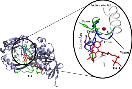
Figure 2.2Schematic representation of the “active site lid” of PaDADH.1
The three dimensional structure of the enzyme is shown in ice blue cartoon display. The four loops (L1-L4) are shown in green. The FAD and Iminoarginine are shown in red and purple sticks respectively. The open and close conformation of the “active site lid” are magnified and colored green and blue respectively. Tyr53 serves as the key amino acid residue of the gate which regulates substrate binding and product release. The water molecule is shown as a red sphere.
Fu and Yuan et al1-3 appointed loop L1 of PaDADH as an “active site lid.” The lid served as a controlling factor for substrate entry and product release of the enzyme. Similar findings concerning other flavin dependent enzymes were also reported in regards to the catalytic involvement of an “active site lid”. For example, a loop region established as an “active site lid”
was proposed in the structure of porcine D-amino acid oxidase (pDAAO).1 The 1.06 Å resolution X-ray crystal structure of PaDADH was reported to have two conformations, in which case, the ligand-free and product-bound configurations were denoted in accordance to the open and close state of its amino acid residue Tyrosine 53 (Tyr53). It is important to note that Tyr53 is also part
L3 L4 L2 L1 Open Close Water Tyr53
Active site lid
of loop L1, which is designated as the “active site lid” of the enzyme. This suggested a critical role for Tyr53 as a gated component in regards to enzyme catalysis. Tyr53 undergoes an open and close conformation which regulates the gated mechanism of PaDADH in respect to substrate binding and product release.
Typically, catalysis involves an enzyme binding to its corresponding substrate of high specificity. However, PaDADH, has a catalytic efficiency for every amino acid except for D-glutamate, D-aspartate, and glycine (achiral). Furthermore, the enzyme has been shown to not bind to L-arginine due to stereochemistry. As a result, the preference for catalysis is solely based on the configuration of D-amino acids. Experimental analyses were carried out to gain further insight on PaDADH’s broad substrate specificity. PaDADH has a proposed catalytic site in
Table 2.1 Steady-state kinetics study of PaDADH with D-amino acids.1
aThese measurements were taken at 25oC, pH 8.7, 1mM PMS in 20mM Tris-HCl. bK
m and kcat values were not recorded due to the inability to saturate the enzyme with the substrate. cKinetics with cysteine was not shown due to its reduction with PMS (phenazine methosulfate). D-leucine was the last ligand with three established kinetic parameters (kcat/Km, kcat, Km).
iminohistidine is suggested to be catalytically competent due to its interactions with Glu87 and the FAD. However, conformation B of iminohistidine is proposed as being incompetent since the critical interactions with Glu87 and the FAD are lost.1
According to Fu et al1, PaDADH’s broad substrate specificity with different D-amino acids may be due to its preference for positively charged side chain ligands. For instance, amino acid residue glutamate 87 of PaDADH (Glu87) was proposed to form strong electrostatic interactions with the substrates D-arginine and D-lysine, hence their high catalytic efficiency with PaDADH during steady-state kinetics analysis.
The pocket entry of the active site of PaDADH is small yet increases in size as it approaches the FAD cofactor.1 Since the 1.06 Å resolution X-ray crystal structure of PaDADH showed both ligand-free and product-bound conformations, the four designated loops (L1- L4), more specifically the “active site lid” (L1, residues 50 – 56) may be involved in the broad
substrate specificity displayed by this enzyme. Thus, an additional suggestion on PaDADH’s broad substrate specificity would be its ability to regulate the entrance and release of substrates at different kinetics based on the fluctuation of its “active site lid”. The same approach could be taken in terms of binding, in which case, the stability of the “active site lid” in the close conformation would reflect PaDAH’s ability to accommodate a variety of substrates.
A similar “loop-and-lid” structure within other glucose-methanol-choline (GMC) family
a rmsd of 2.4 Å for 270 alpha carbons in relative to three-dimensional structure comparisons.41 Thus, by gaining further insight on the catalytic properties of PaDADH, feasible hypotheses may be developed for other members of the GMC family.
Figure 2.3 Distance betweenTyr53 (CZ) and FAD (N5) with water recognition motif.1
The water molecule is shown as a red sphere. The open and close conformations are colored green and blue respectively. In addition, the distances between the zeta carbon (CZ) of Tyr53 and N5 atom of FAD in the open and close conformations are labeled accordingly.
Root-mean-square deviation (RMSD) evaluations were calculated to investigate the fluctuation of PaDADH and its cofactor in comparison to their frame of reference at the beginning of the trajectory. It was a necessity to show a localized FAD structure since its N5 atom was chosen as an arbitrary point to measure the change in distance and conformation of Tyr53 over time. Distance analyses between the FAD N5 atom and the zeta carbon (CZ) of Tyr53 were made apparent to investigate the open (A) and close (B) states of PaDADH (PDB ID 3NYC). Following such results, the probability distribution in regards to the presence and
FAD
Open
Close
Tyr53
N5
14.32 Å
absence of water was plotted in relation to the A and B conformational states of Tyr53 in PaDADH.
2.2 Experimental Procedures
Both PaDADH wild type free in solution and in complex with its D-arginine were constructed from an X-ray crystal structure of the enzyme with a resolution of 1.06 Å (Protein Data Bank (PDB) identification no. 3NYC).1 D-arginine’s partial charges were obtained with the
application of the two-step RESP method45 and Gaussian0346 electrostatic potentials.
AmberTools xleap software was used to construct the appropriate system required for each MD
simulation of PaDADH. The FAD parameters originated from the Gaff amber force field.
Amber14 simulation package47 was used to carry out all simulations with modified force
field parameters of Cornell et al. (1995)48 by Perez et al. (2007),49 The solvation of each system
was defined by a 10 Å TIP3P explicit water model in octahedron box. Once solvated, each system was neutralized accordingly with sodium ions. Both systems were minimized for a 1000
steps with a 100 kcal/mol/Å in harmonic constraints. Then, the systems were equilibrated in three steps at a constant pressure and temperature of 1 bar and 300K respectively. During the
three equilibrated steps, the harmonic constraints were reduced in decreasing order of 50
kcal/mol/Å and 25 kcal/mol/Å, and 0 kcal/mol/Å. Both minimization and equilibration constraints were only applied on the enzyme, its cofactor (FAD), and the substrate when in
complex with PaDADH. The first equilibrated step was carried out for 0.1 nsec with a time step
of 2 fsec. The last two equilibrated steps were separately simulated for 0.2 nsec with also a 2 fsec
time step.
Temperature regulation (300K) was carried out by using the Langevin thermostat with a
collision frequency of 1 psec-1. A 2 fsec time step was established to calculate Newton’s equation of motion. The particle mesh Ewald (PME) method50 was applied to evaluate the effects of
atoms was taking into account by the SHAKE algorithm.51 As a result, each system was
simulated for a total of 1.1 µsec because the equilibration was extended for the first 100 nsec of the run.
Root mean square deviation of DADH was solely evaluated in respect to the backbone heavy atoms of each residue within PaDADH. RMSD of FAD was targeted towards the flavin component, in which case, the N1, N5, and C9 atoms were taken into consideration. This approach was plausible due to the planarity of the flavin component in regards to the N1, N5, and C9 atom positions. Amber14 package47 was used for all distance analyses and to record the residue number of the water molecule that was closest to the water recognition motif in each frame. Then, water molecules that were located 3.5 Å away or less to either two of the carbonyl backbone of Thr50, Val51, and Tyr53 of the motif were taken into account. This particular
strategy was plausible due to the position of the localized water molecule within the water
recognition motif from the X-ray crystal structure of PaDADH and throughout the restart files of
the two simulations. Whenever a water molecule was considered buried within the water
recognition motif, it was in close interaction with either two of the backbone carbonyls of Val51,
2.3 Results & Discussions
[image:44.612.98.506.134.437.2]2.3.1 PaDADH free in solution
Figure 2.4 Dynamics analyses of PaDADH free in solution.
Panel A and B represent the RMSD assessment of PaDADH and FAD respectively. Panel C displays the distance between the Zeta carbon of Tyr53 and N5 of FAD over time. Panel D illustrates the probability distribution of the distance between the zeta carbon of Tyr53 and the N5 atom of FAD.
The root-mean-square deviation (RMSD) of PaDADH and its FAD cofactor in relative to their frame of reference at 0.1 µs indicate that both structures were relatively localized throughout the trajectory. The distances between the PaDADH Tyr53 zeta carbon and the N5 atom of FAD were established at the open and close conformations to be 14.32 Å, and 6.93 Å respectively. The fluctuation in distance between these two atoms over time (Figure 2.4, panel C) indicate that Tyr53 was indeed undergoing two conformational states while the rest of the
C
D
A
B
Time (µs)
Time (µs)
Time (µs)
D
ista
nce
(Å)
RM
SD (
Å)
RM
SD (
Å)
d, Distance (Å)
P
(
enzyme and FAD were low in mobility. Thus, the gated mechanism of PaDADH was supported by the MD simulation of the enzyme free in solution, in which case, ligand-free and product-bound conformations of the “active site lid” were occurring stochastically. The establishment of additional close and open populations in the probability distributions of PaDADH free in solution was due to the translational motion of the “active site lid” throughout the trajectory. According to the probability distribution analysis of PaDADH free in solution, an interesting characteristic of the enzyme was its ability to easily exist between open and close populations of its gated mechanism throughout the simulation.
It is plausible to suggest this particular enzyme to be one of the machineries of
Figure 2.5 Distribution of buried water molecules in the water recognition motif.
The probability distribution of the distance between the zeta carbon (CZ) of Tyr53 in PaDADH and the N5 atom of FAD is illustrated in black. The probability distribution of the presence and absence of water in respect to the distance between PaDADH Tyr53 (CZ) and FAD (N5) is shown in blue and green respectively.
While PaDADH was free in solution, water was established to be buried within the water recognition motif at a percentage of 88%. Under the same conditions, water was defined absent from the enzyme’s water recognition motif pocket 12% of the time. In other words, during the
simulation, water molecules were consistently exchanging within the vacant pocket of the water recognition motif of PaDADH. Even though the percentage of absent water molecules was minimal in comparison to the percentage of water presence, distinct enzymatic characteristics of PaDADH became evident whenever water was not embedded in the motif’s pocket during the simulation. According to Figure 2.5, when water was concluded buried within the water recognition motif, a probability distribution very similar in magnitude and shape to the
d, Distance (Å)
P(d)
probability distribution of the gated mechanism of PaDADH free in solution was established. However, during the period of water absence from the motif’s pocket, the probability distribution of the populations of the gated mechanism increase in intensity in relative to each other. Since Gibb’s free energy is equivalent to the gas constant multiplied by temperature and the natural log
[image:47.612.78.540.239.533.2]of probability, increases in the probability distribution peaks correlate with higher energy barriers between the observed gated populations of PaDADH.
Figure 2.6 Schematic representation of the absence and presence of water.
A 15mer peptide was selected to illustrate the water recognition motif in purple color. Water (not labeled) is represented as red spheres, and PaDADH Tyr53 is shown as sticks, in which case, its open and close conformations were established.
The water recognition motif is the “active site lid” that is involved in the gated mechanism of PaDADH. As previously established, this gate can fluctuate stochastically between the open and close conformations of the enzyme over time. In addition, water within this “active site lid” can be buried or considered absent in both conformations. The key was to
XH2O
H2O Close
Open
Fast Slow
XH2O H2O
Close
establish any given trend(s) between buried water molecules and “active site lid” conformations. Consequently, the analyses of embedded water molecules in the motif pocket during the simulation were evaluated to decrease the energy barriers between the gated populations of PaDADH. Thus, buried water molecules in the water recognition motif over time facilitated the change in conformations of the “active site lid”. However, whenever water molecules were not included in the motif pocket PaDADH underwent a much slower fluctuation in its gated mechanism.
2.3.2 PaDADH in complex with D-arginine
Figure 2.7 Dynamics analyses of PaDADH in complex with D-arginine.
RMSD calculation of PaDADH and FAD are shown in panel A and B respectively. Panel C indicates the probability distribution of the distance between Tyr53 (CZ) and FAD (N5) of PaDADH free in solution and in complex with D-arginine. Panel D shows the distribution of the absence and presence of buried water molecules within the water recognition motif during the conformational changes of Tyr53 while the enzyme is in its close state.
D
C
Time (µs)
Time (µs)
A
B
d, Distance (Å)
d, Distance (Å)
RM
SD (
Å)
RM
SD (
Å)
P
(
d)
P
(
d)
CZ-N5
Present
Absent
Free
RMSD calculations of PaDADH in complex with D-arginine showed localization of the enzyme and its FAD cofactor. Using the same analytical approach of PaDADH free in solution, the probability distribution of the gated mechanism of the enzyme in complex with D-arginine indicated a shift towards its close configurated populations.
2.4 Conclusions
As established with PaDADH free in solution, ligand-free and product-bound conformations of the “active site lid” were occurring via a stochastic distribution over time. The presence of water within the vacant pocket of the water recognition motif facilitated the open and close configurations of the gated mechanism of PaDADH, while the absence of water increased the energy barriers between the gated populations of the enzyme. PaDADH in complex with D-arginine showed a preference of the gated mechanism of the enzyme towards its close conformations. The close gated configuration of the enzyme that was suggested to be catalytically competent showed a preference for water absence in the motif’s pocket, while water
3 PADADH Y53F, S45A, AND A46G MUTANT DYNAMICS
3.1 Introduction
PaDADH is a flavin-dependent enzyme that catalyzes the oxidative deamination of different D-amino acids into their corresponding imino acids. The zwitterionic forms of its substrates are suggested to be the preferred protonation state for ligand-enzyme binding. PaDADH is suggested to deprotonate the backbone amino groups of its various D-amino acid substrates, which in turn causes electrons to delocalize and create a hydride transfer from their alpha carbons to the N5 atom of the FAD cofactor. This reaction is hypothesized to be triggered by a basic catalytic amino acid residue in the enzyme’s active site. A similar reaction is seen with choline oxidase where a hydrogen and proton transfer are proposed to coincide in catalysis.1-3,5 Furthermore, a suggestion concerning the concurrence of a proton and hydride transfer using FAD as a cofactor was proposed from the X-ray crystal structure of D-amino acid oxidase in
Rhodotorula gracilis.52
Scheme 3.1 Enzymatic mechanism of PaDADH.
R COO H N H H H
+
FAD
FADH
2Base
R COO
N
H H
+
D-Amino Acid Imino Acid
Base
pH effects on the kinetic parameters of PaDADH suggested that an unprotonated group with a pKa of 9.6 was the basic catalytic amino acid residue which triggered electron delocalization within the enzyme.5 Such pKa value became apparent to either the backbone amino groups of PaDADH D-amino acid substrates (pKa ~ 9) or a tyrosine side chain residue of the enzyme (pKa ~ 10). The hydroxyl group of Tyr53 from the “active site lid” of PaDADH was observed to be 3.74 Å away from the backbone nitrogen of iminoarginine in the product-bound conformation.5 Thus, PaDADH Tyr53, in close proximity for acid-base chemistry, was proposed to be the catalytic base, which triggered the oxidative deamination reaction of the enzyme.
Table 3.1 Kinetic study of PaDADH Y53F mutant variant.53
As a consequence, a single amino acid mutation of PaDADH was carried out, in which case, Tyr53 was mutated into a phenylalanine (Y53F).53 The goal of the experiment was to render the enzyme nonfunctional since the oxidative deamination reaction would no longer be triggered due to the absence of the hydroxyl group of tyr53. However, PaDADH remained functional with a significant decrease in efficiency.
region (Ser45 – Ala47) remained conserved in other FAD-dependent enzymes.1 Furthermore, members of the glucose-methanol-choline (GMC) family, like PaDADH, shared a similar Ser45 – Ala47 sequence near the si face of the FAD flavin component, in which case, bulkier and
smaller groups within this sequence were suggested to correspond to an enzyme with low and high reactivity with oxygen respectively. For example, the reduced FAD of L-galactono- γ-lactone dehydrogenase increased in reactivity with oxygen by approximately 400-fold during a
single amino acid mutation from Alanine 113 to Glycine.1
Table 3.3 Kinetic study of PaDADH A46G mutant variant.58
Consequently, a similar approach was taken into consideration in regards to the structural stability and enzymatic properties of PaDADH.58 For example, a single amino acid mutation of Ser45 to alanine (S45A) was carried out in hopes of increasing reactivity of the reduced FAD cofactor of the enzyme with oxygen. As a result, PaDADH reactivity with oxygen remained minimal while the enzyme became significantly less efficient catalytically with different substrates. In addition, assessable kinetic parameters of D-leucine from wild type PaDADH could no longer be measured in the S45A mutant variant of the enzyme. Following such approach, PaDADH Alanine 46 was mutated to Glycine (A46G) to increase the reactivity of the enzyme’s reduced FAD with oxygen. Subsequently, PaDADH A46G mutant variant had a
Figure 3.1 Distance betweenTyr53 (CZ) and FAD (N5) with site of mutations.1
Ala46 and Ser45 are located on the si face of the FAD flavin component with Tyr53 positioned on the re face of the structure. The open and close conformations are colored orange and black respectively with their corresponding distances labeled.
In all three cases, as indicated from the kinetic analyses, PaDADH mutant variants remained functional but decreased in catalytic efficiency. All three mutation sites were part of the flexible loop (L1), which included the “active site lid” of the enzyme. MD simulation results of wild type PaDADH free in solution supported a stochastic distribution of both ligand-free and product-bound configurations of the enzyme over time. Thus, carrying MD simulations of single amino acid mutations (Y53F, S45A, and A46F) of PaDADH free in solution would indicate the effect of dynamics on the mechanical properties of the enzyme.
Following each MD simulation, both FAD and apoenzyme RMSDs were evaluated to investigate the fluctuation of PaDADH and its cofactor over time. In addition, a more localized
Tyr53 Open
Close Ala46
Ser45
re face si face
N5
14.32 Å
6.93
Å
3.2 Experimental Procedures
The Y53F, S45A, and A46G mutant variants of PaDADH were derived from a 1.06 Å resolution X-ray crystal structure of the enzyme (PDB ID 3NYC).1 All MD simulated variant
system of PaDADH were constructed via AmberTools xleap software. Gaff amber force field
parameters were used to derive the parameters for the FAD structure.47
All simulations were carried out using amber14 softwares47 under the same force field parameters.48,49 Each system was properly solvated with a 10 Å TIP3P water model octahedron
box, in which case, sodium ions were subsequently added for neutralization. All simulations were minimized for a 1000 steps with harmonic constraints of a 100 kcal/mol/Å on PaDADH and FAD. Following minimization, all systems underwent three rounds of equilibration with
harmonic constraints in decreasing order of 50 kcal/mol/Å, 25 kcal/mol/Å, and 0 kcal/mol/Å on the enzyme and its cofactor. All equilibrations were processed at a constant temperature, and
pressure of 300K, and 1bar respectively. Each equilibration was parameterized with a time step
of 2 fsec, however, the first equilibrated round was carried out for 0.1 nsec, while the last two
rounds were individually processed for 0.2 nsec.
Langevin thermostat with a collision frequency of 1 psec-1 was the applied method to regulate temperature. Newton’s equation of motion was consistently evaluated through a time step of
3.3 Results & Discussions
3.3.1 Y53F
Figure 3.2 Dynamics analyses of PaDADH Y53F mutant variant.
Panel A and B indicate the RMSD results for Y53F PaDADH and FAD respectively. The distance between PaDADH Tyr53 (CZ) and FAD (N5) over time is shown in panel C. The probability distributions of the distance concerning Tyr53 (CZ) – FAD (N5) in respect to both the wild type (WT) PaDADH and the Y53F mutant variant of the enzyme are shown in panel D.
As seen from the wild type enzyme free in solution and in complex with D-arginine, the PaDADH Y53F mutant variant and its FAD cofactor were relatively low in mobility. As established from previous distance approaches, the initial distance between the CZ atom of Tyr53 and the N5 atom of FAD were measured from the PDB structure of PaDADH in complex with iminoarginine (PDB ID 3NYC).1 Based on the fluctuation in distance between these two atoms, the Y53F mutant variant of the enzyme indicated a tendency of the “active site lid” to
Time (µs)
Time (µs)
D
ista
nce
(Å)
RM
SD (
Å)
RM
SD (
Å)
d, Distance (Å)
P
(
d)
Time (µs)
A
B
WT
Y53F
3.3.2 S45A
Figure 3.3 Dynamics analyses of PaDADH S45A mutant variant.
RMSD evaluations of S45A PaDADH and FAD are established in panel A and B respectively. Panel C indicates the change in distance between the zeta carbon (CZ) of Tyr53 and N5 atom of FAD over time. Panel D shows the probability distribution of the Tyr53 (CZ) – FAD (N5) distance in respect to the wild type (WT) and the S45A mutant variant of the enzyme.
This was another case of high localization of PaDADH S45A mutant variant and its FAD cofactor. Tyr53 (CZ) – FAD (N5) distance analysis of S45A PaDADH indicated a preference of the “active site lid” to its open conformation over time (panel C). In addition, comparison of probability distributions involving wild type and S45A PaDADH free in solution (panel D), established a shift towards the ligand-free configuration in the S45A mutation of the enzyme. Similar to the dynamics of the Y53F mutant variant of PaDADH, the gated mechanism of the enzyme was mechanically affected when Serine 45 was mutated into an alanine. The S45A mutant variant of PaDADH did not significantly increase in reactivity with oxygen, however, the
Time (µs)
Time (µs)
Time (µs)
RM
SD (
Å)
RMSD
(Å)
A
B
D
ista
nce
(Å)
P
(
d)
d, Distance (Å)
WT
S45A
3.3.3 A46G
Figure 3.4 Dynamics analyses of PaDADH A46G mutant variant.
Panel A and B refers to the RMSD calculations of A46G PaDADH and FAD respectively. The change in distance between Tyr53 (CZ) and FAD (N5) over time is established in panel C. Panel D illustrates the probability distribution of the distance between the zeta carbon (CZ) of Tyr53 and the N5 atom of FAD in regards to the wild type and the A46G mutant variant of the enzyme. The A46G mutant variant of PaDADH, including its FAD cofactor displayed high localization properties from the RMSD assessments. Mutating PaDADH Alanine 46 to glycine resulted into an “active site lid” that was predominately open over time (panel C). Similar to previous single amino acid mutations of the enzyme, certain mechanical aspects of the “active site lid” were altered in A46G PaDADH. In addition, the kinetic measurements (kcat, Km, kcat/Km) of the A46G mutant variant of PaDADH indicated a decrease in efficiency. Comparison of S45A and A46G results showed noticeable convergences between kinetic and dynamics analyses of the enzyme. For instance, the “active site lid” of A46G had a probability distribution that was more
WT
A46G
RM
SD (
Å)
RM
SD (
Å)
Time (µs)
Time (µs)
Time (µs)
d, Distance (Å)
P
(
d)
D
ista
nce
(Å)
A
B
3.4 Conclusions
4 PADADH TYR53, AND GLU246 PKA SHIFT CALCULATIONS
4.1 Introduction
Bacteria such as Pseudomonas aeruginosa have the ability to grow solely with D-arginine as a carbon and nitrogen source.42,54 For example, Pseudomonas aeruginosa uses DADH in a two-enzyme-coupled system to catalyze the D- to L- conversion of amino acids. PaDADH converts D-arginine into ketoarginine and ammonia, and PaLADH catalyzes the process of 2-ketoarginine to L-arginine.1-3,42
After its oxidative deamination reaction, PaDADH can either form an enzyme-imino acid complex or release the product into its aqueous environment. Thus, hydrolysis of the imino acid can either occur by water molecules within the enzyme’s active site or in the surrounding environment. Previous crystallography reports have shown the X-ray crystal structure of PaDADH in complex with iminoarginine1,3, however, an unexpected 1.07 Å resolution crystal structure of a covalent N(5) flavin adduct was discovered in the active site of the enzyme after crystallization with D-leucine.2 This PaDADH acyl adduct complex was replicated by photoreduction of the enzyme in the presence of ketoleucine. The acyl adduct structure within the active site of PaDADH was suggested to be the product of a nucleophilic substitution reaction, in which case, the N(5) atom of the flavin component of the FAD cofactor was reduced
by the X-ray beam from the crystallization process, and formed a covalent bond with the
-carbon of ketoleucine to generate 4-methly-2-pentanone-FAD and CO2.
Further studies on another flavin-dependent enzymatic structure of the GMC family,