Vol. 26 No. 3 Genes variation of murMN operon in S. pneumoniae:- Kumari N, et al. 97
97
Original Article
Variation of Sequence of Genes Encoding the MurMN
Operon and Cell Wall Composition in Streptococcus
pneumoniae Strains of Different Susceptibility Levels
to Penicillin
Navindra Kumari, Ph.D.*,
Mohd Yasim Yusof, MBBS*,
Siok Yan Ong, MBBS**,
Marzida Bt. Mansor, MBBS**,
Cheng Foh Le, B.Sc.*,
Shamala Devi Sekaran, Ph.D.*
*Department of Medical Microbiology, Faculty of Medicine, University of Malaya 50603 Kuala Lumpur, Malaysia. **Department of Anaesthesiology, Faculty of Medicine, University of Malaya 50603 Kuala Lumpur, Malaysia. Received for publication: June 8, 2009.
Reprint request: Shamala Devi Sekaran, Ph.D., Department of Medical Microbiology, Faculty of Medicine, University of Malaya 50603 Kuala Lumpur, Malaysia.
Email address: shamalamy@yahoo.com
Keywords:Streptococcus pneumonia, muropeptides, murM variants, β-lactam resistance, penicillin-binding proteins. ABSTRACT
It has been reported that there are structural differences in the muropeptides of the cell wall in penicillin-resistant Streptococcus pneumoniae. The cell wall composition and variation of the murMN operon sequence of S. pneumoniae strains with different penicillin susceptibilities were investigated. PCR amplification and sequencing of the murM and murN genes were carried out on three selected strains of S. pneumoniae. The cell wall was then extracted and elucidated using Fourier Transfer InfraRed (FTIR) Spectroscopy, followed by proton Nuclear Magnetic Resonance (NMR) Spectroscopy. The sequences of the murM and murN genes were shown to be highly conserved while FTIR and NMR analysis suggested a branching structure of the cell wall and also the presence of ethanolamine in the resistant strain. The variations in murM and murN genes may have caused modifications in the cell wall structure leading to decreased binding capacity of penicillins and other β-lactam drugs. (J Infect Dis Antimicrob Agents 2009;26:97-108.)
INTRODUCTION
Penicillin resistance in Streptococcus pneu-moniae is due to altered penicillin-binding proteins
(PBPs), which are essential in cell wall synthesis.1-4
-lactam-resistant isolates from South Africa.4 Penicillin
resistance has been documented in many parts of the world. International Surveillance studies by the Asian Network for Surveillance of Resistant Pathogens (ANSORP) have reported varying levels of penicillin resistance in S. pneumoniae: Vietnam (71.4%), Korea (54.0%), Hong Kong (43.2%), Taiwan (38.6%), China (23.4%), Sri Lanka (14.3%), Thailand (26.9%), Singapore (17.1%), Malaysia (29.5%), and Saudi Arabia (10.3%).5 In the United States, the SENTRY
surveillance program documented penicillin-resistant S. pneumoniae in 20 percent of clinical isolates causing bloodstream infections in North America.6 In Europe,
the circulated Spanish 23F penicillin-resistant clone was mainly in Spain and Hungary in the 1980s, however dissemination of this clone to other parts of the world was reported in the 1990s with the highest prevalence rate of penicillin resistance being documented in France (57%) and Spain (40%).7
PBPs are active-site serine peptidases which catalyze the polymerization as well as cross-linking of peptidoglycan precursors in the assembly of bacterial cell walls.8,9 The PBPs of resistant strains have been
shown to have reduced affinities and or binding capacities to β-lactam drugs. It has also been reported that cell wall peptidoglycans of resistant strains have abnormal chemical compositions, indicating differences in the proportion of branched muropeptides.10 The
presence of branched muropeptides, carrying an alanyl-serine or alanyl-alanine substituent on the lysine epsilon amino group of the stem peptide residues has been shown to be higher in resistant strains than in susceptible strains.10 This finding was also demonstrated by other
researchers from South Africa, Hungary, and the Czech Republic.10, 11
Recently, the identification of the murMN operon has shed some light into the mechanisms of
synthesis of muropeptides and the physiological role of branched peptides. Previous studies have shown that inactivation of the murMN operon causes the production of cell wall peptidoglycans composed of linear muropeptides and also causes the complete loss of the resistant phenotype in penicillin-resistant strains.12 It has also been shown that several
penicillin-resistant isolates carry murM genes with unique polymorphic regions.13 The aim of this study
was to analyze the murMN operon from three Malaysian S. pneumoniae strains with varying susceptibilities to penicillin in order to determine any sequence divergence within these strains and its correlation to penicillin resistance. Another aim was to study the variation that may have occurred in the cell wall of these strains using Fourier Transfer Infrared (FTIR) Spectroscopy and Proton Nuclear Magnetic Resonance (NMR) techniques.
MATERIALS AND METHODS
Bacterial strains
Three pneumococcal isolates of different susceptibility levels to penicillin were obtained from clinical specimens processed in the Microbiology Laboratory of University Malaya Medical Centre (UMMC), Kuala Lumpur, Malaysia. The three strains were strain S676 (penicillin-susceptible), strain I81 (penicillin-intermediately susceptible, and strain R98 (penicillin-resistant). Bacterial cultures isolated from these clinical specimens were stored in brain heart infusion broth supplemented with 10 percent glycerol at -80°C without antibiotics until needed. Each stock culture was kept in triplicates to avoid multiple passaging.
Strain identification
Vol. 26 No. 3 Genes variation of murMN operon in S. pneumoniae:- Kumari N, et al. 99
99 susceptibility to ethylhydrocupreine disc (optochin), whereby all the strains showed a diameter measurement of ≥ 14 mm.14 The strains were also shown to be
positive by bile solubility testing and to be catalase negative.
Susceptibility testing
Antibiotic susceptibility of the strains was tested on Mueller Hinton Agar (Oxoid) plates containing 5 percent sheep blood (Oxoid), incubated at 37°C with 5 percent CO2 using the agar dilution method described
by the Clinical and Laboratory Standards Institute.14
The antimicrobial agents used were penicillin, cefotaxime, and ceftriaxone, obtained from Sigma Aldrich (Sigma Chemical Co., St. Louis, Mo., USA). S. pneumoniae ATCC 49619 was used as control.
DNA extraction and PCR amplification of murMN
operon
Genomic DNA was extracted from pure bacterial cultures using a previously described method.15 Bacterial colonies suspended in 15 μL of
H2O containing 50 mg/mL lysostaphin (Sigma Chemical Co., St. Louis, Mo) were incubated at 37°C for 10 minutes. This was followed by addition of 10
μg/mL proteinase K and 0.1 mM Tris HCL pH 7.5 and incubated at 37°C for another 10 minutes. Subsequently, the suspension was boiled for 5 minutes and finally centrifuged at 13,000 rpm for 2 minutes. The supernatant obtained acts as the template in the PCR reaction.
The primers used in this study were as follows: murM (Forward): 5’ – CTG GAG GAAA GAGA GTAG GA -3’ murM (Reverse) : 5’ – CTC TTC TTT CGT GAG TGT AG -3’ murN (Forward) : 5’ – GAC TTG CTC TTG ATTT CCGT -3’ murN (Reverse) : 5’ – TGT CTC TCC ACC TTT CTA GC -3’
The optimal PCR condition for a 50 μL reaction included 1X PCR buffer, 1.5 mM MgCl2 0.2 mM dNTP
mix, 2 U Taq polymerase (Fermentas), and 20 pmol of each primer. The PCR cycling parameters were as follows: an initial denaturation step at 95°C for 5 minutes, 30 cycles of amplification performed as follows: denaturation at 94°C for 5 minutes, annealing temperature at 40°C for 2 minutes and extension temperature at 72°C for 3 minutes and finally completed with an extension at 72°C for 5 minutes. PCR products were purified using the PCR purification kit (Qiagen) and PCR DNA sequencing was carried out using an automated DNA sequencer (an ABI Prism 377 DNA sequencer, Perkin Elmer ABI).
Cell wall preparation
Pneumococcal cell walls were prepared using a previously published procedure with modifications.16
suspended in water and lyophilized.
Fourier transform infrared spectroscopy (FTIR
spectroscopy)
A small amount of bacterial cell wall was placed on a stainless steel plate and spread evenly using the straight edge of a teflon sheet. The evaporated sample was then placed on a potassium bromide window of spectrometer (Bruker, model IFS 66 v/s). The spectrometer was coupled to an infrared microscope equipped with grazing angle reflectance (GAR) objective of 15X magnification and a mercury-cadmium-tellurite detector and potassium bromide beamsplitter. Bacterial cell wall spectra were collected using the OPUS, version 4.2, Bruker software, over a range of 4,000-400 wavenumber (cm-1) by averaging
50 scans with a resolution of 4 cm-1 and absorbance
mode. A background spectrum was obtained before each measurement using a clean stainless steel plate at the same instrumental conditions used for the experiment data acquisition.
NMR spectroscopy
One gram of the lyophilized form of the bacterial cell wall was dissolved in deuterium oxide. The same solvent was used to acquire a proton NMR spectrum. Chemical shifts were established by assigning 4.65 ppm to the resonance from the proton of monodeuterated water.
RESULTS
Sequence analysis of the murM and murN genes
The entire coding region of the murM gene was sequenced from three strains of varying susceptibility levels to penicillin. The highest penicillin minimal inhibitory concentration (MIC) value was 4 μg/mL (R98) representing a penicillin-resistant strain, while
the penicillin-intermediate (I81) and -susceptible strains (S676) had MIC values of 1.0 μg/mL and 0.032 μg/ mL, respectively (Table 1). The sequencing of the murM gene yielded nucleotide sequence homology to different murM variant alleles that have been characterized previously.17,18 The murM gene of the
penicillin-susceptible (S676) and -intermediate (I81) strains was homologous to eight published murM variant alleles (murMB1, murMB3, murMB4, murMB5, murMB6, murMB7, murMB10, and murMB11) (Gene Bank Ascension Numbers: AF281135, AF281136.1, AF281137.1, AF281138.2, AF281139.1, AF281140, DQ100160.1, and DQ100161.1). These alleles were of the strain Pen6 origin (a genetic transformant constructed with the highly penicillin-resistant South African strain 8249 as a DNA donor), which is a resistant phenotype. The penicillin-intermediate strain had a significant level of divergence (29% homology) in the nucleotide sequence to the murM variant alleles murMB10 and murMB11. Interestingly, the resistant strain had an additional murM allele, murMB9 with a homology of 85 percent (Table 1). This variant allele was not detected in the susceptible and intermediate strains. Table 1 also shows that the intermediate and resistant strains had a higher percentage of homology to the alleles (murMB1, murMB3, murMB4, murMB5, murMB6, murMB7, murMB10, murMB11, and murMB9 (only in the resistant strain), as compared to the penicillin-susceptible strain. The murM gene sequence from our isolates had a higher percentage of homology when compared to published sequences of strains R36A (93-99%), while the murN gene showed a higher percentage of homology to both R36A (∼99%) and Pen6 (∼98%).
Variation of cell wall molecular vibration by FTIR
Vol. 26 No. 3
101
Genes variation of
murMN
operon in
S. pneumoniae
:- Kumari N, et al.
[image:5.595.57.752.183.355.2]
101
Table 1. Nucleotide sequence homology of the murM gene.
MIC S676: penicillin-susceptible strain, I81: penicillin-intermediate strain, R98: penicillin-resistant strain
1Breakpoints recommended by the Clinical and Labolatory Standards Institute guidelines, PEN (S : ≤ 0.06, I : 0.12-1.0,and R: ≥ 2.0 mg/L), CRO (S: ≤ 0.5, I :1.0,
and R: ≥ 2.0 mg/L),
CTX (S: ≤ 0.5, I : 1.0, and R: ≥ 2.0 mg/L)
NS: no similarities, PEN: penicillin, CTX: cefotaxime, CRO: ceftriaxone n
i a r t S
C I M
) L / g m ( 1
M r u
m variants ) s e i t i r a l i m i s f o % (
N E
P CTX CRO M 1B M 3B M 4B M 5B M 6B M 7B M 9B MB10 MB11
6 7 6
S 0.032 <0.032 0.064 78 79 89 84 89 87 NS 89 91
1 8
I 1.0 2.0 1.0 97 98 98 98 98 98 NS 29 29
8 9
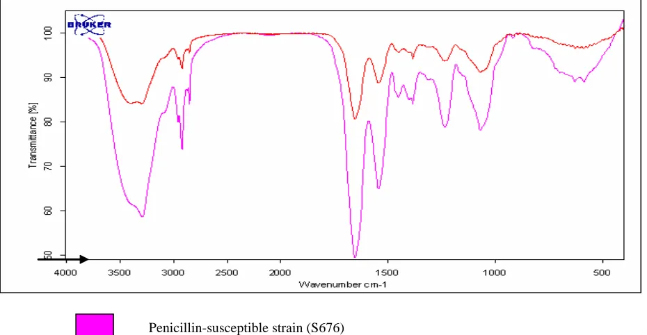
Using the FTIR technique, visually, the spectra obtained from the molecular vibration of the susceptible (S676) and resistant strain (R98) was similar (Figure 1). This indicates that the composition of the cell wall of both strains is similar in chemical properties. However, the transmittance (%) of the vibration differed in both strains. The susceptible strains were seen to have a higher transmittance of vibration, as compared to the resistant strain (indicated as an arrow in Figure 1). This suggests some variations in the bonding structure of the chemical properties. The higher transmittance value also suggests a stronger molecular vibration, hence postulating a muropeptide of a larger mass. In the resistant strain, a lower transmittance value was noted suggesting the
muropeptides of the cell wall to be of a smaller mass. This could be due to the branching of the muropeptide chain. Table 2 shows the assignment of functional groups detected from the FTIR technique. A shoulder peak at 3,293-3,997 wave number/cm indicates the water content of the sample. A functional group detected was the aromatic C=C stretch at 3,299.4 wave number/cm, which was present only in the susceptible strain. The other functional compounds detected were the aliphatic iodo compound (C-I) stretch, CH2
methylene stretch, C-F stretch, and alkenyl C=C stretch. The ratio of the absorbance/ transmittance of the sensitive strain against the resistant strain was highest for the alkenyl C=C stretch whereas the aliphatic Iodo compound was almost equal in both strains.
[image:6.595.60.525.406.647.2]Penicillin-resistant strain (R98) Penicillin-susceptible strain (S676)
Vol. 26 No. 3 Genes variation of murMN operon in S. pneumoniae:- Kumari N, et al. 103
103 t n e m n g i s s A ) s p u o r g l a n o i t c n u f ( 6 7 6
S Absorbance g o l -=
A 10T (a)
8 9
R Absorbance g o l -=
A 10T (b)
o i t a R ) b / a (
H2O 3997.1528 0.976 3293.6280 0.586 1.666
o d o i c i t a h p i l A h c t e r t s I -C d n u o p m o c 8 4 1 7 . 7 8
5 0.965 586.6499 0.890 1.084
H
C 2/methylene/C-H
h c t e r t s c i r t e m m y s / c i r t e m m y s a 6 5 1 7 . 1 2 9
2 0.921 2921.1439 0.738 1.248
h c t e r t s F
-C 1070.6980 0.912 1070.8571 0.781 1.168
C = C c i t a m o r A d n u o p m o c c i n e l y t e c a H -C / h c t e r t s 4 0 2 4 . 9 9 2
3 0.841 - -
-l y n e k l A h c t e r t s C = C 8 5 0 9 . 4 5 6
[image:7.595.56.553.120.331.2]1 0.807 1655.2540 0.494 1.634
Table 2. Assignment of the functional groups from the Fourier Transfer Infrared (FTIR) Spectroscopy analysis.
S676: penicillin-susceptible strain, R98: penicillin-resistant strain, T: transmittance, A: absorbance, C: carbon, H: hydrogen, C=C: double bond between 2 carbons, F: fluoride
Variation in the chemical composition of the cell
wall structure using NMR
The spectral regions 0.5 to 5.0 ppm were compared for the resistant and susceptible strain. Typical spectra obtained are shown in Figures 2(a) and 2(b). These spectra show the specific characteristic resonances of the cell wall extracted from strains of penicillin-resistant and -susceptible S. pneumoniae, respectively. Visually, the spectra in Figures 2(a) and 2(b) appear similar but with differences in the intensity and the frequency of the peak. Detailed spectral analysis identified four significant regions, representing different metabolites (Table 3). Regions of chemical shift at 2.06, 1.12, and 0.75 ppm was found in both the susceptible and resistant strain. The presence of the peak at 0.75 ppm is characteristic of the presence of valine, leucine, and isoleucine residues whereas the presence of a peak at 1.12 ppm is characteristic of a methyl group in these amino acid residues which forms long chain fatty acids. The presence of the peak at 2.06 ppm is characteristic of amino acids such as isoleucine, glutamine, glutamate, methionine, polyamine,
and N-acetyl compounds. An additional peak is observed in the cell wall composition of the resistant strain at a region with a chemical shift at 3.89 ppm. This peak is characteristic of amino acids, betaine, glycerol phosphorylcholine, glycerol phosphoethano-lamine, ethanophosphoethano-lamine, glycerol, and glycerol-3-phosphate.
DISCUSSION
The murMN genes exhibit the mosaicsm phenomena, similar to the PBPs in pneumococci18,19,
and are responsible for the variations in the catalytic function of the PBPs.13,20 Previous studies have shown
that the amount of murM alleles indicates branching of the muropeptides.20 The branching of the muropeptides
(a) Strain R98 (penicillin-resistant strain).
(b) Strain S676 (penicillin-susceptible strain).
Vol. 26 No. 3
105
Genes variation of
murMN
operon in
S. pneumoniae
:- Kumari N, et al.
[image:9.595.112.668.201.412.2]
105
Table 3. Chemical shift of the Proton Nuclear Magnetic Resonance (NMR) analysis.
ppm: parts per minute, R98: penicillin-resistant strain, S676: penicillin-susceptible strain
o
N PPM
n i a r t S e c n a n o s e r h t i w s e t i l o b a t e M 8 9
R S676
y t i s n e t n
I Frequency Intensity Frequency
1 3.89 0.01533 1052.521 Not Identified - Amino acid (non-specific), betaine, glycerolphosphorylcholine, , l o r e c y l g , e n i m a l o n a h t e , e n i m a l o n a h t e o h p s o h p l o r e c y l g e t a h p s o h p -3 -l o r e c y l g
2 2.06 0.01559 556.656 0.02282 557.949 Isoleucine,glutamine, glutamate, methionine, polyamine, s d n u o p m u o c l y t e c a -N
3 1.12 0.05882 300.811 0.07196 302.775 Methylgroup in valine,leucine, isoleucine, terminalmethyl . s d i c a y t t a f n i a h c g n o l n i s p u o r g
peptidoglycans during the cell wall formation. In our study, the additional murM allele in the penicillin-resistant strain could be the cause of reduced binding affinity to the drug. The higher divergence of the penicillin-susceptible strain, compared to the murM variant allele might be due to its difference in the genetic background of the strain. The published murMB variant alleles13 that have been
reported are of the strain Pen6 origin, which is a penicillin-resistant strain. However, the function of this allele should be investigated in order to conclude its role in the development of penicillin resistance in S. pneumoniae.
The FTIR analysis showed that the functional group detected at 3,299 wave number/cm, representing the aromatic structure, was only detected in the susceptible strain. These data suggest the lack of branching of the muropeptides in the susceptible strain whereas the absence of an aromatic structure suggests branching of the peptides into smaller structures. The higher transmittance value in the susceptible strain also postulates the presence of molecular vibration of a larger mass as compared to the resistant strain, which had smaller vibrational energy.
Pneumococci require choline residues for the structure of the cell wall. However, it can also be replaced by ethanolamine, which is a component of the pneumococci teichoic acid (muropeptides). Previous studies have shown that S. pneumoniae becomes resistant to autolysis-inducing agents in the presence of ethanolamine.21 This was also observed in our study,
as the additional peak at 3.89 ppm, representing amino acids, betaine, glycerol phosphorylcholine, glycerol phosphoethanolamine, ethanolamine, glycerol, and glycerol-3-phosphate was only observed in the resistant strain. This suggests that the presence of the
ethonolamine component has reduced the autolytic activity of the strain, causing it to be more tolerant to penicillin. The amino acid composition indicates the presence of disaccharide tetrapeptide units covalently linked to teichoic acid chains.
The variation in the nucleotide sequence of the murMN operon, together with the cell wall analysis using FTIR and NMR suggest that the structure and branching of the cell wall may be the cause of reduced affinity of the cell wall to β-lactam drugs. However, the variation caused by the different serotypes of strains may also contribute to the frequency of divergence of the gene. Other variations of the genetic background such as the source of isolation, horizontal transfer of genes, and environmental stress may also have a role to play in the variation of the structure of the cell walls of these strains. Variations occurring at the cell wall may be an adaptive response of the organism towards environmental changes for survival. To further elucidate, and confirm the role of the murM and murN genes more strains need to be sequenced in order to study the distribution and evolution of the murM and murN genes. It would be an added advantage to quantitate the amount of cell wall components in order to investigate further the specific role of these components towards the development of penicillin resistance.
Vol. 26 No. 3 Genes variation of murMN operon in S. pneumoniae:- Kumari N, et al. 107
107 ACKNOWLEDGEMENT
This work was supported by grant provided by the Ministry of Science and Technology, Malaysia: 36-02-03-6027. There are no conflicts of interest.
References
1. Hakenbeck R, Ellerbrok H, Briese T, Handwerger S,
Tomasz A. Penicillin-binding proteins of
penicillin-susceptible and -resistant pneumococci: immunological
relatedness of altered proteins and changes in peptides
carrying the beta-lactam binding site. Antimicrob
Agents Chemother 1986;30:553-8.
2. Hakenbeck R, Tarpay M, Tomasz A. Multiple changes
of penicillin-binding proteins in penicillin-resistant
clinical isolates of Streptococcus pneumoniae.
Antimicrob Agents Chemother 1980;17:364-71.
3. Laible G, Spratt BG, Hakenbeck R. Interspecies
recombinational events during the evolution of altered
PBP 2x genes in penicillin-resistant clinical isolates of
Streptococcus pneumoniae. Mol Microbiol 1991;5:
1993-2002.
4. Zighelboim S, Tomasz A. Penicillin-binding proteins
of multiply antibiotic-resistant South African strains
of Streptococcus pneumoniae. Antimicrob Agents
Chemother 1980;17:434-42.
5. Song JH, Jung SI, Ko KS, et al. High prevalence of
antimicrobial resistance among clinical Streptococcus
pneumoniae isolates in Asia (an ANSORP study).
Antimicrob Agents Chemother 2004;48:2101-7.
6. Biedenbach DJ, Moet GJ, Jones RN. Occurrence and
antimicrobial resistance pattern comparisons among
bloodstream infection isolates from the SENTRY
Antimicrobial Surveillance Program (1997-2002). Diagn
Microbiol Infect Dis 2004;50:59-69.
7. Jacobs MR, Felmingham D, Appelbaum PC, Gruneberg
RN. The Alexander Project 1998-2000: susceptibility
of pathogens isolated from community-acquired
respiratory tract infection to commonly used
anti-microbial agents. J Antimicrob Chemother 2003;52:
229-46.
8. Holtje JV, Schwarz U. Biosynthesis and growth of
the murein sacculus, molecular cytology of the
Escherichia coli. In: Nanninga N, ed. Molecular
Cytology of Escherichia Coli. London: Academic
Press, 1985:77-119.
9. Waxman DJ, Strominger JL. Penicillin-binding
proteins and the mechanism of action of β-lactam
antibiotics. Annu Rev Biochem 1983;52:825-69.
10. Garcia-Bustos J, Tomasz A. A biological price of
antibiotic resistance: major changes in the
peptido-glycan structure of penicillin-resistant pneumococci.
Proc Natl Acad Sci USA 1990;87: 5415-9.
11. Severin A, Tomasz A. Naturally occurring
peptido-glycan variants of Streptococcus pneumoniae. J
Bacteriol 1996;178:168-74.
12. Filipe SR, Tomasz A. Inhibition of the expression of
penicillin resistance in Streptococcus pneumoniae by
inactivation of cell wall muropeptide branching genes.
Proc Natl Acad Sci U S A 2000;97:4891-6.
13. Filipe SR, Severina E, Tomasz A. Distribution of the
mosaic structured murM genes among natural
populations of Streptococcus pneumoniae. J Bacteriol
2000;182:6798-805.
14. Clinical and Laboratory Standards Institute.
Per-formance Standards for Antimicrobial Susceptibility
Testing; 15th Informational Supplement. CLSI
document M100-S15. Wayne, Pa: Clinical and
Laboratory Standards Institute, 2005.
15. Unal S, Hoskins J, Flokowitsch JE, Wu CY, Preston
DA, Skatrud PL. Detection of methicillin-resistant
staphylococci by using the polymerase chain reaction.
J Clin Microbiol 1992;30:1685-91.
16. Severin A, Figueiredo AM, Tomasz A. Separation of
resistance through genetic transformation of
Strep-tococcus pneumoniae. J Bacteriol 1996;178:1788-92.
17. del Campo R, Cafini F, Morosini MI, et al. Combinations
of PBPs and MurM protein variants in early and
contemporary high-level penicillin-resistant
Strep-tococcus pneumoniae isolates in Spain. J Antimicrob
Chemother 2006;57:983-6.
18. Hakenbeck R, Balmelle N, Weber B, Gardes C, Keck W,
de Saizieu A. Mosaic genes and mosaic chromosomes:
intra- and interspecies genomic variation of
Strep-tococcus pneumoniae. Infect Immun 2001;69:2477-86.
19. Hakenbeck R, Coyette J. Resistant penicillin-binding
proteins. Cell Mol Life Sci 1998;54:332-40.
20. Smith AM, Klugman KP. Alterations in MurM, a cell
wall muropeptide branching enzyme, increase high-level
penicillin and cephalosporin resistance in Streptococcus
pneumoniae. Antimicrob Agents Chemother 2001;45:
2393-6.
21. Tomasz A. Biological consequences of the
replace-ment of choline by ethanolamine in the cell wall of
Pneumococcus: chanin formation, loss of
trans-formability, and loss of autolysis. Proc Natl Acad Sci