R E S E A R C H
Open Access
Systems biology meets stress ecology: linking
molecular and organismal stress responses in
Daphnia magna
Lars-Henrik Heckmann
1,2*, Richard M Sibly
1, Richard Connon
1,3, Helen L Hooper
1, Thomas H Hutchinson
4,5,
Steve J Maund
6, Christopher J Hill
1, Anthony Bouetard
1, Amanda Callaghan
1Abstract
Background:Ibuprofen and other nonsteroidal anti-inflammatory drugs have been designed to interrupt
eicosanoid metabolism in mammals, but little is known of how they affect nontarget organisms. Here we report a systems biology study that simultaneously describes the transcriptomic and phenotypic stress responses of the model crustaceanDaphnia magnaafter exposure to ibuprofen.
Results:Our findings reveal intriguing similarities in the mode of action of ibuprofen between vertebrates and invertebrates, and they suggest that ibuprofen has a targeted impact on reproduction at the molecular, organismal, and population level in daphnids. Microarray expression and temporal real-time quantitative PCR profiles of key genes suggest early ibuprofen interruption of crustacean eicosanoid metabolism, which appears to disrupt signal transduction affecting juvenile hormone metabolism and oogenesis.
Conclusion:Combining molecular and organismal stress responses provides a guide to possible chronic consequences of environmental stress for population health. This could improve current environmental risk assessment by providing an early indication of the need for higher tier testing. Our study demonstrates the advantages of a systems approach to stress ecology, in whichDaphniawill probably play a major role.
Background
Organismal stress responses have been studied for decades in ecology and ecotoxicology to establish the factors that limit species distributions and to investigate the effects of anthropogenic activities [1]. It was not until recently, however, that stress responses were inves-tigated at the genomic level to illuminate underlying mechanisms [2,3]. Studying stress responses individually at just one level of biological organization yield little insight into how the organism deals with stress overall, but integration of responses at different levels promotes a holistic understanding of the whole system. Knowledge of the phenotypic consequences of stress as well as the genomic components (for instance, genes) that are induced or suppressed enables us to identify not only the mode of action (MOA) of the stressor but also
which genomic components affect organismal growth, reproduction and survival, and thus populations. So, increased knowledge of the fundamental interactions between genome and phenotype should enable us to predict population stress responses better.
In genomic nonmodel organisms an overview of global transcriptomic responses may be achieved through using, for example, Gene Ontology (GO) [4] and the Kyoto Encyclopedia of Genes and Genomes (KEGG) [5]. KEGG in particular facilitates a pathway-driven approach, which within a toxicogenomic context -allows identification of general molecular stress response as well as highlighting biochemical pathways that are associated with stressor-specific responses. Recently, in stress ecology, a number ofDaphnia magnaStraus [6-8] and other invertebrate [9] microarray reports have been published, but few of these have integrated transcrip-tome and phenotype to an extent that clarifies the link between these biological levels. This may partly be because many environmental and chemical stressors
* Correspondence: lhh@dmu.dk 1
University of Reading, School of Biological Sciences, Environmental Biology, Philip Lyle Building, Reading, RG6 6BX, UK
Full list of author information is available at the end of the article
have a very complex MOA and ecophysiological impact [10], which diminishes the feasibility of linking molecu-lar and organismal levels. We previously identified the nonsteroidal anti-inflammatory drug (NSAID) ibuprofen as having a targeted impact on reproduction in D.
magnafollowing chronic exposure [11], making
ibupro-fen a good model stressor for integrating genomic and higher level phenotypic stress responses. In mammals, ibuprofen and other NSAIDs operate as reversible com-petitive inhibitors of the enzyme cycloxygenase (COX), which is responsible for metabolism of arachidonic acid (AA), an n-6 fatty acid, to produce eicosanoids (for instance, prostaglandins). Eicosanoids act as autocrine or paracrine signallers (local hormones) and are impor-tant regulators of reproduction, ion flux, and immunity in both vertebrates and invertebrates [12].
Daphniaspp. (Crustacea: Cladocera) have emerged as
leading model invertebrates in ecological genomics (here-after referred to as‘ecogenomics’), especially with the recent progress that has been made in sequencing of the
Daphnia pulex genome [13] and to a lesser extent
D. magna[14].Daphniaspp. have some clear advantages as ecogenomic models compared with other commonly studied invertebrates used in genomics, such as Caenor-habditis elegansandDrosophila melanogaster. It is a key genus in lentic ecosystems, making it ecologically rele-vant, and daphnids are widely used in population studies and environmental risk assessments. Although gene expression of organisms with sexual reproduction varies considerably among individuals of similar age [15], genetic variability should be low inDaphniaspp., which mainly reproduce asexually through parthenogenesis. These characteristics mean thatDaphniaspp. are the only aquatic arthropods that can be considered to be ideal ecogenomic models (sensuFeder and Mitchell-Olds [16]), being supported by a large scientific community and several thousand publications. Thus,Daphniaspp. have great potential in the study of genetic and molecular interactions, particularly in combination with phenotypic responses, because of the feasibility of monitoring changes to life history traits [11].
Here we report a systems biology study that simulta-neously describes the transcriptomic and phenotypic stress responses of D. magna to ibuprofen. To gain insight into the molecular MOA of ibuprofen, and its impact on population health, we conducted a microarray study in conjunction with a chronic population experi-ment to study effects on life history traits and popula-tion dynamics. The chronic study revealed a dramatic reduction in reproduction, resulting in population decline, at the highest concentration of ibuprofen (reported in detail by Heckmann and coworkers [11]). The combined microarray and population study was fol-lowed up here by temporal transcriptomic profiling of
selected genes using real-time quantitative PCR (QPCR), plus a further chronic study aimed at investigating phe-notypic responses related to reproduction such as embryogenesis, moulting, and male production. Using microarrays we identified several interlinked pathways and biological processes in response to acute ibuprofen exposure, such as eicosanoid metabolism, peroxisome proliferator-activated receptor (PPAR) signaling, and oogenesis. This could be further integrated with the observed phenotypic stress response after chronic ibu-profen exposure (reduced fecundity and early arrest of embryogenesis). Temporal transcriptomic profiles of key genes confirmed early inhibition by ibuprofen of crusta-cean eicosanoid metabolism (for instance, the gene encoding leukotriene B4 12-hydroxydehydrogenase [LTB4DH]), which appears to disrupt signal transduc-tion, affecting the Daphniaendocrine system related to juvenile hormone metabolism and oogenesis.
Our approach shows strong links between acute tran-scriptomic and chronic phenotypic stress responses, and shows promise for predicting chronic consequences of environmental stress for population health based on insights from the molecular MOA of the stressor. The results also highlight similarities between the eicosanoid pathways of vertebrates and invertebrates, and add sup-port to the possibility of using MOA to aid in test spe-cies selection for assessment of the environmental safety of chemicals [17].
Results
The microarray experiment consisted of quadruplicates of a control and three concentrations of ibuprofen, namely 20, 40, and 80 mg/l. Neonate (<24 hours old)
D. magna(310 individuals/replicate) were used to facili-tate linkage of acute transcriptomic (24 hours) and chronic effects (14 days) at higher levels throughout the first important part of the daphnid life cycle (developing from neonate to adult). Following 24 hours of exposure, 300 individuals/replicate were preserved for microarray hybridizations (one hybridization per replicate), whereas the remaining ten individuals were left in the test vessels to monitor chronic organismal and populations effects for a total exposure of 14 days (for further details, see Materials and methods [below] and the report by Heck-mann and coworkers [11]).
80 mg/l, respectively. Thus, as ibuprofen stress increased, global gene expression appeared to be reduced, suggesting that nonessential processes were suppressed, perhaps in order to save energy [3].
Following sequence analysis, 183 cDNAs were anno-tated (89 cDNAs had nonsignificant matches; see Addi-tional data file 1). Removal of redundant sequences (same annotation or belonging to the same DaphniaBase sequence contig [18]) resulted in a final gene list of 96 unique genes. About 45% of these genes were more than twofold differentially expressed at one or more of the ibuprofen concentrations compared with control (see Additional data file 2). This revealed an overall strong molecular response to the treatment, considering that the transcriptomic data were based on whole organ-ism homogenates. Genes were assigned to functional categories using GO (50 genes) and KEGG (46 genes), as shown in Table 1.
Global transcriptomic response to ibuprofen stress
Twenty-three ribosome encoding and translation-related genes were affected by ibuprofen, with the vast majority induced (Table 1 [section 2.2]). This differs from pre-vious global general stress responses in, for example, budding yeast, in which translation-related genes were mainly downregulated after application of several types of stress (for example, heat shock and oxidative stress) [3]. However, in agreement with previous work on gen-eral stress responses [10], there were sevgen-eral indications of proteolysis and homeostatic insult (Table 1 [sections 1.6 and 4.5]). Data from the same microarray on D.
magna of similar age that were exposed (24 hours) to
cadmium (a fundamentally different stressor) [19] revealed a number of common transcriptomic stress responses when compared with ibuprofen-stressed daph-nids. This included, for instance, induction of glycolytic, proteolytic, homeostatic, and heat shock protein genes, as well as interruption of several genes that are involved in oxidative phosphorylation (energy metabolism) and translation.
Stressor-specific responses were also apparent. Ibupro-fen and other NSAIDs are known anti-inflammatory agents; we therefore expected responses in genes such as CLECT (encoding C-type lectin like) that are involved in the immune system (Table 1 [section 4.4]). More importantly, a number of genes associated with the mammalian MOA of ibuprofen, such asLip (triacylgly-cerol lipase) and Ltb4dh(leukotriene B4 12-hydroxyde-hydrogenase), were significantly upregulated (Table 1 [section 1.3]), representing a highly specific response. The enzyme encoded by Lip has been shown to be important for releasing AA for eicosanoid metabolism in mammals [20], thus representing a key precursor step. Ltb4dh is directly associated with eicosanoid
metabolism, comprising one of the downstream steps of the lipoxygenase (LOX) pathway [12]. AlthoughLtb4dh
responded on the microarray, the fluorescent emission levels were below the set detection criteria. This was perhaps an artefact of studying whole organism homo-genates that inevitably dilute tissue-specific expression, becauseLtb4dhis known to be induced in a concentra-tion-dependent manner [21].
One of the most markedly suppressed genes, JHE
(juvenile hormone esterase), plays an important role in vitellogenesis (yolk formation), which comprises an important part of invertebrate oogenesis [22] (Table 1 [section 4.3]). The encoded enzyme is a key regulator of insect juvenile hormone (JH) [23], and the equivalent crustacean JH, methyl farnesoate, is known to regulate daphnid vitellogenesis by suppressing expression of
DmagVTG1(vitellogenin 1) expression through binding
to upstream JH-responsive elements [22]. Our microar-ray data did not confirm suppression of DmagVTG1
after 24 hours of exposure to ibuprofen (Table 1 [sec-tion 4.3]); they rather indicated upregula[sec-tion (see Addi-tional data file 2), but this may be a matter of timing (see Genes related to eicosanoid metabolism show early response to ibuprofen [below]). LeBlanc and colleagues [24] reported that JH co-regulates the production of both hemoglobin and male offspring in D. magna; thus,
dmHb2, containing a JH-responsive element in its pro-moter region, is strongly upregulated by JH and JH ana-logs (JHAs) [25]. Ibuprofen induced dmHb2 at low effect concentrations in the present study (Table 1 [sec-tion 4.5]), but there was no phenotypic evidence of either increased production of hemoglobin (daphnids becoming distinctly red) or male offspring (see results presented below).
Real-time quantitative PCR validation of microarray data
Six genes -CLECT,DmagVTG1,GPX (glutathione per-oxidase),JHE, LipandUbn(ubinuclein) - covering dif-ferent GOs were selected to validate the global expression profile (see Additional data file 3). Expression levels of the selected microarray responding genes were compared with QPCR results fromD. magnaexposed in a comparable independent experiment. Individual R2 values ranged between 0.87 and 1.00 for the tested genes exceptUbn, for which the R2value was 0.56 (see Additional data file 3). Overall, these QPCR responses validate the use of our microarray data.
Genes related to eicosanoid metabolism show early response to ibuprofen
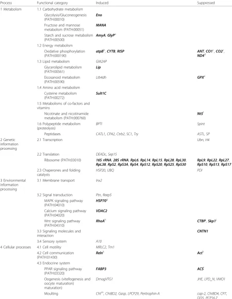
Table 1 Functional categorization ofDaphnia magnagenes responding to acute ibuprofen exposure
Process Functional category Induced Suppressed
1 Metabolism 1.1 Carbohydrate metabolism Glycolysis/Gluconeogenesis (PATH:00010)
Eno
Fructose and mannose metabolism (PATH:00051)
MANA
Starch and sucrose metabolism (PATH:00500)
AmyA,GlyPa
1.2 Energy metabolism Oxidative phosphorylation (PATH:000190)
atpBb,CYTB,RISP ANT,CO1c,CO2c,
ND4d 1.3 Lipid metabolism GM2AP
Glycerolipid metabolism (PATH:00561)
Lip
Eicosanoid metabolism (PATH:00590)
Ltb4dh GPXe
1.4 Amino acid metabolism Cysteine metabolism (PATH:00272)
Sult1C
1.5 Metabolisms of co-factors and vitamins
Nicotinate and nicotinamide metabolism (PATH:000760)
Nt5f
1.6 Polypeptide metabolism (proteolysis)
BPTI Spint
Peptidases CATL1,CPA2,Ctrb2,SC1,Try ASTL,SP
2 Genetic information processing
2.1 Transcription Ubn,H4
2.2 Translation DEADc,Sep15
Ribosome (PATH:03010) 16S rRNA,28S rRNA,RpL6,RpL14,RpL15,RpL28,RpL30,
RpL38,RpS2,RpS3A,RpS4,RpS12,RpS20,RpS25,RpS30
RpL9,RpL22,RpL27,
RpS10,RpS13,RpS17 2.3 Chaperones and folding
catalysts
HSP20,UBQ PDI
3 Environmental information processing
3.1 Membrane transport Inx2
3.2 Signal transduction Ptn,Reep5
MAPK signaling pathway (PATH:04010)
HSP70g
Calcium signaling pathway (PATH:04020)
VDAC2
Wnt signaling pathway (PATH:04310)
RhoAh CTBPi
,Skp1j
3.3 Signaling molecules and interaction
CNTN1
3.4 Sensory system A10
4 Cellular processes 4.1 Cell motility MRLC2,Tm1
4.2 Cell communication (PATH:01430)
Relnk Acth
4.3 Endocrine system PPAR signaling pathway (PATH:03320)
FABP3 ACSl
Oogenesis (vitellogenesis and oocyte maturation)
maturation)
DmagVTG1 JHE,LPD_N,VMO1
Moulting Chtm,ChtBD2,Gasp,LPCP29,Peritrophin-A cap-2,ChtBD4,CP7,
in order to investigate further the expression of key genes using QPCR. The treatments (control and 80 mg/ l ibuprofen) were replicated four times for every time point (2, 4, 8, 24, and 48 hours) and ten genes were ana-lyzed (for further details, see Materials and methods [below]). Four linked to eicosanoid metabolism (Lip,
Ltb4dh, CTP [choline-phosphate cytidylyltransferase], andCOX), and six genes were associated with signal transduction and endocrine functions (Cht[chitinase],
DmagVTG1,FABP3[fatty acid binding protein 3], JHE,
RXR[retinoid × receptor], and VMO1 [vitelline outer layer membrane protein 1]).COXwas included to clarify the interruption of eicosanoid metabolisms because it represents a key component of the MOA of ibuprofen in mammals. RXR was included because recently reported evidence shows that JHAs can change the expression of this receptor in D. magna[26]. CTPwas employed as a ‘negative control’, because this gene is involved in a part of the glycerophospholipid metabo-lism that is less relevant to eicosanoid metabometabo-lism.
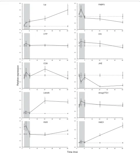
With the exception ofLip, the temporal expression of all of the analyzed genes fluctuated during early expo-sure (2 to 8 hours) to ibuprofen (Figure 1). We suggest that this fluctuation reflects a general homeostatic response. This could be an after effect of handling stress, but it may also show that daphnids are attempt-ing to regulate toxicity durattempt-ing early stages of exposure. This early variation disappears by the classic ecotoxico-logical exposure time points of 24 and 48 hours, empha-sizing the feasibility and importance of applying the latter.
The earliest genes to change expression levels signifi-cantly were the eicosanoid-related genesLipandLtb4dh
(2 hours onward), with Lip being consistently upregu-lated throughout the exposure (Figure 1). As expected, the expression of the ‘negative control’ CTP was unchanged compared with controls. However, the tem-poral expression of COXwas not significantly different from that of the controls, although there was a near
significant (P = 0.088) upregulation after 24 hours of exposure, which may reflect COX inhibition (Figure 1).
Global gene expression data showed that Cht, encod-ing a key moultencod-ing fluid enzyme secreted durencod-ing apolysis [27], and several cuticle-related genes responded differ-entially to ibuprofen stress after 24 hours of exposure (Table 1 [section 4.3]). However, the temporal expres-sion profile revealed that Cht was only significantly induced at 2 hours of exposure, after which there was no difference in expression between exposed and con-trol daphnids (Figure 1). In arthropods, JH is involved in regulating both moulting (of sexually immature instars) and vitellogenesis [23], but there was no strong evidence that ibuprofen (or indirectly JH) affected moulting in the present study based on the temporal expression of
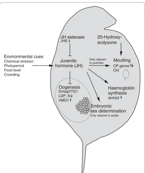
Cht(Figure 1) and phenotypic results (see below). Figure 2 provides an overview of the potential biological inter-actions of JH (methyl farnesoate) in D. magna and related genes responding to ibuprofen stress. Evidence of elevated JH levels was strongly supported by the tem-poral suppression of JHE and late induction of RXR
(Figure 1), suggesting that JH levels increase in exposed
D. magna over time. JHA pyriproxyfen has been shown
to suppress DmagVTG1 expression in 1-hour-old neo-nates after a 96-hour exposure [22]. However, a 48-hour exposure was too short to show a similar suppression of
DmagVTG1in older neonates (24 hours old), although
there was a nonsignificant tendency toward suppression (Figure 1). D. magnastart to ovulate (release mature oocytes into the brood chamber) when they are 5 to 6 days old at 20°C (Heckmann L-H, personal observa-tions). Transcriptomic changes in vitellogenesis may therefore not be noticeable or relevant before daphnids become adolescent. Thus, it is likely that a decrease in
DmagVTG1 expression would have been observed in
the exposed 24-hour-old neonates if the temporal expression profile had been extended beyond 72 hours.
[image:5.595.57.536.104.137.2]The microarray findings revealed that VMO1was sup-pressed at 80 mg/l ibuprofen after 24 hours of exposure.
Table 1 Functional categorization ofDaphnia magnagenes responding to acute ibuprofen exposure(Continued)
4.4 Immune system CLECT,CUB,GNBP
4.5 Inorganic ion transport and metabolism
AT1A,CRIP,Fer,Fer1HCH,dmHb2,VGCa dmHb1,Sfat,Znf_AN1
Genes presented in bold are directly linked to a Kyoto Encyclopedia of Genes and Genomes pathway, whereas other genes are listed with their functional category or subcategory based on Gene Ontology (see Additional data file 2).a
GlyPis also associated with the insulin signaling pathway (PATH:04910).b
atpBis also associated with the type III secretion system (PATH:03070), flagellar assembly (PATH:02040), and epithelial cell signaling inHelicobacter pyloriinfection (PATH:05120).c
CO1andCO2are also associated with the vascular endothelial growth factor signaling pathway (PATH:04370).d
ND4is also associated with ubiquinone biosynthesis (PATH:00130).e
GPXis also associated with glutathione metabolism (PATH:00480).f
Nt5is also associated with purine metabolism (PATH:00230) and pyrimidine metabolism (PATH:00240).g
HSP70is also associated with antigen processing and presentation (PATH:004612) related to the immune system.h
ActandRhoAare involved in eight and nine different pathways, respectively. Here they have been associated with one that is already represented, but because of this multi-alignment they have not been considered any further.iCTBPis also associated with the notch signaling pathway (PATH:04330).j
Skp1is also associated with the cell cycle (PATH:04110), ubiquitin-mediated proteolysis (PATH:04120), and transforming growth factor-bsignaling pathway (PATH:04350), which all interlink through the Wnt signaling pathway.k
Relnis also associated with focal adhesion (PATH:04510) and extracellular matrix receptor interaction (PATH:04512).l
ACSis also associated with the adipocytokine signaling pathway (PATH:04920) and fatty acid metabolism (PATH:00071).
m
However, the temporal expression of VMO1 was strongly upregulated after 24 hours of exposure and onward (Figure 1). In crustaceans, VMO1 proteins are synthesized outside the ovaries and are then transported via the hemolymph to developing oocytes. The major role of the vitelline membrane is to avoid mixing of yolk and albumen [28]. Expression ofVMO1 seems to
pre-cede DmagVTG1, possibly revealing important
func-tional insights into the timing ofD. magnaoogenesis.
Ibuprofen reduces fecundity and arrests early embryogenesis
Although previous studies [11,29] showed that ibuprofen concentrations of 20 mg/l or greater suppress reproduc-tion, questions remain as to whether ibuprofen acts on oogenesis or embryogenesis before hatching. A chronic experiment (8 days) was therefore conducted on adult 14-day-oldD. magna(one individual/replicate) with five replicates of a control and three concentrations of ibu-profen, namely 20, 40 and 80 mg/l (for further details, see Materials and methods [below]). As expected expo-sure to more than 20 mg/l ibuprofen reduced fecundity, but it did not delay brood release or affect associated moulting (Table 2). The broods released after exposure to 80 mg/l ibuprofen had few viable neonates and consisted almost entirely of under-developed embryos (Table 2). Microscopic investigation showed that embry-ogenesis was arrested before completion of the first third of embryonic development - stage 2sensu Kast-Hutche-son and coworkers [30] (see Additional data file 4).
Minor differences between the results presented here and those of our previous chronic studies, using adoles-cent [29] and neonate [11] individuals exposed for 10 and 14 days, respectively, suggest an ontogenetic shift in ibuprofen stress response, with fecundity being less affected in older individuals. This implies that suscept-ibility to the stressor (ibuprofen) decreases with age/size, which appears to be a common phenomenon in ecotoxi-cology [31].
Continued culturing of fourth and fifth brood neo-nates to adulthood in uncontaminated media did not reveal any induction of male offspring as a result of maternal exposure (Table 2). Comparing the number of fifth brood offspring, produced by adults of the control and 20 mg/l ibuprofen treatments, with the number of embryos aborted at 80 mg/l ibuprofen showed that on average there were 20 eggs fewer in the highest ibupro-fen treatment (Table 2). This response was also observed in previous studies [11]. Fewer viable oocytes may have been deposited during ovulation possibly because of impaired ovarian maturation; while under-developed oocytes may have been re-absorbed by the mothers, a response was also found in stressedD. mela-nogasterfollowing starvation [9]. Generally, it appears
that ibuprofen primarily affects oogenesis and that embryogenesis in viable oocytes is arrested at high concentrations.
A putative molecular mode of action of ibuprofen in
Daphniaspp
Based on our microarray (Table 1) and temporal QPCR expression data (Figure 1), we constructed a diagram showing how the genes responding to ibuprofen expo-sure inD. magnacan be tied into a pathway that links the putative molecular MOA of ibuprofen with carbohy-drate metabolism, lipid metabolism, signal transduction, and two main biological target processes, namely oogen-esis and the immune system (Figure 3). Our experimen-tal design was not intended to allow study of phenotypic immune responses, even though eicosanoids play a vital role in invertebrate immune systems [12]. However, future studies may clarify whether ibuprofen-stressed crustaceans are more susceptible to infections because of the apparent repression of their immune system.
The temporal expression data indicated that lipid metabolism was initially affected (for example,Lip and
Ltb4dh), with subsequent effects on carbohydrate meta-bolism and signal transduction ultimately affecting oogenesis (Figure 3); the latter was likewise evident from our phenotypic experiments (Table 2). The obvious genetic link betweenLip(glycerolipid metabo-lism) and Ltb4dh (eicosanoid metabolism) would be
PLA2(phospholipase A2), which encodes a key enzyme that is responsible for hydrolyzing phosphatidylcholine into AA (Figure 3), comprising one of the first steps in eicosanoid metabolism [12]. Unfortunately, the DNA sequence of PLA2 was not available to us, but future studies should aim to identify this key gene in
D. magna.
Discussion
We found a strong link between transcriptomic and phenotypic stress responses inD. magna by integrating data on the molecular MOA of ibuprofen with ecophy-siological effects observed at higher biological levels. Furthermore, to our knowledge, this is among the first studies to investigate the global transcriptomic stress response of an invertebrate exposed to a NSAID. Pre-vious findings in Bacillus megaterium[32] suggest that NSAIDs mimic endogenous fatty acids and may interact with transcriptional regulation of eicosanoid target genes.
Time (hrs)
Relativ
e e
xpression
0 8
0.0
00 88 16 24 32 40 48
0.5 1.0 1.5 2.0
0.0
00 88 16 24 32 40 48
0.5 1.0 1.5 2.0 0.0
00 88 16 24 32 40 48
0.5 1.0 1.5 2.0
0.0
00 88 16 24 32 40 48
0.5 1.0 1.5 2.0
0.0
00 88 16 24 32 40 48
0.5 1.0 1.5 2.0
0.0
00 88 16 24 32 40 48
0.5 1.0 1.5 2.0 0.0
00 88 16 24 32 40 48
0.5 1.0 1.5 2.0
0.0
0 8 16 24 32 40 48
2.0 4.0 6.0 0.0
0 8 16 24 32 40 48
2.0 4.0 6.0
*
*
* * *
0.0
0 8 16 24 32 40 48
2.0 4.0 6.0 8.0
*
Ltb4dh
*
*
*
RXR Lip
CTP
COX
*
* FABP3
Cht
*
JHE
* *
dmagVTG1
VMO1 *
*
[image:7.595.58.538.86.611.2]*
Figure 1Temporal expression profiles ofDaphnia magnagenes after ibuprofen exposure. Shown are the temporal expression profiles of
D. magna(<24 hours old) genes after 2 to 48 hours of exposure to ibuprofen (mean ± standard error). Gene expression was measured using quantitative PCR. Dotted and solid lines represent control and exposed (80 mg/l ibuprofen) expression, respectively. Target gene expression was calculated using DART-PCR [59] and normalized to a geNorm [60] estimated normalization factor based on the geometric mean ofAct(actin),
GAPDH(glyceraldehyde-3-phosphate dehydrogenase) andUBC(ubiquitin conjugating enzyme). Exposed expression levels are shown relative to controls at corresponding duration of exposure (note the different y-axes). The temporal‘gray zones’generally reveal fluctuating expression patterns perhaps reflecting homeostatic instability. Asterisks (*) denote a significant (P< 0.05, Student’st-test) difference from controls. TheLip
(which encodes triacylglycerol lipase),CTP(choline-phosphate cytidylyltransferase),Ltb4dh(leukotriene B412-hydroxydehydrogenase) andCOX
(cycloxygenase) genes are related to lipid metabolism, whereasRXR(retinoid × receptor),JHE(juvenile hormone esterase),DmagVTG1
based MOA in crustaceans, because Ltb4dh was responding. Alternatively, it may reflect ontogenetic dif-ferences, whereby the COX pathway is less important or not activated in neonates. This idea is supported by dif-ferences in fecundity between organisms exposed as either neonates or adults. Individuals exposed as adults had a higher fecundity than those exposed as neonates [11,29]. Adult daphnids may have a relatively higher content of eicosanoids (and phospholipids) in the ovar-ies that may increase their tolerance by buffering the impact of ibuprofen stress on eicosanoid metabolism. Nevertheless,Lipexpression was increased in ibuprofen-stressed neonates, indicating potential recruitment of AA [20] that may buffer the competitive inhibition of COX or LOX by increasing substrate availability. However, increased release of AA could affect signal transduction through the calcium signaling pathway (Figure 3), because AA has been shown to be involved in embryonic calcium signaling [33].
In mammals, prostaglandins and leukotrienes act as ligands at distinct transmembrane G-protein-coupled receptors and nuclear PPARs [34]. PPARs are transcrip-tion factors that form heterodimers with retinoid × receptor (upregulated in this study) and bind to target genes involved in, for instance, controlling prenatal and postnatal development [35,36]. Retinoid × receptor (encoded by RXR) also forms heterodimer complexes with other nuclear receptors and is known to bind JH in
D. magna [26]. It remains unknown whether JH and
eicosanoids interact directly in daphnids or whether the upregulation of RXR relates to PPAR and thus eicosa-noid metabolism, rather than being involved with JH. The PPAR-apathway is activated by leukotriene (LT)B4 in mammals [34], indicating that this could constitute the main signal transduction cut-off in
Juvenile hormone (JH)
Oogenesis Environmental cues
Chemical stressor Photoperiod Food level Crowding
Haemoglobin synthesis dmhb2
Moulting CP genes Cht 20-Hydroxy-ecdysone JH esterase
JHE
Only relevant in juveniles
Only relevant in adults
DmagVTG1 LDP_N VMO1
[image:8.595.57.291.86.364.2]Embryonic sex determination
Figure 2Overview of potential biological interactions of JH (methyl farnesoate) inDaphnia magna. Expression of relevant target genes in ibuprofen-stressed daphnids (24 to 48 hours of exposure) is indicated by small arrows quantified by either microarrays (normal font) or quantitative PCR (bold font). Note that 20-hydroxyecdysone is the main hormone controlling moulting inDaphnia, whereas juvenile hormone (JH; in arthropods) prevents sexual maturation between moults in juveniles. (There is currently no evidence from daphnids on this role.) Large arrows signify synthesis or induction of the particular product or process, whereas end bars denote inhibition. Abbreviations are as in Additional data file 1 and Figure 1;CPgenes signify cuticle protein genes (see text for further details).
Table 2 Reproduction of 14-day-oldDaphnia magnaexposed for 8 days to ibuprofen
Brood Control 20 mg/l ibuprofen 40 mg/l ibuprofen 80 mg/l ibuprofen
Brood release and adult moultinga(age [days]) 3rd 15.0 ± 0.00 15.2 ± 0.20 15.2 ± 0.20 15.2 ± 0.20 4th 18.2 ± 0.20 18.4 ± 0.24 18.6 ± 0.24 18.4 ± 0.24 5th 21.2 ± 0.20 21.4 ± 0.24 21.4 ± 0.24 21.2 ± 0.20 Fecundity (average number of offspring/brood) 3rd 22.8 ± 1.59 19.6 ± 2.42 18.8 ± 2.82 24.6 ± 3.14 4th 28.4 ± 1.40 30.0 ± 0.95 8.20 ± 1.43* 0.00 ± 0.00* 5th 42.0 ± 1.64 41.4 ± 2.25 6.60 ± 1.54* 1.00 ± 0.45*
Embryo abortion (average number of abortions/brood) 3rd Nil Nil Nil Nil
4th Nil Nil Observedb Observedb
5th 0.20 ± 0.20 0.20 ± 0.20 7.20 ± 3.20 20.6 ± 7.08*
Production of male offspringc 3rd Na NA NA NA
4th Nil Nil Nil Nil
5th Nil Nil Nil Nil
Values are expressed as mean ± standard error (n= 5). Asterisks (*) denote a significant (P< 0.05, analysis of variance) difference from controls.a
Moulting follows brood release in mature female daphnids and was assessed by counting shed carapaces (after moulting new eggs are deposited from the ovaries into the brood chamber with the incubation period corresponding to the intermoult period).b
Aborted embryos were not quantified, but a few were observed in 40 mg/l ibuprofen, and considerable numbers in 80 mg/l ibuprofen, comparable to 5th
brood.c
[image:8.595.58.538.527.687.2]Eicosanoid metabolism
GPX COX ( )
Ltb4dh
Glycerolipid metabolism
Lip
Glycero-phospholipid metabolism
CTP
AA / L TB
4
1,2 Diacylglycerol
Phosphatidylcholine Carbohydrate
metabolism
Lipid metabolism
Signal transduction
Target process
Fatty acid metabolism
ACS
Oogenesis
JHE
LDP_N
VMO1
DmagVTG1
Immune system
CLECT
CUB GNBP
MAPK signalling pathway
HSP70
Glycolysis
Eno
Adipo-cytokine signalling pathway
ACS
RXR
Insulin signalling pathway
GlyP
Calcium signalling pathway
VDAC2
PPAR signalling pathway
ACS
FABP3 RXR
Starch and sucrose metabolism
GlyP amyA
Fructose and mannose metabolism
[image:9.595.58.536.88.638.2]MANA
stressedD. magna. This is further supported by ibupro-fen suppression of other PPAR-related genes, such as
ACSandFABP3(Figure 3); the latter of these two genes encodes fatty acid binding protein 3, which is involved in transporting PPAR ligands to the nucleus [35]. The LTB4DH enzyme, encoded byLtb4dh, inactivates LTB4, and also catalyzes the degradation of the prostaglandin (PG)E2 and PGF2a [37]. Mammalianin vitro research has demonstrated that LTB4DH activity is strongly sup-pressed by the NSAIDs diclofenac and indomethacin, whereas ibuprofen has only a moderate effect [38]. Thus, induced expression of Ltb4dh in ibuprofen-stressed daphnids could reflect inhibition of LTB4DH activity, which may affect the catabolism of relevant eicosanoids. In relation to the reduced fecundity observed in this study, LTB4 has been shown to play an important role in yolk uptake during oogenesis in insects [39], as well as being an agonist for calcium sig-naling regulating mitosis in echinoderm eggs and embryos [40].
Decreased fecundity and induction of male offspring have been identified when exposingD. magnato JH and JHAs [41]. We have revealed a concentration-dependent reduction in fecundity after ibuprofen exposure [11], but the chronic follow-up experiment indicated that ibupro-fen did not result in production of male offspring. Our transcriptomic data (JHEand RXR) strongly indicate that there was a higher than normal presence of JH, but why were no male offspring being produced in response to elevated JH levels? In many studies on daphnid and crustacean endocrinology (for review, LeBlanc [42]) indi-viduals are exposed to high concentrations of potent JHAs, thus potentiating the normal JH signal that may lead to male induction in daphnids. If JH levels are ele-vated in ibuprofen-stressed daphnids, then we propose that the endocrine signal produced by JH was sufficient to reduce fecundity but insufficient to initiate produc-tion of male offspring. This suggests that JH signal transduction is tiered, firstly initiating a reduction in fecundity and secondly - if the signal is maintained or increased - causing ontogenetic sex change among the embryos. This type of signal transduction is robust and would make ecological sense.Daphniamainly reproduce through cyclic parthenogenesis, but males are produced after strong environmental cues (for instance, fading light levels causing algae production to cease) to allow sexual reproduction and the formation of diapausing eggs (ephippia).
In summary, based on our genetic and phenotypic data, we suggest that the MOA of ibuprofen in D. magna functions as follows. Initially, ibuprofen inter-rupts eicosanoid metabolism, which is evident from the early response ofLip andLtb4dh. This impairs normal signal transduction possibly through the PPAR and/or
the calcium signaling pathway, which leads to disruption of the endocrine system related to JH metabolism (JHE
and RXR) and oogenesis (DmagVTG1 and VMO1; Figure 3). The phenotypic response links strongly with reproduction showing reduced fecundity. We assume that fecundity is affected by disruption of normal JH metabolism caused by elevated JH levels in ibuprofen-stressed daphnids, or alternatively that ibuprofen mimicks JH, which halts vitellogenesis and thus oogen-esis. Suppressed vitellogenesis (DmagVTG1) and under-development of the vitelline membrane (VMO1) would result in poor accumulation of protein, lipids, and other nutrients in the oocytes, as well as incomplete division of yolk and albumen. This would lead to failing oogen-esis, with abnormal oocytes possibly being re-absorbed [9], or eventually arrested embryogenesis caused by nutrient deficiency. Ye and coworkers [43] showed that downregulation of COX-2 reduced the levels of PGE2 and PGI2, leading to delayed development and death of mice embryos. PGE2 has likewise been shown to play a key role during crustacean reproduction (vitellogenesis), possibly controlling ovulation [44,45]. Future proteomic investigations of LTB4 and PGE2 may further elucidate the role of these eicosanoids in daphnid reproduction. Furthermore, a recent study conducted in queen bees [46] showed that JH affects the expression ofvitellogenin
and insulin/insulin-like growth factor-1 signaling genes in opposite directions. In the present study, we found indications of repressed expression ofDmagVTG1 coin-ciding with a consistent upregulation of genes that are involved in carbohydrate metabolism, and especially gly-colysis, which is closely related to insulin signaling in both vertebrates and invertebrates [47] (Figure 3). How-ever, the link between insulin, vitellogenin and JH, and the consequence of this interaction for daphnid repro-duction remain to be unraveled.
Conclusion
Our systems biology approach to stress ecology has proved fruitful in linking transcriptomic data with eco-physiological stress responses at higher biological levels. This reveals considerable promise for using acute mole-cular responses as a guide to possible chronic impact on populations of environmental stress. Ultimately, this could improve current environmental risk assessment through providing early ‘signposts’ (sensu Hutchinson and coworkers 2006 [48]) to the need for higher tier testing or other appropriate actions.
Materials and methods
Microarray experiment
D. magna were obtained from the Water Research
experiment. Full details of culturing methods were reported by Hooper and coworkers [49]. Tests were conducted in 5 l glass aquaria (height 22 cm, internal diameter 18.5 cm, and thickness 5 mm; Harzkristall GmbH, Derenburg, Germany) at 20 ± 1°C and a 16:8 light:dark photoperiod. During the first 24 hours the aquaria contained an inner exposure vessel (height 13 cm and diameter 9 cm) with a nylon mesh bottom to allow free movement of the test media between the two vessels. Quadruplicates were assigned in a randomized block design and initiated with 310 fourth brood neo-nates (<24 hours old) that were exposed to a control or one of three ibuprofen concentrations (20, 40, and 80 mg/l ibuprofen), applied as ibuprofen-sodium (Sigma-Aldrich, Gillingham, UK; CAS number 31121-93-4; batch number 64K0892) in reconstituted water. Follow-ing 24 hours of exposure, ten individuals were trans-ferred to the outer aquarium for a chronic population study, described in detail by Heckmann and coworkers [11], while the inner vessel with the remaining 300 neo-nates was removed. These neoneo-nates were stored in RNA
later
®
(Ambion, Warrington, UK) at -80°C forsubse-quent RNA extractions. A reference pool of approxi-mately 6,000D. magna that were under 48 hours old was obtained from the same brood as those exposed. Ibuprofen was sampled (1.5 ml) for quantification from every replicate of each treatment at time zero and at 24 hours. Subsequent analysis, using UV spectrophotometry [21], revealed that the difference between nominal and measured concentrations was under 10%, except for one replicate of 20 mg/l ibuprofen, which was under 20% at 24 hours. Further details on water chemistry (conductiv-ity, dissolved oxygen, and pH) are available in the report by Heckmann and coworkers [11].
Microarray hybridization
Hybridization followed a reference pool design in which each experimental sample was hybridized against a com-mon reference pool sample. Total RNA was extracted using the RNeasy Mini kit with on-column DNase treat-ment (Qiagen, Crawley, UK) in order to remove any traces of genomic DNA, following the manufacturer’s instructions. RNA concentrations were determined by spectrophotometry using GeneQuant Pro (Biochrom, Cambridge, UK), and RNA integrity was verified using a BioAnalyzer 2100 (Agilent Technologies, Stockport, UK). cDNA was synthesized from 17.5 μg total RNA (treatment and reference pool material, respectively) and labeled with Alexa Fluor
®
dyes (two-colour reference design: Alexa Fluor®
647 and Alexa Fluor®
555 for experimental and reference pool samples, respectively) using SuperScript™
Plus Indirect cDNA Labeling System(Invitrogen, Paisley, UK). Slides were pre-hybridized in a solution containing 50% vol/vol de-ionized formamide, 5× sodium chloride-sodium citrate, 0.1% sodium dodecyl sulfate and 1% weight/vol bovine serum albumin (Sigma-Aldrich, Warrington, UK), and incubated at 42°C in a Techne HB-1 Hybridiser (Techne Ltd, Stone, UK) for 1 h.
A 45 μl hybridization probe solution was prepared with 22.5μl de-ionized formamide, 5× sodium chloride-sodium citrate, the labeled cDNA mix (combined experimental sample and reference pool cDNA), and a hybridization block mix containing 0.1% sodium dodecyl sulfate, 0.5 mg/ml polyA RNA (Sigma-Aldrich, Warring-ton, UK), 0.5 mg/ml yeast tRNA, 0.5 mg/ml salmon sperm DNA, and 25μg/ml human and 25 μg/ml mouse
Cot-1 DNA (Invitrogen Paisley, UK). The probes were
hybridized to individual microarray slides (one hybridi-zation was performed for each slide; n = 16) under a 2560 lifterslip
™
(Implen, Southend on Sea, UK). The slides were hybridized in batches of four slides corre-sponding to the control and respective ibuprofen treat-ments within a biological replicate. The slides were then placed in an airtight plastic box and incubated at 42°C in a Techne HB-1 Hybridiser (Techne Ltd, Stone, UK) for 16 hours. Details of pre-hybridization and post-hybridization washes, and construction of the microar-ray are described in Additional data file 5.Microarray analysis
Microarray slides were scanned using a GenePix 4200A microarray scanner (Axon Instruments, Inverurie, UK) installed with GenePix
®
Pro 5.0. The data were normal-ized per slide to the median of ratios using spots with a regression ratio above 0.7, a sum of medians above 500, a saturation value below 3, and a signal to noise ratio of 3 or greater [50]. Overall, some 15% of the spots per chip were flagged as ‘present’ based on these criteria, and they were utilized to calculate normalization factors [50]. Regrettably, one slide failed (80 mg/l ibuprofen; replicate 2) and was omitted from further analysis. Only spots flagged as present and/or marginal in 80% of the arrays were analyzed (7,135 spots).treatments within each biological replicate were normal-ised to the control sample of the same biological repli-cate;n = 4). MA plots of raw and normalized data are available in Additional data file 6, which shows data quality before and after normalization. Following data normalization, spots with expression levels between 0.714 and 1.4 in all conditions (4,912 spots) were removed from further analysis using GeneSpring filters (resulting in 2,223 spots) [51]. Two sample independent
t-tests (equal variances assumed) were carried out on log2 ratios between control and ibuprofen treatments. This filtering step ensured that only spots that changed in at least one concentration were subjected to further analysis. The resultingt-test gene lists were then merged (827 spots) and subjected to a one-way analysis of var-iance (equal varvar-iances not assumed) with no multiple testing corrections, resulting in a list of 272 spots. For all statistical tests, a significance level of 5% was applied.
Annotation
Basic local alignment search tool (BLAST) analyses were conducted between August 2006 and March 2007 on fragments that responded significantly to the exposure treatment. Sequences were annotated according to BLASTX homology search against GenBank [52], Uni-Prot [53], and InterPro [54]. Sequences were only anno-tated if they had a BLAST hit with an expect value (E value) below 10-5and a score above 50. GeneBank/Uni-Prot accession number and species’match were recorded with each annotation (see Additional data file 1).
MIAME (minimum information about a microarray experiment) compliance
AvailableD. magnasequences can be found at Daphnia-Base [55] and from the website of theDaphniaresearch group of the University of Reading [56]. Microarray images and data are accessible through the public repo-sitory Array Express at the European Bioinformatics Institute (accession number: E-MAXD-20). Microarray images and normalized expression data were also catalo-gued on our website [56].
Follow-up experiment assessing chronic phenotypic responses
The experiment was based on a randomized block design with five replicates of a control and three treat-ments with ibuprofen-sodium (Sigma-Aldrich, Warring-ton, UK: CAS number 31121-93-4; batch number 64K0892) containing 20, 40 and 80 mg/l ibuprofen, respectively. Each replicate consisted of one adult (14 days old) placed in a 1,000 ml glass beaker contain-ing 1 l reconstituted freshwater (see Hooper and
coworkers [49]), with or without the addition of ibupro-fen. The test vessels were kept in a 20 ± 1°C tempera-ture-controlled room with a light:dark regimen of 16:8 hours. Adults were exposed to ibuprofen for 8 days and were fed daily with equal amounts of green algae Chlor-ella vulgarisvar viridis(equivalent to 1.00 mg/day car-bon). Measured biological end-points are displayed in Table 2. To assess the potential induction of male off-spring caused by maternal exposure to ibuprofen, a total of 20 fourth and fifth brood offspring from each treat-ment were transferred to 2 l plastic beakers with 1.2 l of uncontaminated culture media, except in the 80 mg/l ibuprofen treatment were zero, and five offspring were produced in each of the fourth and fifth broods. Fourth and fifth brood neonates were reared like normal cul-tures (see Hooper and coworkers [49]) until they reached sexual maturity (approximately 9 days). No males were present in either of the treatments. However, 60% mortality was observed among fifth brood neonates that had been maternally exposed to 80 mg/l ibuprofen. There was no mortality among the other neonates or during the exposure of adults.
Ibuprofen was sampled (1.5 ml) from each replicate on days 0 and 8 (adult exposure only), and subsequent quantifications revealed that the difference between nominal and measured concentrations was under 10%. Water temperature was checked daily and averaged 19.6 ± 0.2°C (mean ± standard error; n= 60) throughout the experimental period. Other measured water chemistry parameters are available in Additional data file 7. Corre-sponding with our previous studies [11], both pH and conductivity were slightly but significantly (P < 0.05, analysis of variance) increased with increasing ibuprofen concentration.
Follow-up experiment assessing temporal expression of key genes
n= 8) throughout the experimental period. Other water chemistry parameters were measured at every time point from pooled samples of the same treatment (see Additional data file 7).
Following exposure (2, 4, 8, 24, and 48 hours), neonates were immediately transferred to 0.2 ml RNA later
®
(Ambion, Warrington, UK) using our recently developed methodology [57]. Samples were stored at -80°C and total RNA was subsequently extracted and processed as previously described [21]. cDNA was synthesized from 1
μg total RNA and diluted 10-fold, resulting in total RNA concentrations of 5 ng/μl, and stored at -20°C. Primers were designed using Primer3 [58] and synthesized by MWG (Ebersberg, Germany; see Additional data file 8). QPCR was conducted on the GeneAmp 5700 Sequence Detection System (Applied Biosystems) using ABsolute
™
QPCR SYBR®
Green ROX (500 nmol/l) mix (ABgene, Epsom, UK). Each reaction was run in duplicate and con-tained 2.5μl cDNA template (equivalent to 12.5 ng total RNA) along with 900 nmol/l primers in a final volume of 25μl. Cycling parameters were 95°C for 15 minutes to activate the DNA polymerase, then 40 cycles of 95°C for 15 seconds and 60°C for 1 minute. Melting curves were performed by using dissociation curve Sequence Detec-tion System software version 1.3 (Applied Biosystems) to verify that only a single product with no primer-dimers was amplified. QPCR data processing and statistical ana-lysis were performed as previously reported [21] using DART-PCR [59] and geNorm [60].Additional data files
The following additional data are available with the online version of this paper. Additional data file 1 lists all of the cDNAs (annotated) that responded to ibupro-fen treatment on the D. magnamicroarray. Additional data file 2 shows the relative expression and GO of the unique D. magnagenes responding to acute ibuprofen exposure. Additional data file 3 displays QPCR confir-mation of selectedD. magna genes responding on the cDNA microarray. Additional data file 4 shows an image of aD. magnaembryo arrested at developmental stage 1 to 2 after maternal exposure to ibuprofen. Addi-tional data file 5 provides supplementary methods on microarray hybridization and microarray construction. Additional data file 6 shows MA plots of raw and nor-malized microarray data. Additional data file 7 shows water chemical parameters measured during the follow-up experiments. Additional data file 8 lists technical data on QPCR (for example, primers and amplification efficiency) from the follow-up experiment assessing tem-poral expression of key genes responding to ibuprofen.
Additional material
Additional data file 1: cDNAs responding to ibuprofen treatmentPresented is a table listing all the cDNAs (annotated) that responded to ibuprofen treatment on theD. magnamicroarray.
Additional data file 2: Expression and GO of uniqueD. magna
genes responding to acute ibuprofenPresented is a table showing the relative expression and GO of the uniqueD. magnagenes responding to acute ibuprofen exposure.
Additional data file 3: QPCR confirmationPresented is a table displaying QPCR confirmation of selectedD. magnagenes responding on the cDNA microarray.
Additional data file 4:D. magnaembryo arrested at developmental stage 1 to 2Presented is an image of aD. magnaembryo arrested at developmental stage 1 to 2 after maternal exposure to ibuprofen.
Additional data file 5: Supplementary methods on microarray hybridization and microarray constructionPresented is a document with supplementary methods on microarray hybridization and microarray construction.
Additional data file 6: MA plots of raw and normalized microarray dataPresented are MA plots of raw and normalized microarray data
Additional data file 7: Water chemical parametersPresented is a table showing water chemical parameters measured during the follow-up experiments.
Additional data file 8: Technical data on QPCRPresented is a table listing technical data on QPCR (for instance, primers and amplification efficiency) from the follow-up experiment assessing temporal expression of key genes responding to ibuprofen.
Abbreviations
AA, arachidonic acid; BLAST, basic local alignment search tool; COX, cycloxygenase; GO, Gene Ontology; JH, juvenile hormone; JHA, juvenile hormone analog; KEGG, Kyoto Encyclopedia of Genes and Genomes; LOX, lipoxygenase; LT, leukotriene; LTB4DH, leukotriene B4
12-hydroxydehydrogenase; MOA, mode of action; NSAID, nonsteroidal anti-inflammatory drug; PCR, polymerase chain reaction; PG, prostaglandin; PPAR, peroxisome proliferator-activated receptor; QPCR, quantitative PCR.
Acknowledgements
We gratefully acknowledge the financial support of AstraZeneca, Syngenta, NERC (project NER/D/S/2002/00413‘The population and molecular stress responses of an ecotoxicology indicator species’) and The Research Endowment Trust Fund of the University of Reading. We are grateful to Dr Hajime Watanabe, Professor Taisen Iguchi, Dr Anneleen Soetaert, and Professor Wim De Coen for generously providingD. magnaclones for our microarray; Dr Fei-Ling Lim, Dr David J Moore, and Dr Jonathan G Moggs for assisting in printing the microarrays; Katie Cook for advice on bioinformatics; Yuya Hayashi for stimulating discussions; Stephen Pountney for help on microscopy; and Dr Viacheslav Bolshakov for advice on microarray analysis. Finally, we thank two anonymous reviewers for their valuable comments on the manuscript.
Author details
1
University of Reading, School of Biological Sciences, Environmental Biology, Philip Lyle Building, Reading, RG6 6BX, UK.2University of Aarhus, National Environmental Research Institute, Department of Terrestrial Ecology, Vejlsøvej, DK-8600, Silkeborg, Denmark.3University of California, School of Veterinary Medicine, Department of Anatomy, Physiology and Cell Biology, Davis, California 95616, USA.4AstraZeneca Global SHE, Brixham
Authors’contributions
All authors were involved in designing the experiments. RC, HLH, CJH, and AB assisted LHH in conducting parts of the practical experimental work. LHH performed the statistical analyses and drafted the main manuscript under the supervision of AC, RMS, THH, and SJM. Methods on microarray hybridization, and analysis and supplementary methods on microarray construction were drafted by LHH and RC. All authors read, contributed intellectually toward, and approved the final manuscript.
Received: 20 September 2007 Revised: 15 November 2007 Accepted: 21 February 2008 Published: 21 February 2008
References
1. Walker CH, Hopkin SP, Sibly RM, Peakall DB:Principles of Ecotoxicology.3 edition. Boca Raton, FL: Taylor & Francis; 2006.
2. Chen DR, Toone WM, Mata J, Lyne R, Burns G, Kivinen K, Brazma A, Jones N, Bahler J:Global transcriptional responses of fission yeast to
environmental stress.Mol Biol Cell2003,14:214-229.
3. Gasch AP, Spellman PT, Kao CM, Carmel-Harel O, Eisen MB, Storz G, Botstein D, Brown PO:Genomic expression programs in the response of yeast cells to environmental changes.Mol Biol Cell2000,11:4241-4257. 4. Ashburner M, Ball CA, Blake JA, Botstein D, Butler H, Cherry JM, Davis AP,
Dolinski K, Dwight SS, Eppig JT, Harris MA, Hill DP, Issel-Tarver L, Kasarskis A, Lewis S, Matese JC, Richardson JE, Ringwald M, Rubin GM, Sherlock G:Gene Ontology: tool for the unification of biology.Nat Genet2000,25:25-29. 5. Kanehisa M, Goto S:KEGG: Kyoto Encyclopedia of Genes and Genomes.
Nucleic Acids Res2000,28:27-30.
6. Poynton HC, Varshavsky JR, Chang B, Cavigiolio G, Chan S, Holman PS, Loguinov AV, Bauer DJ, Komachi K, Theil EC, Perkins EJ, Hughes O, Vulpe CD:Daphnia magnaecotoxicogenomics provides mechanistic insights into metal toxicity.Environ Sci Technol2007,41:1044-1050. 7. Soetaert A, Moens LN, van der Ven K, van Leemput K, Naudts B, Blust R, De
Coen WM:Molecular impact of propiconazole onDaphnia magnausing a reproduction-related cDNA array.Comp Biochem Physiol C Toxicol Pharmacol2006,142:66-76.
8. Watanabe H, Takahashi E, Nakamura Y, Oda S, Tatarazako N, Iguchi T:
Development of aDaphnia magnaDNA microarray for evaluating the toxicity of environmental chemicals.Environ Toxicol Chem2007,
26:669-676.
9. Terashima J, Bownes M:A microarray analysis of genes involved in relating egg production to nutritional intake inDrosophila melanogaster. Cell Death Differ2005,12:429-440.
10. Korsloot A, van Gestel CAM, van Straalen NM:Environmental Stress and Cellular Response in ArthropodsBoca Raton, FL: CRC Press; 2004. 11. Heckmann L-H, Callaghan A, Hooper HL, Connon R, Hutchinson TH,
Maund SJ, Sibly RM:Chronic toxicity of ibuprofen toDaphnia magna: effects on life history traits and population dynamics.Toxicol Lett2007,
172:137-145.
12. Stanley DW:Eicosanoids in Invertebrate Signal Transduction SystemsNew Jersey: Princeton University Press; 2000.
13. JGI Genome Portal.[http://www.jgi.doe.gov/Daphnia].
14. Shaw JR, Pfrender ME, Eads BD, Klaper R, Callaghan A, Colson I, Gilbert DG, Colbourne JK:Daphniaas an emerging model for toxicological genomics.InAdvances in Experimental Biology in Toxicogenomics.Edited by: Kille P, Hogstrand C. Amsterdam, The Netherlands: Elsevier; .
15. Oleksiak MF, Roach JL, Crawford DL:Natural variation in cardiac metabolism and gene expression inFundulus heteroclitus.Nat Genet
2005,37:67-72.
16. Feder ME, Mitchell-Olds T:Evolutionary and ecological functional genomics.Nat Rev Genet2003,4:651-657.
17. Hutchinson TH:Small is useful in endocrine disrupter assessment - four key recommendations for aquatic invertebrate research.Ecotoxicology
2007,16:231-238.
18. Watanabe H, Tatarazako N, Oda S, Nishide H, Uchiyama I, Morita M, Iguchi T:Analysis of expressed sequence tags of the water fleaDaphnia magna.Genome2005,48:606-609.
19. Connon R, Hooper HL, Sibly RM, Lim FL, Heckmann L-H, Moore DJ, Watanabe H, Soetaert A, Cook K, Maund SJ, Hutchinson TH, Moggs J, De Coen W, Iguchi T, Callaghan A:Linking molecular and population stress responses ofDaphnia magnaexposed to cadmium.Environ Sci Technol
2008.
20. Fujimoto Y, Shimada S, Fujikawa T, Sakuma S, Fujita T:Triacylglycerol lipase mediated release of arachidonic acid for prostaglandin synthesis in rabbit kidney medulla microsomes.Prostaglandins Leukot Essent Fatty Acids1991,42:251-256.
21. Heckmann L-H, Connon R, Hutchinson TH, Maund SJ, Sibly RM, Callaghan A:
Expression of target and reference genes inDaphnia magnaexposed to ibuprofen.BMC Genomics2006,7:175.
22. Tokishita S, Kato Y, Kobayashi T, Nakamura S, Ohta T, Yamagata H:
Organization and repression by juvenile hormone of a vitellogenin gene cluster in the crustacean,Daphnia magna.Biochem Biophys Res Commun
2006,345:362-370.
23. Gilbert LI, Granger NA, Roe RM:The juvenile hormones: historical facts and speculations on future research directions.Insect Biochem Mol Biol
2000,30:617-644.
24. Rider CV, Gorr TA, Olmstead AW, Wasilak BA, Leblanc GA:Stress signaling: coregulation of hemoglobin and male sex determination through a terpenoid signaling pathway in a crustacean.J Exp Biol2005,208:15-23. 25. Gorr TA, Rider CV, Wang HY, Olmstead AW, LeBlanc GA:A candidate
juvenoid hormone receptor cis-element in theDaphnia magnahb2 hemoglobin gene promoter.Mol Cell Endocrinol2006,247:91-102. 26. Wang YH, Wang GR, LeBlanc GA:Cloning and characterization of the
retinoid × receptor from a primitive crustaceanDaphnia magna.Gen Comp Endocrinol2007,150:309-318.
27. Merzendorfer H, Zimoch L:Chitin metabolism in insects: structure, function and regulation of chitin synthases and chitinases.J Exp Biol
2003,206:4393-4412.
28. Sricharoen S, Kim JJ, Tunkijjanukij S, Soderhall I:Exocytosis and proteomic analysis of the vesicle content of granular hemocytes from a crayfish. Dev Comp Immunol2005,29:1017-1031.
29. Hayashi Y, Heckmann L-H, Callaghan A, Sibly RM:Reproduction recovery of the crustaceanDaphnia magnaafter chronic exposure to ibuprofen. Ecotoxicology2008.
30. Kast-Hutcheson K, Rider CV, LeBlanc GA:The fungicide propiconazole interferes with embryonic development of the crustaceanDaphnia magna.Environ Toxicol Chem2001,20:502-509.
31. Heckmann L-H, Maraldo K, Krogh PH:Life stage specific impact of dimethoate on the predatory miteHypoaspis aculeiferCanestrini (Gamasida: Laelapidae).Environ Sci Technol2005,39:7154-7157. 32. English N, Hughes V, Wolf GR:Induction of cytochrome P-450(BM-3) (CYP
102) by non-steroidal anti-inflammatory drugs inBacillus megaterium. Biochem J1996,316:279-283.
33. Erriquez J, Gilardino A, Ariano P, Munaron L, Lovisolo D, Distasi C:Calcium signals activated by arachidonic acid in embryonic chick ciliary ganglion neurons.Neurosignals2005,14:244-254.
34. Funk CD:Prostaglandins and leukotrienes: advances in eicosanoid biology.Science2001,294:1871-1875.
35. Feige JN, Gelman L, Michalik L, Desvergne B, Wahli W:From molecular action to physiological outputs: peroxisome proliferator-activated receptors are nuclear receptors at the crossroads of key cellular functions.Prog Lipid Res2006,45:120-159.
36. Mark M, Ghyselinck NB, Chambon P:Function of retinoid nuclear receptors: lessons from genetic and pharmacological dissections of the retinoic acid signaling pathway during mouse embryogenesis.Annu Rev Pharmacol Toxicol2006,46:451-480.
37. Hori T, Yokomizo T, Ago H, Sugahara M, Ueno G, Yamamoto M, Kumasaka T, Shimizu T, Miyano M:Structural basis of leukotriene B-4 12-hydroxydehydrogenase/15-oxo-prostaglandin 13-reductase catalytic mechanism and a possible Src homology 3 domain binding loop.J Biol Chem2004,279:22615-22623.
38. Clish CB, Sun Y-P, Serhan CN:Identification of dual cyclooxygenase-eicosanoid oxidoreductase inhibitors: NSAIDs that inhibit PG-LX reductase/LTB4 dehydrogenase.Biochem Biophys Res Commun2001,
288:868-874.
39. Medeiros MN, Mendonca LH, Hunter AL, Paiva-Silva GO, Mello FG, Henze IP, Masuda H, Maya-Monteiro CM, Machado EA:The role of lipoxygenase products on the endocytosis of yolk proteins in insects: participation of cAMP.Arch Insect Biochem Physiol2004,55:178-187.
41. Tatarazako N, Oda S, Watanabe H, Morita M, Iguchi T:Juvenile hormone agonists affect the occurrence of maleDaphnia.Chemosphere2003,
53:827-833.
42. LeBlanc GA:Crustacean endocrine toxicology: a review.Ecotoxicology
2007,16:61-81.
43. Ye XQ, Hama K, Contos JJA, Anliker B, Inoue A, Skinner MK, Suzuki H, Amano T, Kennedy G, Arai H, Aoki J, Chun J:LPA(3)-mediated lysophosphatidic acid signalling in embryo implantation and spacing. Nature2005,435:104-108.
44. Spaziani EP, Hinsch GW, Edwards SC:Changes in prostaglandin E2and F2-alpha during vitellogenesis in the Florida crayfishProcambarus paeninsulanus.J Comp Physiol B1993,163:541-545.
45. Sagi A, Silkovsky J, Fleisher-Berkovich S, Danon A, Chayoth R:Prostaglandin E2in previtellogenic ovaries of the prawnMacrobrachium rosenbergii: synthesis and effect on the level of cAMP.Gen Comp Endocrinol1995,
100:308-313.
46. Corona M, Velarde RA, Remolina S, Moran-Lauter A, Wang Y, Hughes KA, Robinson GE:Vitellogenin, juvenile hormone, insulin signaling, and queen honey bee longevity.Proc Natl Acad Sci USA2007,104:7128-7133. 47. Kimura KD, Tissenbaum HA, Liu YX, Ruvkun G:daf-2, an insulin
receptor-like gene that regulates longevity and diapause inCaenorhabditis elegans.Science1997,277:942-946.
48. Hutchinson TH, Ankley GT, Segner H, Tyler CR:Screening and testing for endocrine disruption in fish: biomarkers as‘signposts’, not‘traffic lights’, in risk assessment.Environ Health Perspect2006,114:106-114.
49. Hooper HL, Connon R, Callaghan A, Maund SJ, Liess M, Duquesne S, Hutchinson TH, Moggs JG, Sibly RM:The use of image analysis methods to estimate population growth rate inDaphnia magna.J Appl Ecol2006,
43:828-834.
50. Verdnik D:Guide to Microarray EnalysisUnion City, CA: Axon Instruments; 2004.
51. GeneSpring.[http://www.chem.agilent.com/scripts/pds.asp?lpage=27881]. 52. GenBank.[http://www.ncbi.nlm.nih.gov/Genbank/].
53. UniProt.[http://www.ebi.ac.uk/uniprot]. 54. InterPro.[http://www.ebi.ac.uk/interpro/]. 55. DaphniaBase.[http://daphnia.nibb.ac.jp].
56. Daphniaresearch group of the University of Reading.[http://www.biosci. rdg.ac.uk/Research/eb/daphnia.htm].
57. Heckmann L-H, Bouetard A, Hill CJ, Sibly RM, Callaghan A:A simple and rapid method for preserving RNA of aquatic invertebrates for ecotoxicogenomics.Ecotoxicology2007,16:445-447.
58. Primer3.[http://frodo.wi.mit.edu/cgi-bin/primer3/primer3_www.cgi]. 59. Peirson SN, Butler JN, Foster RG:Experimental validation of novel and
conventional approaches to quantitative real-time PCR data analysis. Nucleic Acids Research2003,31:e73.
60. Vandesompele J, De Preter K, Pattyn F, Poppe B, Van Roy N, De Paepe A, Speleman F:Accurate normalization of real-time quantitative RT-PCR data by geometric averaging of multiple internal control genes.Genome Biol2002,3:RESEARCH0034.
doi:10.1186/gb-2008-9-2-r40
Cite this article as:Heckmannet al.:Systems biology meets stress ecology: linking molecular and organismal stress responses inDaphnia magna.Genome Biology20089:R40.
Submit your next manuscript to BioMed Central and take full advantage of:
• Convenient online submission
• Thorough peer review
• No space constraints or color figure charges
• Immediate publication on acceptance
• Inclusion in PubMed, CAS, Scopus and Google Scholar
• Research which is freely available for redistribution