C A S E R E P O R T
Open Access
End stage renal disease caused by
thromboangiitis obliterans: a case report
Hyo-Jin Yun
1, Dong-Il Kim
1, Kyung-Ho Lee
1, Seong-Joo Lim
2, Won-Min Hwang
1, Sung-Ro Yun
1and Se-Hee Yoon
1,3*Abstract
Introduction:Thromboangiitis obliterans or Buerger’s disease is a nonatherosclerotic, segmental, inflammatory vasculitis that is strongly associated with tobacco products and commonly affects the small- and medium-sized arteries of the upper and lower extremities. However, the disease can, rarely, involve large central or visceral arteries. We report here the case of end stage renal disease due to renal artery thrombosis caused by thromboangiitis obliterans.
Case presentation:A 51-year-old Korean man who had previously required amputation of both great toes due to thromboangiitis obliterans presented with left flank pain and oliguria. Both his renal arteries were occluded on contrast-enhanced abdominal computed tomography and abdominal angiography. He also had abdominal angina. He had no risk factor of thromboembolism from cardiac origin, atherosclerosis except for tobacco abuse, collagen diseases or hypercoagulable disorders. Renal failure and mesenteric ischemia associated with thromboangiitis obliterans progression was diagnosed.
Conclusions:Renal failure due to renal artery thrombosis and mesenteric ischemia represents an unusual manifestation of thromboangiitis obliterans. But once it occurs, it can be life-threatening. When we care for a patient with thromboangiitis obliterans, we should pay attention to this rare disease course, and encourage cessation of the smoking of tobacco products.
Keywords:End stage renal disease, Infarction, Kidney, Mesenteric ischemia, Thromboangiitis obliterans
Introduction
Thromboangiitis obliterans (TAO) or Buerger’s disease is a nonatherosclerotic, segmental, inflammatory vasculitis that is strongly associated with tobacco products and commonly affects the small- and medium-sized arteries of the upper and lower extremities. It was first described and established in the English literature in 1908 as a clinico-pathologic entity distinct from atherosclerosis [1]. TAO usually occurs in young male patients and is associated with tobacco consumption; it presents with a highly cellu-lar thrombus with relative sparing of the blood vessel wall and an absence of elevated acute-phase reactants or
immunological markers. It is reported that the prevalence of TAO among all patients with peripheral arterial disease is higher in East Asia (16−66%) than Western Europe (0.5
−5.6%) [2]. Although it is rare, the disease can involve large central or visceral arteries and cause intestinal ische-mia or renal infarction.
We report a case of end stage renal disease caused by renal artery thrombosis and abdominal angina associated with TAO.
Case presentation
A 51-year-old Korean man was admitted to our hospital because of severe left flank pain, hematuria, and oliguria for 3 days. Additional complaints included epigastric dis-comfort and generalized weakness, but he denied fever or emesis. He had a medical history of hypertension for 1 year and TAO for 10 years with intermittent claudication. He had undergone amputation of both of his great toes 10 years prior because of gangrenous change due to TAO. At
* Correspondence:sehei@hanmail.net
1Division of Nephrology, Department of Internal Medicine, Konyang University College of Medicine, 158 Gwanjeo-dong-ro, Seo-gu, Daejeon 302-718, South Korea
3
Konyang University Myunggok Medical Research Institute, Daejeon, South Korea
Full list of author information is available at the end of the article
JOURNAL OF MEDICAL
CASE REPORTS
© 2015 Yun et al.Open AccessThis article is distributed under the terms of the Creative Commons Attribution 4.0 International License (http://creativecommons.org/licenses/by/4.0), which permits unrestricted use, distribution, and reproduction in any medium, provided you give appropriate credit to the original author(s) and the source, provide a link to the Creative Commons license, and indicate if changes were made. The Creative Commons Public Domain Dedication waiver (http://creativecommons.org/publicdomain/zero/1.0/) applies to the data made available in this article, unless otherwise stated.
that time, lower extremity angiography showed that the flow of the right distal portion of the popliteal artery and the proximal portion of the tibiofibular artery were re-markably decreased by occlusion. The left superficial fem-oral artery was also occluded from its origin, at which collateral arteries had developed (Fig. 1). He took bera-prost for TAO but had not stopped smoking tobacco products. He had smoked approximately 1 pack per day for 30 years. Four years later, he underwent repeat angiog-raphy of his abdominal aorta and lower extremities be-cause of worsening claudication. Occlusion of his left superficial femoral artery, bilateral tibial, and peroneal ar-teries had progressed. He had never been diagnosed with diabetes mellitus, collagen disease or cardiac disease.
Upon presentation, his blood pressure was 180/ 100mmHg, and his body temperature was 36.4°C. He com-plained of severe tenderness in his left costovertebral angle area. Raynaud’s toe, skin nodules and phlebitis were not observed.
Laboratory findings showed the following: white blood cell count (WBC) 9700/uL, hemoglobin (Hb) 12.7g/dL, platelets 201×103/uL, serum creatinine 14.02mg/dL, cre-atinine clearance 3.6ml/minute/1.73m2 according to the Chronic Kidney Disease Epidemiology Collaboration (CKD-EPI) formula, β2 microglobulin 19.80mg/L, phos-phorus 5.48mg/dL, intact parathyroid hormone (PTH) 329.5pg/mL, creatine phosphokinase (CPK) 74U/L and lactate dehydrogenase (LDH) 1687IU/L. Urine sediment contained 0 to 2 WBC and 3 to 5 red blood cells (RBC) per field. Urine protein electrophoresis revealed no para-protein bands. The blood lipid profile, coagulation tests, protein C, protein S activity, complement fractions, antinuclear antibodies, rheumatoid factor, anti-Scl-70, anticardiolipin and antiphospholipid, and antineutrophil cytoplasmic antibodies were all negative or within normal
limits. Electrocardiography and echocardiography were normal.
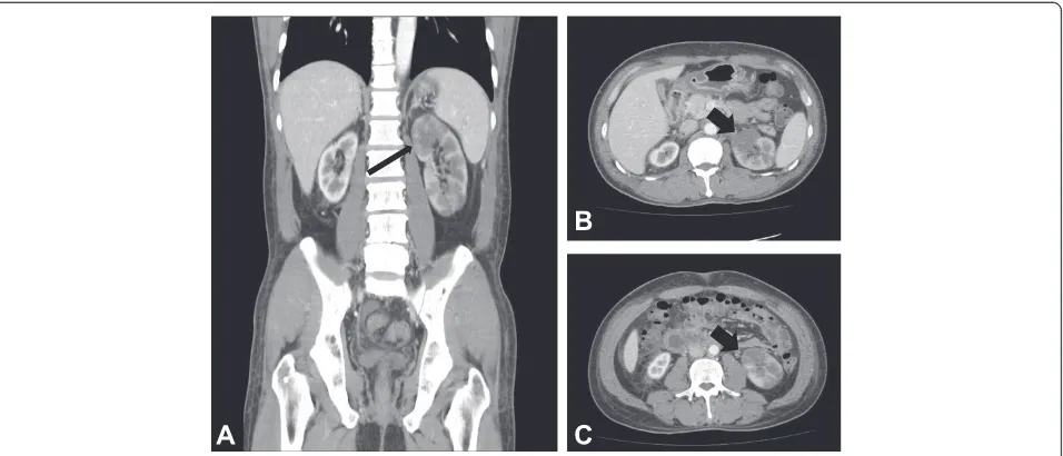
Contrast-enhanced abdominal computed tomography (CT) demonstrated left kidney enlargement (9.3cm) with a multifocal infarcted area and a shrunken right kidney (7.6cm). Neither renal artery was visualized (Fig. 2).
Abdominal and lower extremity angiography was per-formed to examine his abdominal aorta and lower extrem-ity arteries. The vascular status in both lower limbs and the viscera had worsened. His superior mesenteric artery, inferior mesenteric artery, both renal arteries, left com-mon iliac artery, and left superficial femoral artery were not visualized, and the arteries below both his knees were occluded. Collateral vessels were well developed in his lower extremities. During examination, a stent was inserted into his left common iliac artery (Fig. 3). Upper extremity angiographic CT showed no abnormal findings. His ankle-brachial index was 0.82 on the right and 0.61 on the left.
[image:2.595.57.545.91.255.2]medications and an increase in body weight, his pain resolved.
Discussion
Many kinds of diseases have been identified as causes of renal infarction, the most common of which are cardio-genic thromboembolism and atheromatous disease. Other less frequent causes include trauma, hypercoagulable state, cocaine abuse, and neoplastic and renal vascular
disease [3]. Even though TAO commonly affects the small- and medium-sized arteries of the upper and lower extremities, a few cases of kidney infarction and throm-bosis of visceral vessels have been reported [4–9].
In some of these cases, visceral damage is more likely to be a result of atherosclerosis or other diseases, so a broad differential diagnosis is important. The present patient had symptoms typical of TAO. Clinical criteria for the diagnosis of TAO include a cigarette smoking history, Fig. 2Contrast-enhanced abdominal computed tomography.aCoronal and (b, c) transverse scans showed left kidney enlargement with a multifocal infarcted area (arrows). Neither renal artery was traced from the proximal part on computed tomography
Fig. 3Abdominal and lower extremity angiography (2014).aRenal angiography could not identify either renal artery due to total occlusion.b Lower extremity angiography showed a chronic total obstruction lesion of the left common iliac artery due to progression of chronic thrombosis. cA stent was deployed at the site of occlusion of the left common iliac artery (arrow).dFlow was recovered.eObstruction of the left superficial femoral artery and abnormal corkscrew collateral blood supply from the left deep femoral artery was similar to that seen in 2004 (arrows).fThe left tibioperoneal trunk was occluded (arrow), and blood flow below the knee was supplied by collateral vessels
[image:3.595.61.539.89.294.2] [image:3.595.57.539.463.676.2]onset before age 50, infrapopliteal arterial occlusive dis-ease, either upper limb involvement or phlebitis migrans, and the absence of atherosclerotic risk factors other than cigarette smoking [10]. The present patient had intermit-tent claudication and an ischemic ulcer beginning at 42 years of age. He had no atherosclerotic risk factors except for tobacco abuse. He denied any drug ingestion other than an antihypertensive agent (nifedipine) and beraprost. Although hypertension was diagnosed 1 year prior, it seemed likely that this was a result of renal artery occlu-sion caused by TAO. No serologic data suggested collagen disease or anticoagulation disorder. An electrocardiogram did not show atrial fibrillation that could cause distal embolization. An angiogram of his lower limb showed abrupt occlusion and a tree root pattern, findings typical of TAO. Although histological examination of arterial tis-sue was not possible in the present study, aortography demonstrated no atheromatous plaques in the remaining portion of the aorta. So thrombosis in his visceral arteries
including both renal arteries, associated with TAO pro-gression was diagnosed.
[image:4.595.57.541.88.265.2]We comprehensively reviewed the English literature reporting renal involvement in TAO in either abstract or full text form [4–9]. The clinical characteristics of six patients with TAO who showed renal artery involvement with TAO, including the present case, are summarized in Table 1. Among the seven cases, one was excluded be-cause it was not an English report [7]. The mean age was 40.3 years, and all patients were male. TAO was diagnosed in five of six cases before renal artery involvement, except one in which the medical history could not be found in the literature. The average duration from initial diagnosis of TAO to involvement of the renal arteries was 11 years (range 7–15 years). Severe hypertension was the most common symptom, followed by flank pain caused by renal infarction. In five cases, visceral arteries including the de-scending aorta were involved. None of the patients stopped smoking tobacco products after they were diagnosed with Fig. 4Contrast-enhanced abdominal CT and abdominal aorta CT angiography. Contrast-enhanced abdominal CT demonstrated colitis of the (a) hepatic flexure and (b) transverse colon, most likely due to ischemic colitis.cAbdominal aorta CT angiography showed total occlusion of both renal arteries (white arrows). Superior and inferior mesenteric arteries cannot be seen because the arteries were occluded from their origins
Table 1Reported cases of renal artery involvement of thromboangiitis obliterans
Case Age/ sex
Diagnosed TAO before (duration)
Symptom Affected visceral artery Treatment Reference
(reported year)
1 34/
M
Yes Severe hypertension Right renal artery Right
nephrectomy
Malisoff and Macht (1951) [4]
2 30/
M
Yes (15 years) Diffuse back and muscle pain
Descending aorta, celiac axis, iliac artery, femoral artery, coronary artery, left renal artery
Fleshet al. (1977) [5]
3 42/
M
Yes (12 years) Severe hypertension Left renal artery, aorta below the level of renal artery
Antihypertensive medication
Gomiet al. (1978) [6]
4 51/
M
Yes Severe hypertension,
respiratory distress
Both renal arteries, descending aorta, celiac trunk, superior mesenteric artery
Hepatorenal artery bypass
Stillaertet al. (2003) [8]
5 37/
M
Yes (7 years) Right flank pain, weakness, fever
Intrarenal branches of the right renal artery Conservative care
Goktaset al. (2006) [9]
6 52/
M
Yes (10 years) Left flank pain, anuria, weakness
Descending aorta, both renal arteries, superior mesenteric artery, common iliac artery
Hemodialysis This case
[image:4.595.55.540.564.726.2]TAO, highlighting the importance of tobacco smoking ces-sation to prevent the progression of TAO.
In the presented case, the patient complained of post-prandial abdominal pain after starting peritoneal dialysis. Intestinal TAO has been rarely reported. Kobayashiet al. [11] reported a case of TAO with intestinal ischemia and reviewed the literature. They summarized 26 cases of vis-ceral TAO including their case. The mean age of patients was 39.1, and all but two patients were male. The predom-inant symptom was abdominal pain, and 20 of 26 patients underwent digestive organ resection. The perioperative mortality rate was 30%, and only three patients underwent conservative treatment. The present patient experienced improvement in postprandial abdominal pain after avoid-ing dehydration and switchavoid-ing from peritoneal dialysis to hemodialysis. It is known that hemodialysis is more sus-ceptible to intestinal ischemia than peritoneal dialysis be-cause of its more unstable hemodynamics [12]. The patient in this case underwent peritoneal dialysis for this reason; however, even though he was not dehydrated and his blood pressure was not low while receiving peritoneal dialysis, he experienced severe abdominal pain that got worse after starting peritoneal dialysis. His pain was relieved after stopping peritoneal dialysis. We presumed that hyperglycemia of the peritoneal cavity induced several changes, including leukostasis, vasoconstriction, and a pro-inflammatory state that caused aggravation of intes-tinal hypoxia [13].
Conclusions
Although visceral involvement, including renal and intes-tinal arteries, is rare in TAO, once an internal organ is affected, the disease becomes life-threatening and usually cannot be cured. When treating a patient with TAO, we have to carefully observe unusual symptoms such as an-uria, flank pain, uncontrolled hypertension, and abdom-inal pain and strongly encourage cessation of tobacco smoking.
Consent
Written informed consent was obtained from the patient for the publication of this case report and any accompany-ing images. A copy of the written consent is available for review by the Editor-in-Chief of this journal.
Abbreviations
CAPD:Continuous ambulatory peritoneal dialysis; CT: Computed
tomography; TAO: Thromboangiitis obliterans; WBC: White blood cell count.
Competing interests
The authors declare that they have no competing interests.
Authors’contributions
HJY, DIK, KHL and SHY were the physicians who treated the patient in this report. SJL performed the radiology studies. The manuscript was prepared by HJY, WMH, SRY, and SHY. All authors participated in discussions about the manuscript and approved the final version.
Acknowledgments
This study was supported by Konyang University Research Fund of 20.
Author details
1Division of Nephrology, Department of Internal Medicine, Konyang University College of Medicine, 158 Gwanjeo-dong-ro, Seo-gu, Daejeon 302-718, South Korea.2Department of Radiology, Konyang University College of Medicine, Daejeon, South Korea.3Konyang University Myunggok Medical Research Institute, Daejeon, South Korea.
Received: 24 March 2015 Accepted: 20 July 2015
References
1. Buerger L. Thromboangiitis obliterans: a study of the vascular lesions leading to presenile spontaneous gangrene. Am J Med Sci. 1908;136:567–80.
2. Vijayakumar A, Tiwari R, Kumar PV. Thromboangiitis obliterans (Buerger’s disease)–current practices. Int J Inflam. 2013;2013:1–9.
3. Huang CC, Chen WL, Chen JH, Wu YL, Shiao CJ. Clinical characteristics of renal infarction in an Asian population. Ann Acad Med Singapore. 2008;37:416–20.
4. Malisoff S, Macht MB. Thromboangitic occlusion of the renal artery with resultant hypertension. J Urol. 1951;65:371–9.
5. Flesh LH, Kihm RH, Ciccio SS. Radionuclide imaging of aortic involvement in Buerger’s disease: case report. J Nucl Med. 1977;18:125–7.
6. Gomi T, Ikeda T, Yuhara M. Renovascular hypertension due to Buerger’s disease. Jpn Heart J. 1978;19:308–14.
7. Keller F, Gotzen R. A rare case: thromboangiitis obliterans in renal artery stenosis. Med Klin Prax. 1982;77:58–62.
8. Stillaert P, Louagie Y, Donckier J. Emergency hepato-renal artery bypass using a PTFE graft. Acta Chir Belg. 2003;103:524–7.
9. Goktas S, Bedir S, Bozlar U, Ilica AT, Seckin B. Intrarenal arterial stenosis in a patient with thromboangiitis obliterans. Int J Urol. 2006;13:1243–4. 10. Dimmick SJ, Goh AC, Cauzza E, Steinbach LS, Baumgartner I, Stauffer E, et al.
Imaging appearances of Buerger’s disease complications in the upper and lower limbs. Clin Radiol. 2012;67:1207–11.
11. Kobayashi M, Kurose K, Kobata T, Hida K, Sakamoto S, Matsubara J. Ischemic intestinal involvement in a patient with Buerger disease: case report and literature review. J Vasc Surg. 2003;38:170–4.
12. Zier M, Hupp T, Wiesel M, Rambausek M, Ritz E. Non-occlusive intestinal ischemia as a complication of hemodialysis treatment. Dtsch Med Wochenschr. 1993;118:1020–4.
13. Arden GB, Sivaprasad S. Hypoxia and oxidative stress in the causation of diabetic retinopathy. Curr Diabetes Rev. 2011;7:291–304.
Submit your next manuscript to BioMed Central and take full advantage of:
• Convenient online submission
• Thorough peer review
• No space constraints or color figure charges
• Immediate publication on acceptance
• Inclusion in PubMed, CAS, Scopus and Google Scholar
• Research which is freely available for redistribution
Submit your manuscript at www.biomedcentral.com/submit